

Routine application of lung ultrasonography in the neonatal intensive care unit
- PMID: 28079811
- PMCID: PMC5266173
- DOI: 10.1097/MD.0000000000005826
Routine application of lung ultrasonography in the neonatal intensive care unit
Abstract
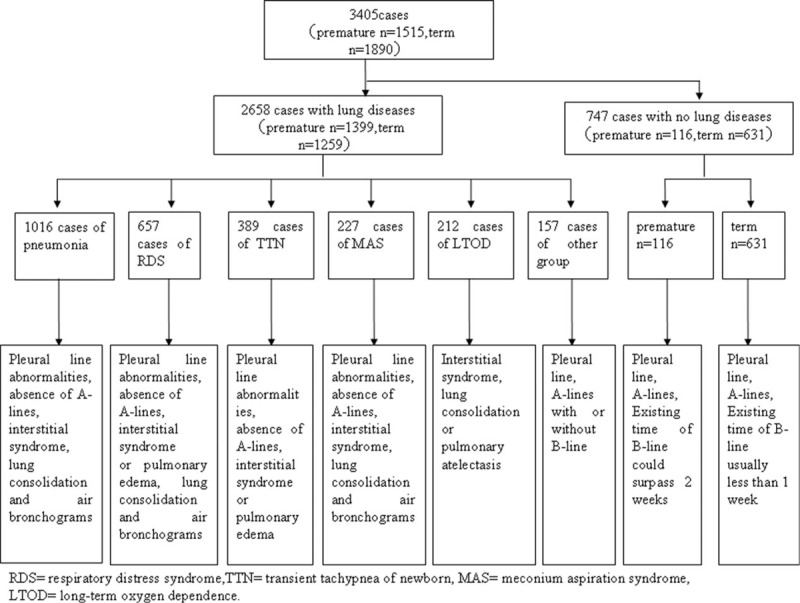
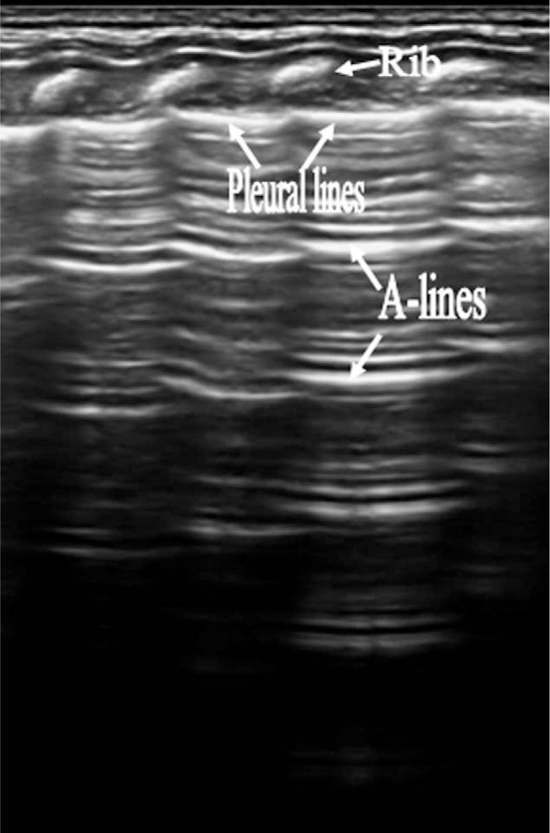
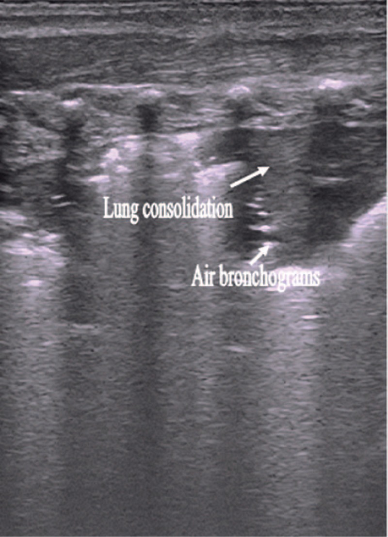
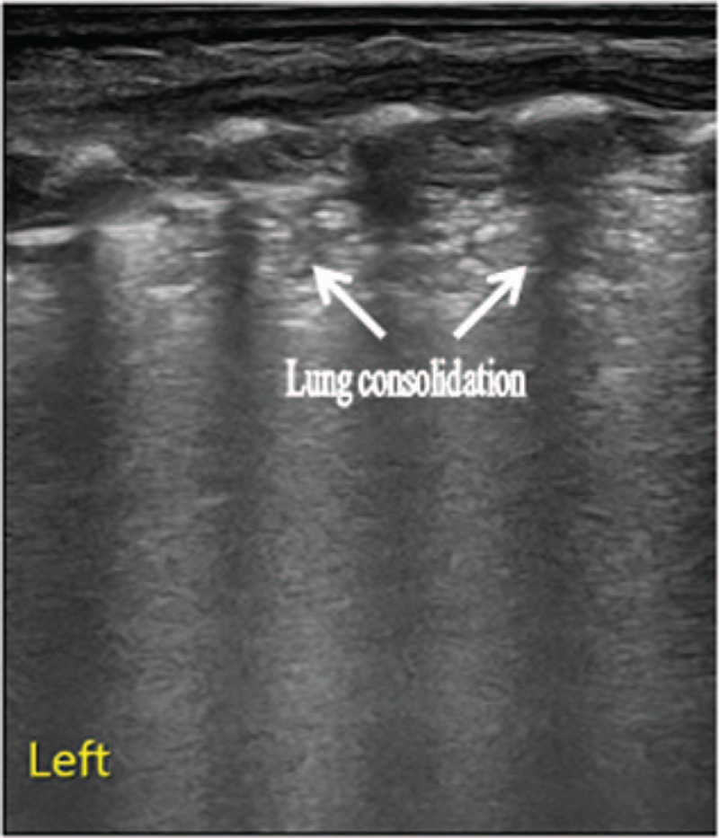
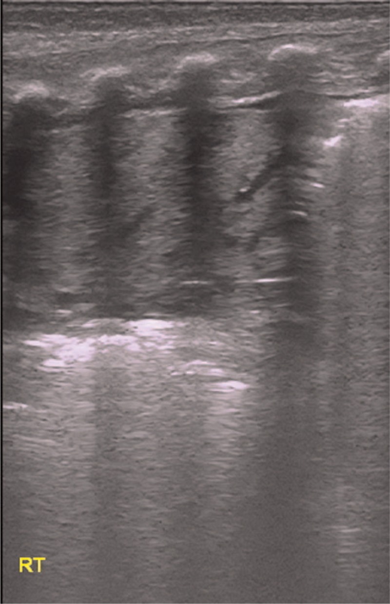
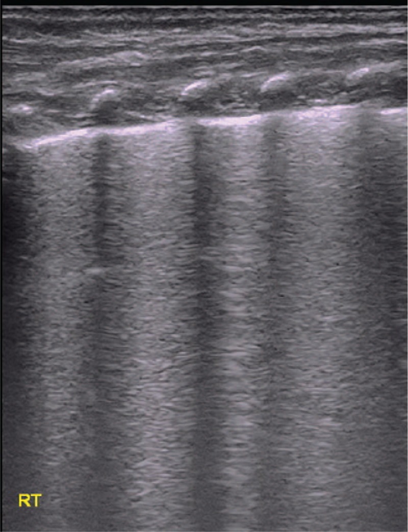
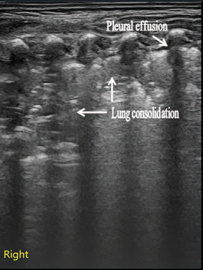
The aim of this study was to study the features of lung ultrasonography (LUS) in lung disease and to evaluate the usefulness of LUS in the neonatal intensive care unit (NICU).All of 3405 neonates included in this study underwent an LUS examination. Diagnoses were based on medical history, clinical manifestation, laboratory examination, and signs on chest radiography (CR) and/or computed tomography (CT). A single expert physician performed all LUS examinations.There were 2658 cases (78.9%) with lung disease and 747 cases (21.9%) without lung disease. The main signs of neonates with lung disease on LUS were as follows: absence of A-lines, pleural-line abnormalities, interstitial syndrome, lung consolidation, air bronchograms, pulmonary edema, and lung pulse. These abnormal signs were reduced or eliminated on LUS as patient conditions improved. There were 81 cases that could not be diagnosed as lung disease by CR but were discovered as pneumonia, respiratory distress syndrome (RDS), or transient tachypnea of newborn (TTN) on LUS. Likewise, 23 cases misdiagnosed as RDS by CR were diagnosed as TTN on LUS. Among 212 cases of long-term oxygen dependence (LTOD) that failed to yield signs of pulmonary edema and lung consolidation on CR, 103 cases showed abnormal signs on LUS. Among 747 cases without lung disease, B-lines of 713 neonates (95.4%) could be found within 3 days after birth, and 256 neonates (34.3%) could be observed from 3 days to 1 week after birth. B-lines of 19 cases could be detected from 1 to 2 weeks after birth. The longest time at which B-lines could still be observed was 19 days after birth.LUS has clinical value for the diagnosis of lung disease and the discrimination of causes of LTOP in premature infants, particularly for the diagnosis and identification of RDS and TTN. Moreover, LUS has additional advantages, including its lack of radiation exposure and its ability to noninvasively monitor treatment progress. Therefore, LUS should be routinely used in the NICU.
Conflict of interest statement
The authors report no conflicts of Interest.
Figures
Similar articles
-
Lung ultrasound in early diagnosis of neonatal transient tachypnea and its differentiation from other causes of neonatal respiratory distress.J Neonatal Perinatal Med. 2018;11(3):281-287. doi: 10.3233/NPM-181796. J Neonatal Perinatal Med. 2018. PMID: 30040751
-
Diagnosis of neonatal transient tachypnea and its differentiation from respiratory distress syndrome using lung ultrasound.Medicine (Baltimore). 2014 Dec;93(27):e197. doi: 10.1097/MD.0000000000000197. Medicine (Baltimore). 2014. PMID: 25501071 Free PMC article. Clinical Trial.
-
Lung Ultrasonography to Diagnose Transient Tachypnea of the Newborn.Chest. 2016 May;149(5):1269-75. doi: 10.1016/j.chest.2015.12.024. Epub 2016 Jan 11. Chest. 2016. PMID: 26836942
-
Lung ultrasound: its role in neonatology and pediatrics.Early Hum Dev. 2013 Jun;89 Suppl 1:S17-9. doi: 10.1016/S0378-3782(13)70006-9. Early Hum Dev. 2013. PMID: 23809341 Review.
-
Accuracy and Reliability of Lung Ultrasound to Diagnose Transient Tachypnoea of the Newborn: Evidence from a Meta-analysis and Systematic Review.Am J Perinatol. 2022 Jul;39(9):973-979. doi: 10.1055/s-0040-1721134. Epub 2020 Nov 26. Am J Perinatol. 2022. PMID: 33242910
Cited by
-
[Guideline on lung ultrasound to diagnose pulmonary diseases in newborn infants].Zhongguo Dang Dai Er Ke Za Zhi. 2019 Feb;21(2):105-113. doi: 10.7499/j.issn.1008-8830.2019.02.001. Zhongguo Dang Dai Er Ke Za Zhi. 2019. PMID: 30782270 Free PMC article. Chinese. No abstract available.
-
Lung Ultrasound for the Diagnosis of Neonatal Respiratory Distress Syndrome: A Meta-analysis.Ultrasound Q. 2020 Jun;36(2):102-110. doi: 10.1097/RUQ.0000000000000490. Ultrasound Q. 2020. PMID: 32511203 Free PMC article.
-
The Role of Lung Ultrasound in the Management of the Critically Ill Neonate-A Narrative Review and Practical Guide.Children (Basel). 2021 Jul 24;8(8):628. doi: 10.3390/children8080628. Children (Basel). 2021. PMID: 34438519 Free PMC article. Review.
-
Lung ultrasound: a new basic, easy, multifunction imaging diagnostic tool in children undergoing pediatric cardiac surgery.J Thorac Dis. 2017 Jun;9(6):1396-1399. doi: 10.21037/jtd.2017.05.71. J Thorac Dis. 2017. PMID: 28740641 Free PMC article. No abstract available.
-
Ultrasound in neonatal lung disease.Quant Imaging Med Surg. 2018 Jun;8(5):535-546. doi: 10.21037/qims.2018.06.01. Quant Imaging Med Surg. 2018. PMID: 30050788 Free PMC article. Review.
References
-
- Wardlaw T, Salama P, Johansson EW, et al. Pneumonia: the leading killer of children. Lancet 2006;368:1048–50. - PubMed
-
- Elia F, Verhovez A, Molino P, et al. Lung ultrasound in the reexpansion of pulmonary atelectasis. Intern Emerg Med 2011;6:461–3. - PubMed
-
- Reissig A, Gramegna A, Aliberti S. The role of lung ultrasound in the diagnosis and follow-up of community- acquired pneumonia. Eur J Intern Med 2012;23:391–7. - PubMed
-
- Caiulo VA, Gargani L, Caiulo S, et al. Lung ultrasound characteristics of community-acquired pneumonia in hospitalized children. Pediatr Pulmonol 2013;48:280–7. - PubMed
-
- Cortellaro F, Colombo S, Coen D. Lung ultrasound is an accurate diagnostic tool for the diagnosis of pneumonia in the emergency department. Emerg Med J 2012;29:19–23. - PubMed
Publication types
MeSH terms
LinkOut - more resources
Full Text Sources
Other Literature Sources
Medical
