

Induction of Antibodies Directed Against Branched Core O-Mannosyl Glycopeptides-Selectivity Complimentary to the ConA Lectin
- PMID: 28079948
- PMCID: PMC5548291
- DOI: 10.1002/chem.201605627
Induction of Antibodies Directed Against Branched Core O-Mannosyl Glycopeptides-Selectivity Complimentary to the ConA Lectin
Erratum in
-
Corrigendum: Induction of Antibodies Directed Against Branched Core O-Mannosyl Glycopeptides-Selectivity Complimentary to the ConA Lectin.Chemistry. 2019 Mar 12;25(15):3963. doi: 10.1002/chem.201900093. Epub 2019 Feb 21. Chemistry. 2019. PMID: 30861233 No abstract available.
Abstract
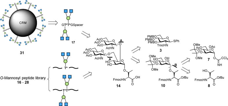
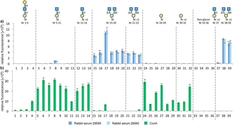
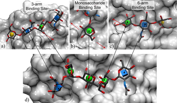
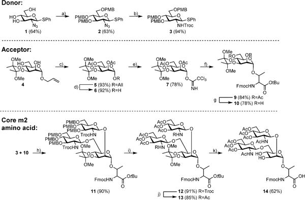
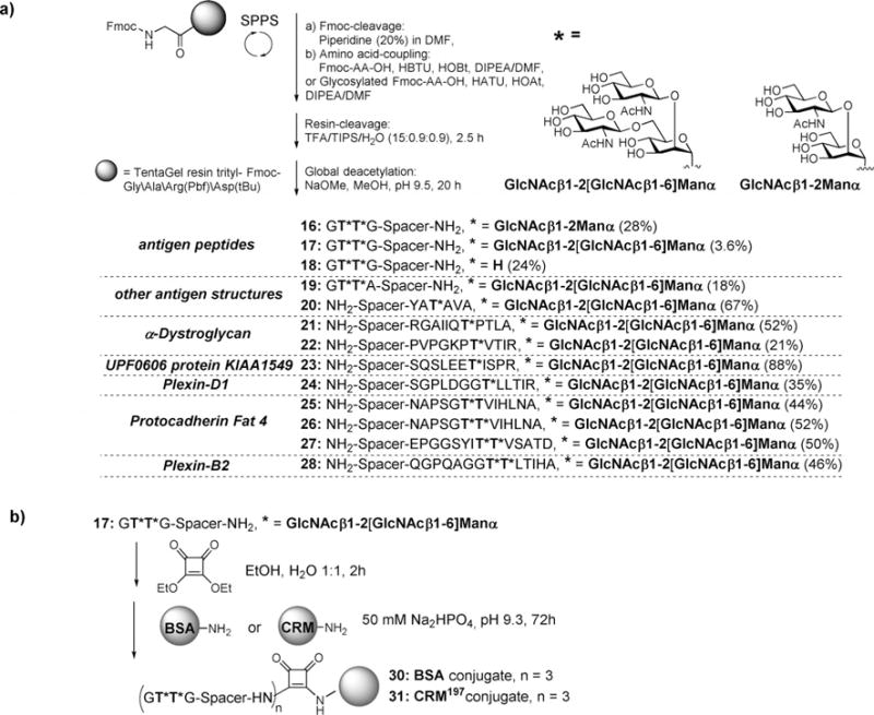
Mammalian protein O-mannosylation, initiated by attachment of α-mannopyranose to Ser or Thr residues, comprise a group of post-translational modifications (PTMs) involved in muscle and brain development. Recent advances in glycoproteomics methodology and the "SimpleCell" strategy have enabled rapid identification of glycoproteins and specific glycosylation sites. Despite the enormous progress made, the biological impact of the mammalian O-mannosyl glycoproteome remains largely unknown to date. Tools are still needed to investigate the structure, role, and abundance of O-mannosyl glycans. Although O-mannosyl branching has been shown to be of relevance in integrin-dependent cell migration, and also plays a role in demyelinating diseases, such as multiple sclerosis, a broader understanding of the biological roles of branched O-mannosyl glycans is lacking in part due to the paucity of detection tools. In this work, a glycopeptide vaccine construct was synthesized and used to generate antibodies against branched O-mannosyl glycans. Glycopeptide microarray screening revealed high selectivity of the induced antibodies for branched glycan core structures presented on different peptide backbones, with no cross-reactivity observed with related linear glycans. For comparison, microarray screening of the mannose-binding lectin concanavalin A (ConA), which is commonly used in glycoproteomics workflows to enrich tryptic O-mannosyl peptides, showed that the ConA lectin did not recognize branched O-mannosyl glycans. The binding preference of ConA for short linear O-mannosyl glycans was rationalized in terms of molecular structure using crystallographic data augmented by molecular modeling. The contrast between the ConA binding specificity and that of the new antibodies indicates a novel role for the antibodies in studies of protein O-mannosylation.
Keywords: antibodies; carbohydrates; glycopeptides; lectins; vaccines.
© 2017 Wiley-VCH Verlag GmbH & Co. KGaA, Weinheim.
Figures
References
-
- Abbott KL, Matthews RT, Pierce M. J Biol Chem. 2008;283:33026–33035. - PMC - PubMed
- Inamori KI, Endo T, Gu J, Matsuo I, Ito Y, Fujii S, Iwasaki H, Narimatsu H, Miyoshi E, Honke K, Taniguchi N. J Biol Chem. 2004;279:2337–2340. - PubMed
- Lee JK, Matthews RT, Lim JM, Swanier K, Wells L, Pierce JM. J Biol Chem. 2012;287:28526–28536. - PMC - PubMed
MeSH terms
Substances
Grants and funding
LinkOut - more resources
Full Text Sources
Other Literature Sources
Miscellaneous
