Clinical Trial
. 2017 Jan 12;12(1):e0169346.
doi: 10.1371/journal.pone.0169346. eCollection 2017.

Human Lacrimal Gland Gene Expression

Affiliations
Clinical Trial

Human Lacrimal Gland Gene Expression

Vinay Kumar Aakalu et al. PLoS One. .

Abstract

Background: The study of human lacrimal gland biology and development is limited. Lacrimal gland tissue is damaged or poorly functional in a number of disease states including dry eye disease. Development of cell based therapies for lacrimal gland diseases requires a better understanding of the gene expression and signaling pathways in lacrimal gland. Differential gene expression analysis between lacrimal gland and other embryologically similar tissues may be helpful in furthering our understanding of lacrimal gland development.

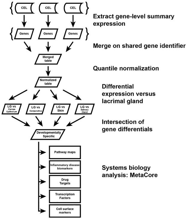
Methods: We performed global gene expression analysis of human lacrimal gland tissue using Affymetrix ® gene expression arrays. Primary data from our laboratory was compared with datasets available in the NLM GEO database for other surface ectodermal tissues including salivary gland, skin, conjunctiva and corneal epithelium.

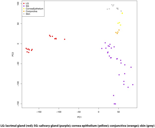
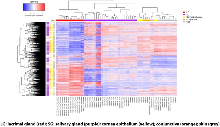
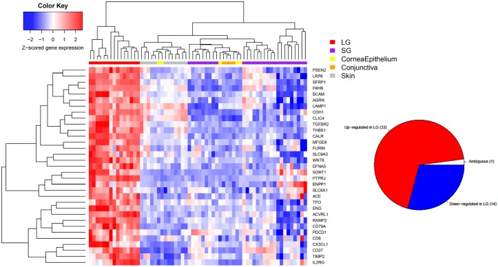
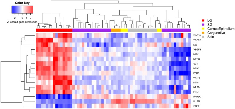
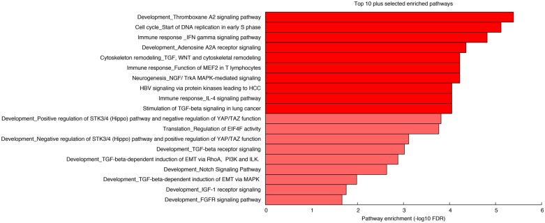
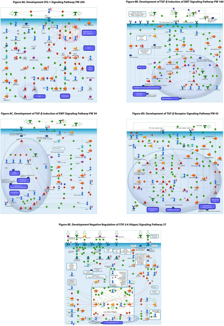
Results: The analysis revealed statistically significant difference in the gene expression of lacrimal gland tissue compared to other ectodermal tissues. The lacrimal gland specific, cell surface secretory protein encoding genes and critical signaling pathways which distinguish lacrimal gland from other ectodermal tissues are described.

Conclusions: Differential gene expression in human lacrimal gland compared with other ectodermal tissue types revealed interesting patterns which may serve as the basis for future studies in directed differentiation among other areas.


Conflict of interest statement

The authors have declared that no competing interests exist.

Figures

Fig 1
Fig 1. Schematic Illustration of Experimental Work-Flow.
Fig 2
Fig 2. Principal Component Analysis of Tissue Transcriptomes.
Fig 3
Fig 3. Heat-Map of Gene Expression Analysis of Lacrimal Gland and Other Ectodermal Tissues.
Heat-Map showing genes with significant differential expression (FDR<0.01 for both t-test and U test) for all genes studied. The heat-map shows all the genes that were significant in at least one of the comparisons, which was 7787 total. The number significant in the LG vs all other comparisons (LG-specific genes) was 1855. The tissue types are color coded: bars along the top indicate which tissue each sample belongs to; bars down the left-hand side indicate which tissues were significantly differentially expressed with respect to LG for each gene.
Fig 4
Fig 4. Heat Map of Top-50 Genes Expressed in Lacrimal Gland Versus Other Ectodermal Tissues.
Heat-map of the most differentially expressed genes in lacrimal gland versus other tissue types (top 50). Only genes that have FDR <0.01 in all pairwise comparisons and prioritized by average fold change either up or down regulated between lacrimal gland and each other tissue. The inset pie chart shows the total number of differentially expressed genes which are up, down or ambiguously differentially expressed.
Fig 5
Fig 5. Heat-Map of Cell Surface Genes in Lacrimal Gland Versus Other Ectodermal Tissues.
Heat-map of the up-regulated lacrimal gland-specific cell surface genes in lacrimal gland versus other tissue types. The inset pie chart shows the total number of cell surface genes which are up, down or ambiguously differentially expressed.
Fig 6
Fig 6. Heat-Map of Genes for Secreted Proteins Expressed in Lacrimal Gland Versus Other Ectodermal Tissues.
Heat-map of differentially expressed genes that encode for secreted proteins in lacrimal gland versus other tissue types.
Fig 7
Fig 7. Differentially Regulated Signaling Pathways in Lacrimal Gland.
Bar plot showing the top 10 differentially regulated developmental signaling pathways in lacrimal gland in addition to 9 (depicted in a lighter shade of red) pathways of potential interest in lacrimal gland development.
Fig 8
Fig 8. (A-E) Selected signaling pathway schematics demonstrating increase (red bar) or decrease (blue) bar and relative amplitude (height of bar) of differentially expression in each gene and their roles in the signaling pathway depicted.
Green arrows indicate positive interactions or activation, red arrows indicate negative interactions or inhibition, while grey arrows indicate an unknown interaction. More details of symbol meanings can be found in the supplemental tables in the Metacore ® quick reference guide.
Fig 9
Fig 9. List of Drugs and Their Action Targeting Differentially Expressed Genes in Lacrimal Gland.
Drugs categorized by differentially expressed targeted genes in lacrimal gland and their action (inhibition blue, activation red).

References

    1. The epidemiology of dry eye disease: report of the Epidemiology Subcommittee of the International Dry Eye WorkShop. Ocul Surf. 2007;5(2):93–107. - PubMed
    1. Tiwari S, Ali MJ, Vemuganti GK. Human lacrimal gland regeneration: Perspectives and review of literature. Saudi J Ophthalmol. 2014;28(1):12–8 10.1016/j.sjopt.2013.09.004 - DOI - PMC - PubMed
    1. Ubels JL, Gipson IK, Spurr-Michaud SJ, Tisdale AS, Van Dyken RE, Hatton MP. Gene expression in human accessory lacrimal glands of Wolfring. Invest Ophthalmol Vis Sci. 2012;53(11):6738–47. 10.1167/iovs.12-10750 - DOI - PMC - PubMed
    1. Shah D, Ali M, Pasha Z, Jaboori AJ, Jassim SH, Jain S, Aakalu VK. Histatin-1 Expression in Human Lacrimal Epithelium. PLoS One. 2016;11(1):e0148018 10.1371/journal.pone.0148018 - DOI - PMC - PubMed
    1. Voronov D, Gromova A, Liu D, Zoukhri D, Medvinsky A, Meech R, et al. Transcription factors Runx1 to 3 are expressed in the lacrimal gland epithelium and are involved in regulation of gland morphogenesis and regeneration. Invest Ophthalmol Vis Sci. 2013;54(5):3115–25. 10.1167/iovs.13-11791 - DOI - PMC - PubMed

Publication types