. 2017 Jan 12;12(1):e0169397.
doi: 10.1371/journal.pone.0169397. eCollection 2017.

Dermal γδ T Cells Do Not Freely Re-Circulate Out of Skin and Produce IL-17 to Promote Neutrophil Infiltration during Primary Contact Hypersensitivity

Affiliations

Dermal γδ T Cells Do Not Freely Re-Circulate Out of Skin and Produce IL-17 to Promote Neutrophil Infiltration during Primary Contact Hypersensitivity

Xiaodong Jiang et al. PLoS One. .

Abstract

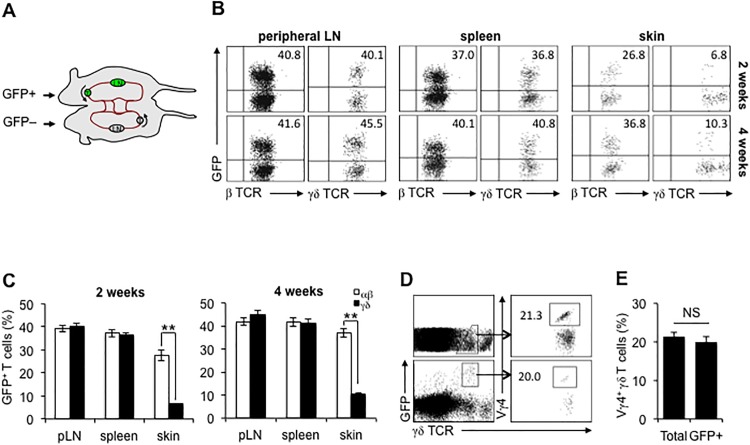
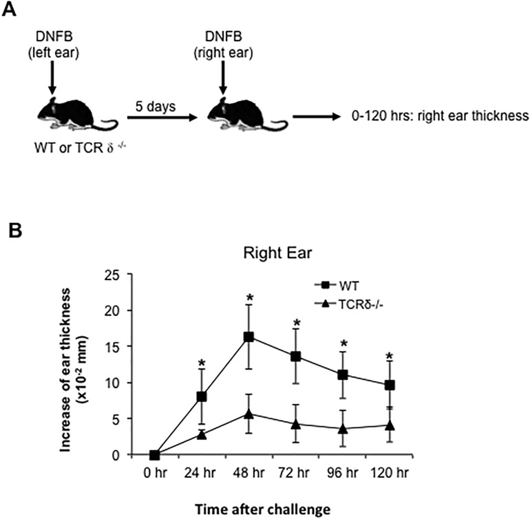
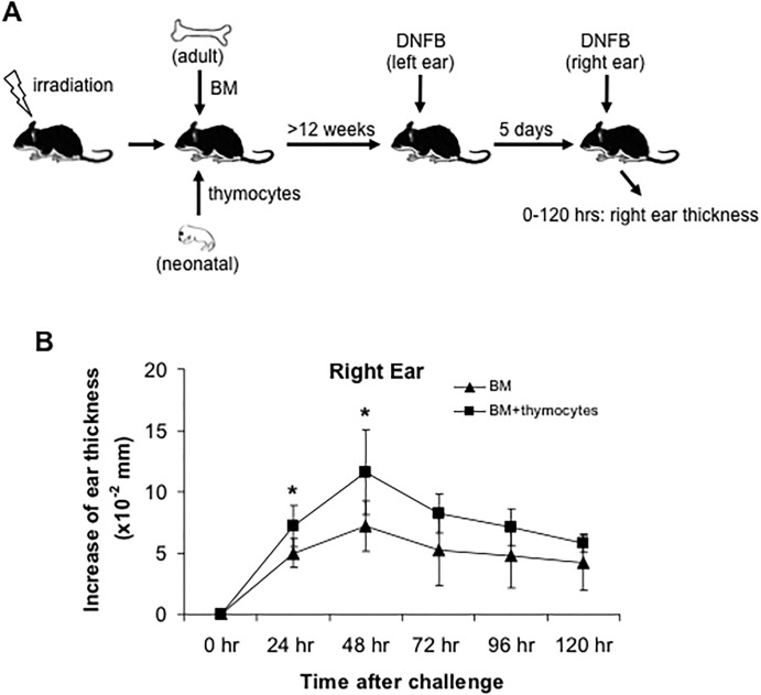
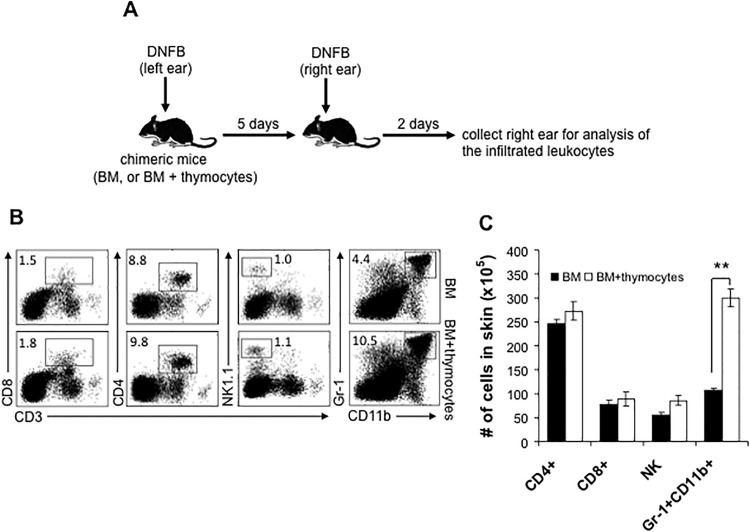
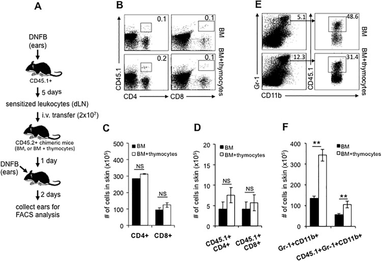
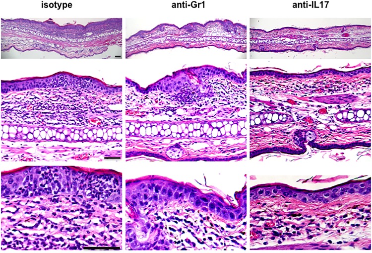
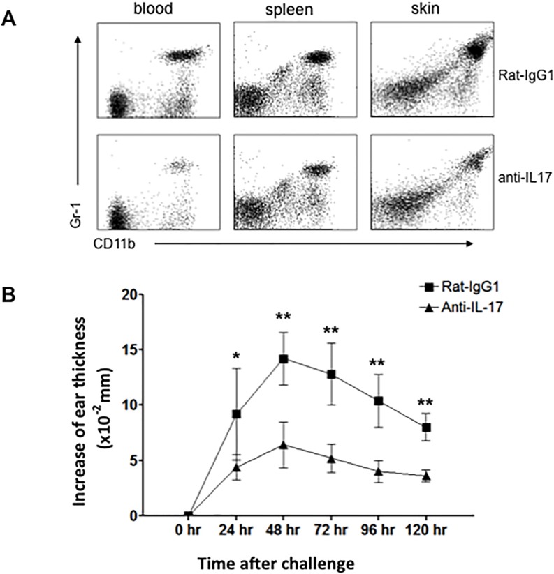
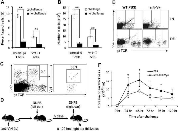
The role of mouse dermal γδ T cells in inflammatory skin disorders and host defense has been studied extensively. It is known that dendritic epidermal T cells (DETC) have a monomorphic γδ T cell receptor (TCR) and reside in murine epidermis from birth. We asked if dermal γδ cells freely re-circulated out of skin, or behaved more like dermal resident memory T cells (TRM) in mice. We found that, unlike epidermal γδ T cells (DETC), dermal γδ cells are not homogeneous with regard to TCR, express the tissue resident T cell markers CD69 and CD103, bear skin homing receptors, and produce IL-17 and IL-22. We created GFP+: GFP- parabiotic mice and found that dermal γδ T cells re-circulate very slowly-more rapidly than authentic αβ TCR TRM, but more slowly than the recently described dermal αβ TCR T migratory memory cells (TMM). Mice lacking the TCR δ gene (δ-/-) had a significant reduction of 2,4-dinitrofluorobenzene (DNFB)-induced contact hypersensitivity (CHS). We created mice deficient in dermal γδ T cells but not DETC, and these mice also showed a markedly reduced CHS response after DNFB challenge. The infiltration of effector T cells during CHS was not reduced in dermal γδ T cell-deficient mice; however, infiltration of Gr-1+CD11b+ neutrophils, as well as ear swelling, was reduced significantly. We next depleted Gr-1+ neutrophils in vivo, and demonstrated that neutrophils are required for ear swelling, the accepted metric for a CHS response. Depletion of IL-17-producing dermal Vγ4+ cells and neutralization of IL-17 in vivo, respectively, also led to a significantly reduced CHS response and diminished neutrophil infiltration. Our findings here suggest that dermal γδ T cells have an intermediate phenotype of T cell residence, and play an important role in primary CHS through producing IL-17 to promote neutrophil infiltration.


Conflict of interest statement

The authors have declared that no competing interests exist.

Figures

Fig 1
Fig 1. The circulation of dermal γδ T cells is limited at steady state.
A, parabiotic mice were created by surgically joining the flank and tying the limbs of age- and sex-matched GFP+ and GFP mice. B and C, at 2 or 4 weeks after surgery, peripheral LNs (pLN), spleen, and skin were collected from GFP parabiotic mice and the frequencies of GFP+ αβ or γδ T cells were analyzed. D and E, at 4 weeks after surgery, the skin of GFP parabiotic mice was harvested and the frequency of Vγ4+ cells in total or GFP+ dermal γδ T cells was determined. The number in the quadrant represents the percentage. **: P<0.01. NS: no significant. FACS results are representative of three independent parabiosis experiments (4 parabiotic pairs for each time point).
Fig 2
Fig 2. CHS response is remarkably reduced in TCR δ-/- mice.
WT or TCR δ-/- mice were sensitized with 0.25% DNFB on left ears for 2 consecutive days. 5 days later, the right ears were challenged with one dose of 0.25% DNFB. The ear thickness was measured at 0–120 hours after challenge (A and B). *: P<0.05. 5 mice for each group were tested. Results are representative of two independent experiments.
Fig 3
Fig 3. CHS response is significantly reduced in dermal γδ T cell-deficient chimeric mice.
A, Generation of chimeric mice. 5–10 x 106 neonatal thymocytes from newborn C57BL/6 mice (day 0–1 after birth) were transferred to half of the irradiated mice. 24 hours later, 5 x 10⁶ bone marrow (BM) cells from adult naïve C57BL/6 mice were intravenously transferred to all irradiated mice. At least 12 weeks later, these BM or BM + thymocytes chimeric mice were sensitized with 0.25% DNFB on left ears at first two days. 5 days later, the right ears were challenged with one dose of 0.25% DNFB. The ear thickness was measured at 0–120 hours after challenge (B). *: P<0.05. 5 mice for each group were tested. Results are representative of two independent experiments.
Fig 4
Fig 4. The recruitment of Gr-1+ CD11b+ neutrophils, not the sensitized CD4+ or CD8+ T cells and NK cells, to DNFB-challenged skin is significantly influenced in dermal γδ T cell-deficient chimeric mice.
A, The DNFB-induced CHS model using chimeric mice was established as described in Fig 3. 48 hours after DNFB challenge, right ears were collected and digested to prepare single cell suspensions. After washed with cold PBS, skin cells were stained with fluorescence-conjugated antibodies for flow cytometry. B, The frequencies of CD4+ or CD8+ T cells, NK cells, and Gr-1+ CD11b+ neutrophils in the challenged skin. C, The numbers of skin infiltrated CD4+ or CD8+ T cells, NK cells, and Gr-1+ CD11b+ neutrophils. **: P<0.01. Results are representative of two independent experiments.
Fig 5
Fig 5. The sensitized CD4+ or CD8+ T cells infiltrate into DNFB-treated skin normally after intravenous transfer while Gr-1+ CD11b+ neutrophils are greatly reduced in dermal γδ T cell-deficient chimeric mice.
A, Wild type CD45.1+ C57BL/6 mice were sensitized with DNFB at first 2 days. 5 days later, dLNs were collected to prepare cell suspension. After washed with PBS, 2 x 10⁷ CD45.1+ sensitized leukocytes were intravenously transferred to CD45.2+ BM or BM+thymocytes chimeric mice. 1 day after transfer, the ears of chimeric mice were treated with 0.25% DNFB. 48 hours later, the treated ears were collected for flow cytometry analysis. B, The frequency of total or donor T cells. C and D are the numbers of total or donor T (CD4+ or CD8+) cells in the skin, respectively. E and F are the frequency and number of total or donor Gr-1+CD11b+ neutrophils in the skin, respectively. NS: no significant. **: P<0.01. Results are representative of two independent experiments.
Fig 6
Fig 6. CHS response is reduced significantly after depletion of Gr-1+ neutrophils in vivo.
Naive C57BL/6 mice were treated with DNFB as Fig 2 to generate DNFB-CHS model. 1 day prior to DNFB challenge, mice were i.p. injected with 100 μg anti-Gr-1 or 100 μg isotype antibodies (rat-IgG2b). Neutrophils (CD11b+ Gr-1+) in blood, spleen, and challenged skin site at 48 hours after DNFB challenge were analyzed by FACS (A). The ear thickness was measured at 0–120 hours after challenge (B). *: P<0.05. 5 mice for each group were tested. Results are representative of three independent experiments.
Fig 7
Fig 7. H&E staining of DNFB-challenged skin.
Naive C57BL/6 mice were treated with DNFB as Fig 2 to generate DNFB-CHS model. 1 day prior to DNFB challenge, mice were i.p. injected with 100 μg anti-Gr-1, 100 μg anti-IL-17, or 100 μg isotype antibodies. 48 hours after DNFB challenge, the challenged skin site was harvested for H&E staining. 5 mice for each group were tested. Results are representative of two independent experiments. The bar represents 100 μm.
Fig 8
Fig 8. CHS response is dramatically reduced after neutralization of IL-17 in vivo.
Naive C57BL/6 mice were treated with DNFB as Fig 2 to generate DNFB-CHS model. 1 day prior to DNFB challenge, mice were i.p. injected with 100 μg anti-IL-17 or 100 μg isotype antibodies (rat-IgG1). Neutrophils (CD11b+ Gr-1+) in blood, spleen, and challenged skin site at 48 hours after DNFB challenge were analyzed by FACS (A). The ear thickness was measured at 0–120 hours after challenge (B). **: P<0.01. 5 mice for each group were tested. Results are representative of three independent experiments.
Fig 9
Fig 9. Dermal γδ T cells rapidly increase in situ and produce large amounts of IL-17 after DNFB challenge.
The DNFB-induced CHS model using wild type C57BL/6 mice was established as described in Fig 2. 24 hours after DNFB challenge, right ears were collected and the frequency and number of dermal γδ T cells were analyzed by flow cytometry (A: of total skin cells; and B: per ear). In parallel, parts of skin cells were incubated in RPMI 1640 media supplemented with 5% FCS, 1% P/S, 0.25% DNFB and Brefeldin A at 37°C. 6–7 hours later, IL-17 production of total dermal γδ T cells and the proportion of IL-17-producing Vγ4+ cells were analyzed by intracellular staining (C). D, Vγ4+ γδ T cells were depleted by i.v. injection of anti-Vγ4 antibody or isotype antibody for 3 consecutive days. DNFB-induced CHS model was created as described in Fig 2. Ear swelling was measured during 0–120 hours after challenge. E: Vγ4+ cells were depleted in LN and skin after 3 consecutive days of anti-Vγ4 injection. F: The increase of ear thickness after DNFB challenge. *: P<0.05. **: P<0.01. 5 mice for each group were tested. Results are representative of three independent experiments.

References

    1. Nanno M, Shiohara T, Yamamoto H, Kawakami K, Ishikawa H. gammadelta T cells: Firefighters or fire boosters in the front lines of inflammatory responses. Immunol Rev. 2007;215:103–113. - PubMed
    1. Macleod AS, Havran WL. Functions of skin-resident γδ T cells. Cell Mol Life Sci. 2011;68:2399–2408. 10.1007/s00018-011-0702-x - DOI - PMC - PubMed
    1. Gray EE, Suzuki K, Cyster JG. Identification of a motile IL-17-producing gammadelta T cell population in the dermis. J Immunol. 2011;186:6091–6095. 10.4049/jimmunol.1100427 - DOI - PMC - PubMed
    1. Sumaria N, Roediger B, Ng LG, Qin J, Pinto R, Cavanagh LL, et al. Cutaneous immunosurveillance by self-renewing dermal gammadelta T cells. J Exp Med. 2011;208:505–518. 10.1084/jem.20101824 - DOI - PMC - PubMed
    1. Cai Y, Shen X, Ding C, Qi C, Li K, Li X, et al. Pivotal role of dermal IL-17-producing γδ T cells in skin inflammation. Immunity. 2011;35:596–610. 10.1016/j.immuni.2011.08.001 - DOI - PMC - PubMed

MeSH terms