. 2017 Jan 12;13(1):e1006470.
doi: 10.1371/journal.pgen.1006470. eCollection 2017 Jan.

Mutations in HYAL2, Encoding Hyaluronidase 2, Cause a Syndrome of Orofacial Clefting and Cor Triatriatum Sinister in Humans and Mice

Affiliations

Mutations in HYAL2, Encoding Hyaluronidase 2, Cause a Syndrome of Orofacial Clefting and Cor Triatriatum Sinister in Humans and Mice

Martina M A Muggenthaler et al. PLoS Genet. .

Abstract

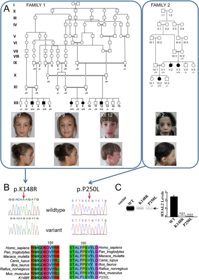
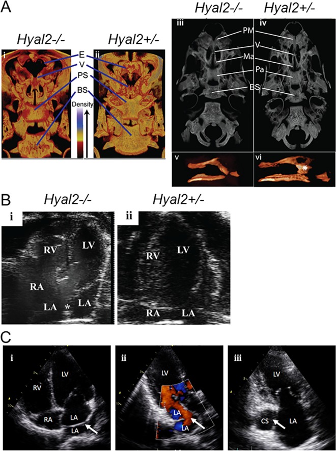
Orofacial clefting is amongst the most common of birth defects, with both genetic and environmental components. Although numerous studies have been undertaken to investigate the complexities of the genetic etiology of this heterogeneous condition, this factor remains incompletely understood. Here, we describe mutations in the HYAL2 gene as a cause of syndromic orofacial clefting. HYAL2, encoding hyaluronidase 2, degrades extracellular hyaluronan, a critical component of the developing heart and palatal shelf matrix. Transfection assays demonstrated that the gene mutations destabilize the molecule, dramatically reducing HYAL2 protein levels. Consistent with the clinical presentation in affected individuals, investigations of Hyal2-/- mice revealed craniofacial abnormalities, including submucosal cleft palate. In addition, cor triatriatum sinister and hearing loss, identified in a proportion of Hyal2-/- mice, were also found as incompletely penetrant features in affected humans. Taken together our findings identify a new genetic cause of orofacial clefting in humans and mice, and define the first molecular cause of human cor triatriatum sinister, illustrating the fundamental importance of HYAL2 and hyaluronan turnover for normal human and mouse development.


Conflict of interest statement

The authors have declared that no competing interests exist.

Figures

Fig 1
Fig 1. Pedigrees, clinical features of individuals homozygous for HYAL2 mutation and identified HYAL2 mutations, expression of wild type (WT), K148R-HYAL2 and P250L-HYAL2.
(A) Pedigree diagrams and facial phenotype of individuals (Amish Family 1: XII:7; XII:9; XII:12 and Saudi Family 2: VI:2) with HYAL2 deficiency. Note the craniofacial similarities including frontal bossing, hypertelorism, widened nasal bridge, flattened broad nasal tip and cupped ears/overfolding of the superior helices. Consent for publication of these photographs was obtained (B) Electropherograms showing the identified c.443A>G & c.749C>T mutations and conservation of protein sequence across species.(C) Expression of wild type (WT), K148R and P250L-HYAL2. Western blots were performed on lysates prepared from MEFs deficient in HYAL2 that were transfected with empty vector, pCMV6-HYAL2, pCMV6-HYAL2K148R, or pCMV6-HYAL2P250L. An arrow indicates HYAL2. HYAL2 levels shown in the graph were quantified by imaging the chemiluminescent signal using a BioRad ChemiDoc. The columns represent the average level (x 106 light units) of HYAL2 ± SEM (n = 4). Significance was determined using the student’s T test. *** indicates p<0.0001.
Fig 2
Fig 2
Micro-CT images of the skulls of Hyal2-/- and Hyal2+/- mice, transthoracic echocardiography images of Amish individuals homozygous for HYAL2 mutation c.443A>G, cardiac analysis in Hyal2-/- and Hyal2+/+ mice (A) 3D Images of Hyal2-/- mice that died at P1, and the control (Hyal2+/-) which was sacrificed at P2, were created using CTvox software. All images were scanned and reconstructed using identical parameters and the dynamic range for the grey scale was held constant (i and ii). Dorsal views from the rostral end of the skull were prepared using a transfer function that colorized the grey scale such that areas of the bone with lower density are red (see density scale in image). (iii and iv). Ventral grey scale views of the skulls without the lower jaw. (v and vi). Space filled images of the vomer. * indicates the vomer head. Similar findings were evident in three additional Hyal2-/- mice found dead at P1. These images are not to scale. E, ethmoid; V, vomer; PS, presphenoid; BS, basisphenoid; PM, premaxilla; MA, maxilla; PA, palantine. (B) (i & ii) Apical 4-chamber image of the heart is shown with right atrium (RA), left atrium (LA), right ventricle (RV), and left ventricle (LV) labelled. Presence of membranous tissue (*) in the LA of Hyal2-/- mouse representing cor triatriatum sinister. (C) (i) Apical four chamber view of Amish individual XII:3: arrow indicates the membrane across the left atrium dividing the pulmonary venous confluence from the body of the left atrium (cor triatriatum sinister). (ii) Apical three chamber view with color. Doppler of individual XII:3: arrow indicates the anterior fenestration in the membrane dividing the left atrium, the laminar flow through the fenestration implies the non-obstructive nature of the membrane. (iii) Apical two chamber view of individual XII:9: arrow indicates the dilated coronary sinus indicative of a persistent left superior vena cava; RV: right ventricle, RA: right atrium, LV: left ventricle, LA: left atrium, CS: coronary sinus.

References

    1. Mossey PA, Little J, Munger RG, Dixon MJ, Shaw WC. Cleft lip and palate. Lancet. 2009;374(9703):1773–85. Epub 2009/09/15. 10.1016/S0140-6736(09)60695-4 - DOI - PubMed
    1. Wyszynski DF, Lewanda AF, Beaty TH. Phenotypic discordance in a family with monozygotic twins and non-syndromic cleft lip and palate. Am J Med Genet. 1996;66(4):468–70. 10.1002/(SICI)1096-8628(19961230)66:4<468::AID-AJMG17>3.0.CO;2-S - DOI - PubMed
    1. Rahimov F, Jugessur A, Murray JC. Genetics of nonsyndromic orofacial clefts. Cleft Palate Craniofac J. 2012;49(1):73–91. PubMed Central PMCID: PMCPMC3437188. 10.1597/10-178 - DOI - PMC - PubMed
    1. Marazita ML. The evolution of human genetic studies of cleft lip and cleft palate. Annu Rev Genomics Hum Genet. 2012;13:263–83. PubMed Central PMCID: PMCPMC3760163. 10.1146/annurev-genom-090711-163729 - DOI - PMC - PubMed
    1. Luijsterburg AJ, Rozendaal AM, Vermeij-Keers C. Classifying common oral clefts: a new approach after descriptive registration. Cleft Palate Craniofac J. 2014;51(4):381–91. 10.1597/12-088 - DOI - PubMed

MeSH terms

LinkOut - more resources