. 2017 Jan 12;12(1):e0169849.
doi: 10.1371/journal.pone.0169849. eCollection 2017.

Human Extravillous Trophoblasts Penetrate Decidual Veins and Lymphatics before Remodeling Spiral Arteries during Early Pregnancy

Affiliations

Human Extravillous Trophoblasts Penetrate Decidual Veins and Lymphatics before Remodeling Spiral Arteries during Early Pregnancy

Nannan He et al. PLoS One. .

Abstract

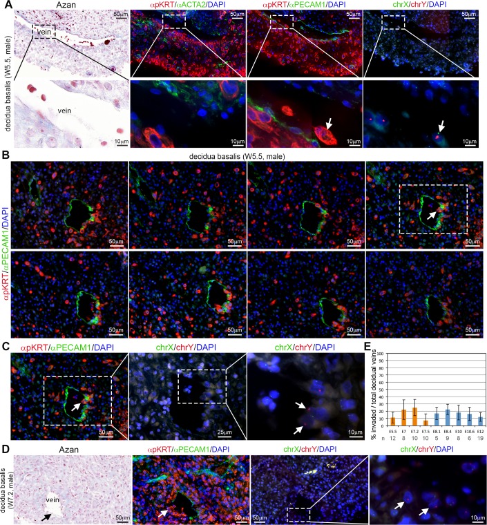
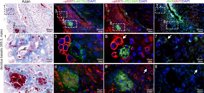
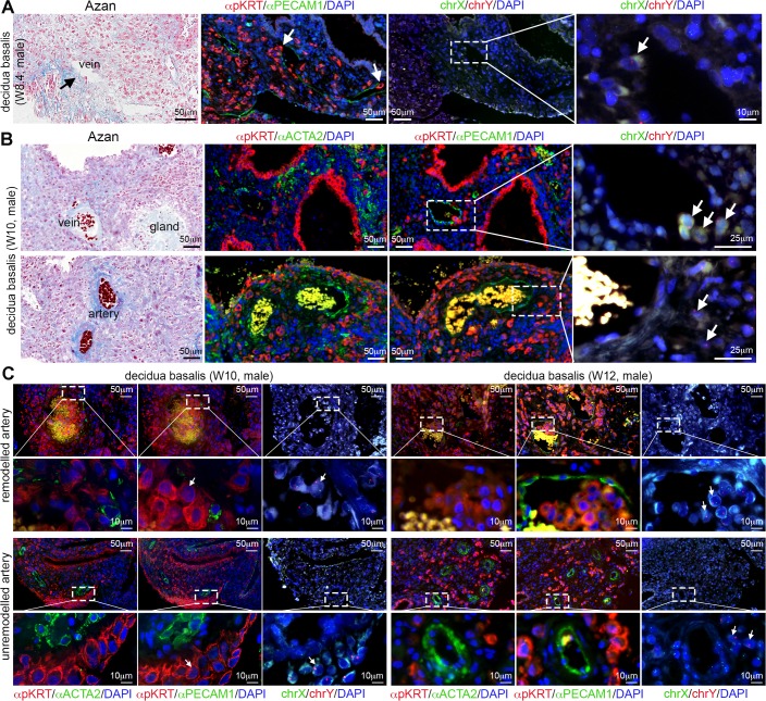
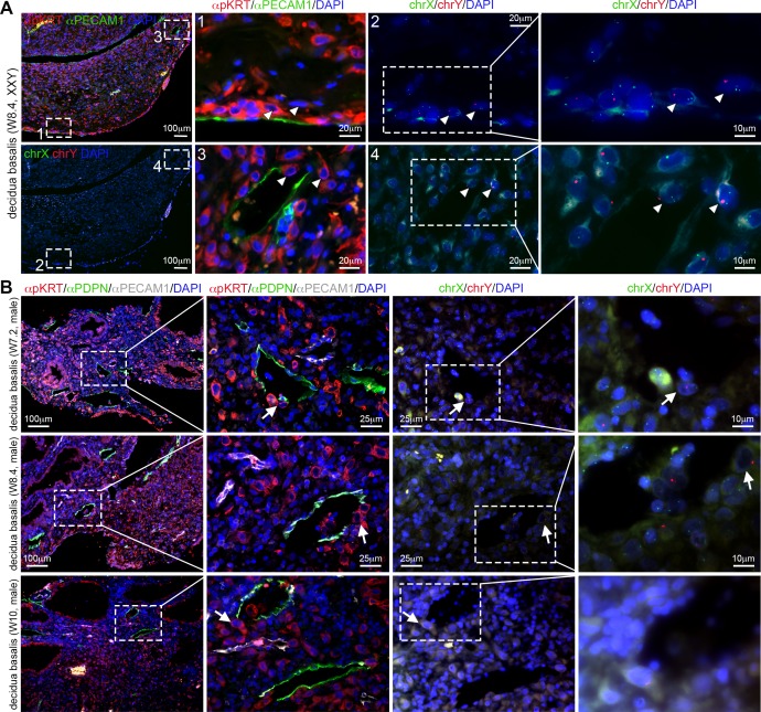
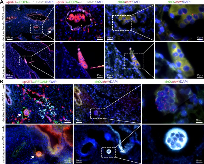
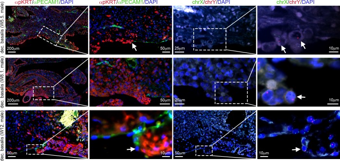
In humans, the defective invasion of the maternal endometrium by fetal extravillous trophoblasts (EVTs) can lead to insufficient perfusion of the placenta, resulting in pregnancy complications that can put both mother and baby at risk. To study the invasion of maternal endometrium between (W)5.5-12 weeks of gestation by EVTs, we combined fluorescence in situ hybridization, immunofluorescence and immunohistochemistry to determine the presence of (male) EVTs in the vasculature of the maternal decidua. We observed that interstitial mononuclear EVTs directly entered decidual veins and lymphatics from W5.5. This invasion of decidual veins and lymphatics occurred long before endovascular EVTs remodelled decidual spiral arteries. This unexpected early entrance of interstitial mononuclear EVTs in the maternal circulation does not seem to contribute to the materno-placental vascular connection directly, but rather to establish (and expand) the materno-fetal interface through an alternative vascular route.


Conflict of interest statement

The authors have declared that no competing interests exist.

Figures

Fig 1
Fig 1. Human EVTs penetrate decidual veins early during first trimester (W5.5-W7.5).
(A) Histological sections of decidua basalis at W5.5 used for Azan staining (left panel), immunostained for pKRT and ACTA2 (left-middle panel) and immunostained for pKRT and PECAM1 (right-middle panel). FISH for chrX/chrY (right) was performed in the pKRT/PECAM1-stained sections. White arrows indicate male EVTs invading veins. The bottom row shows magnifications of the dashed boxes in the top row. (B) Consecutive sections of W5.5 decidua basalis were immunostained for pKRT/PECAM1. (C) FISH for chrX and chrY (right panels) magnifications are shown for the section in dashed box. White arrows point to male EVTs penetrating a decidual vein. (D) Histological sections of decidua basalis at W7.2 used for Azan staining (left panel), immunostained for pKRT and PECAM1 (left-middle panel). FISH for chrX/chrY (right panels) was performed in the pKRT/PECAM1-stained sections. The most right panel shows a magnification of the dashed box. White arrows indicate male EVTs invading veins. (E) Percentage of invaded decidual veins per total veins encountered per histological section (n) between W5.5-W12. Results are sown as mean ± standard deviation. All scale bars are depicted.
Fig 2
Fig 2. Human EVTs in the vicinity of arteries early during first trimester (W5.5).
Histological sections of W5.5 decidua basalis showing decidual arteries. Consecutive sections were used for Azan staining (left panel), immunostained for pKRT and ACTA2 (left-middle panel) and immunostained for pKRT and PECAM1 (right-middle panel). FISH for chrX and chrY (right panel) was performed in the pKRT/PECAM1-stained sections. The (middle and) bottom rows show magnifications of the (numbered) dashed boxes in the top rows. White arrows depict male EVTs. All scale bars are depicted.
Fig 3
Fig 3. Human extravillous trophoblast cells in decidua between W8-W12.
(A) Histological sections of decidua basalis at W8.4 used for Azan staining (left panel), immunostained for pKRT and PECAM1 (left-middle panel). FISH for chrX/chrY (right panels) was performed in the pKRT/PECAM1-stained sections. The most right panel shows a magnification of the dashed box. White arrows indicate male EVTs invading veins. (B) Histological sections of decidua basalis at W10 used for Azan staining (left panel), immunostained for pKRT and ACTA2 (left-middle panel) and immunostained for pKRT and PECAM1 (right-middle panel). FISH for chrX/chrY (right) was performed in the pKRT/PECAM1-stained sections. White arrows indicate male EVTs. Low magnifications are shown in S2 Fig. (C) Histological sections of W10 and W12 decidua basalis showing remodeled and unremodelled arteries. Consecutive sections were immunostained for pKRT and ACTA2 (left panels) and immunostained for pKRT and PECAM1 (middle panels). FISH for chrX and chrY (right panels) was performed in the pKRT/PECAM1-stained sections. The bottom rows show magnifications of the dashed boxes in the top rows. All scale bars are depicted.
Fig 4
Fig 4. Human extravillous trophoblast cells invade decidual veins and lymphatics early during first trimester.
(A) Histological sections of decidua basalis from a (mosaic) Klinefelter syndrome (47,XXY and 46, XY) pregnancy at W8.4. Left panels show low magnifications of same section immunostained for pKRT and PECAM1 (top) and chrX/chrY FISH (bottom). Middle-left and middle-right panels are magnifications of the (numbered) dashed boxes in the left panels. Right panels are magnifications of the dashed boxes in the middle-right panels. White arrowheads point to XXY fetal EVTs. (B) Histological sections of decidua basalis at W7.2, W8.4 and W10 immunostained for pKRT, PDPN and PECAM1 (left panels). FISH for chrX and chrY (right) was performed in the in the pKRT/PDPN/PECAM1-stained sections. White arrows point to EVTs invading the lymphatic vessels. All scale bars are depicted.
Fig 5
Fig 5. EVT aggregates and syncytial knots in the decidua basalis and parietalis.
(A) Histological sections of decidua basalis at W5.5 and W7.5 immunostained for pKRT, PDPN and PECAM1 (lefts panels) showing the presence of mononuclear EVT aggregates (top row) and syncytial knots (bottom row). FISH for chrX and chrY (right panels) was performed in pKRT/PDPN/PECAM1-stained sections. White arrow indicates PDPN-positive lymphatics. (B) Histological sections of W10.6 decidua basalis (top row) and W8.1 decidua parietalis (bottom row) immunostained for pKRT and PECAM1 showing the presence of syncytial knots in veins. FISH for chrX and chrY (right panels) was performed in pKRT/PECAM1-stained sections. All scale bars are depicted.
Fig 6
Fig 6. Allo-epi-endothelial surface of the decidua basalis during first trimester.
Histological sections of decidua basalis containing the basal plate of the decidua basalis immunostained for pKRT and PECAM1 at W5.5, W8.1 and W12. FISH for chrX/chrY was also performed on the pKRT/PECAM1-stained sections. Magnifications of the dashed boxes are shown. White arrows point to EVTs bordering with maternal PECAM1-positive endothelial cells in the basal plate of the maternal decidua basalis. All scale bars are depicted.

References

    1. Brosens JJ, Pijnenborg R, Brosens IA. The myometrial junctional zone spiral arteries in normal and abnormal pregnancies: a review of the literature. American journal of obstetrics and gynecology. 2002;187(5):1416–23. - PubMed
    1. Knofler M. Critical growth factors and signalling pathways controlling human trophoblast invasion. The International journal of developmental biology. 2010;54(2–3):269–80. PubMed Central PMCID: PMC2974212. 10.1387/ijdb.082769mk - DOI - PMC - PubMed
    1. Lyall F, Robson SC, Bulmer JN. Spiral artery remodeling and trophoblast invasion in preeclampsia and fetal growth restriction: relationship to clinical outcome. Hypertension. 2013;62(6):1046–54. 10.1161/HYPERTENSIONAHA.113.01892 - DOI - PubMed
    1. Harris LK. Review: Trophoblast-vascular cell interactions in early pregnancy: how to remodel a vessel. Placenta. 2010;31 Suppl:S93–8. - PubMed
    1. Byrne S, Challis E, Williams JL, Pringle JH, Hennessy JM, Ockleford CD. A mosaic cell layer in human pregnancy. Placenta. 2010;31(5):373–9. 10.1016/j.placenta.2010.02.005 - DOI - PubMed