. 2017 Apr;124(4):496-504.
doi: 10.1016/j.ophtha.2016.11.039. Epub 2017 Jan 9.

Capturing the Moment of Fusion Loss in Intermittent Exotropia

Affiliations

Capturing the Moment of Fusion Loss in Intermittent Exotropia

John R Economides et al. Ophthalmology. 2017 Apr.

Abstract

Purpose: To characterize eye movements made by patients with intermittent exotropia when fusion loss occurs spontaneously and to compare them with those induced by covering 1 eye and with strategies used to recover fusion.

Design: Prospective study of a patient cohort referred to our laboratory.

Participants: Thirteen patients with typical findings of intermittent exotropia who experienced frequent spontaneous loss of fusion.

Methods: The position of each eye was recorded with a video eye tracker under infrared illumination while fixating on a small central near target.

Main outcome measures: Eye position and peak velocity measured during spontaneous loss of fusion, shutter-induced loss of fusion, and recovery of fusion.

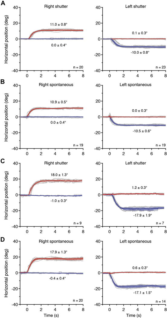
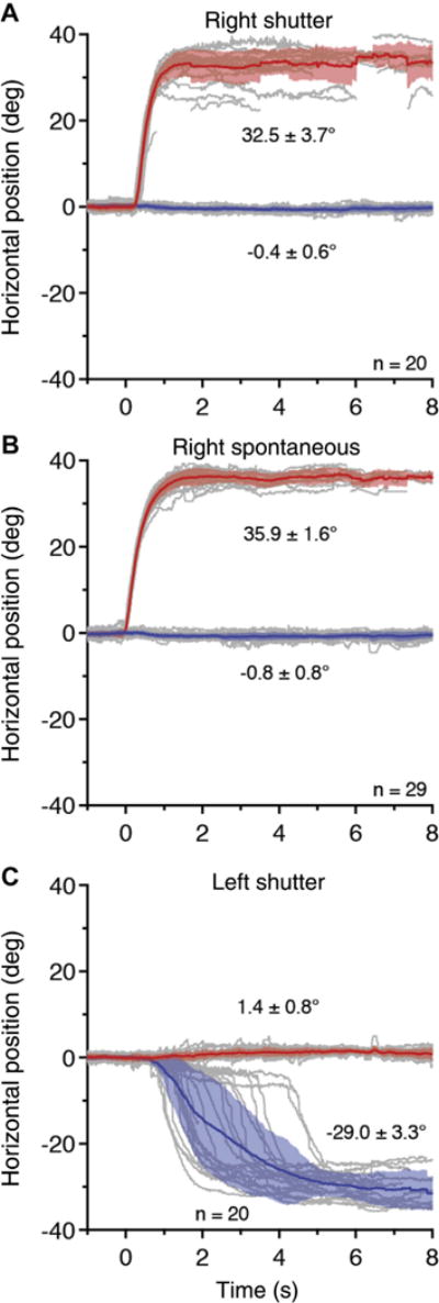
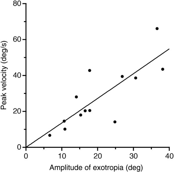
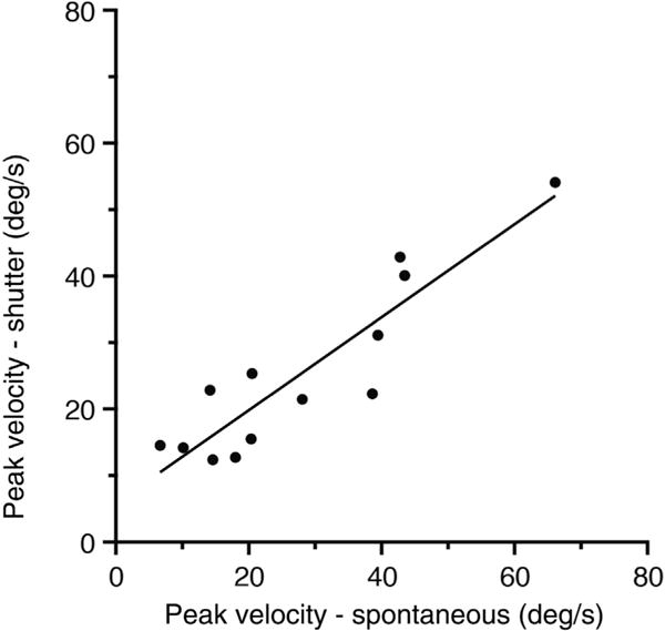
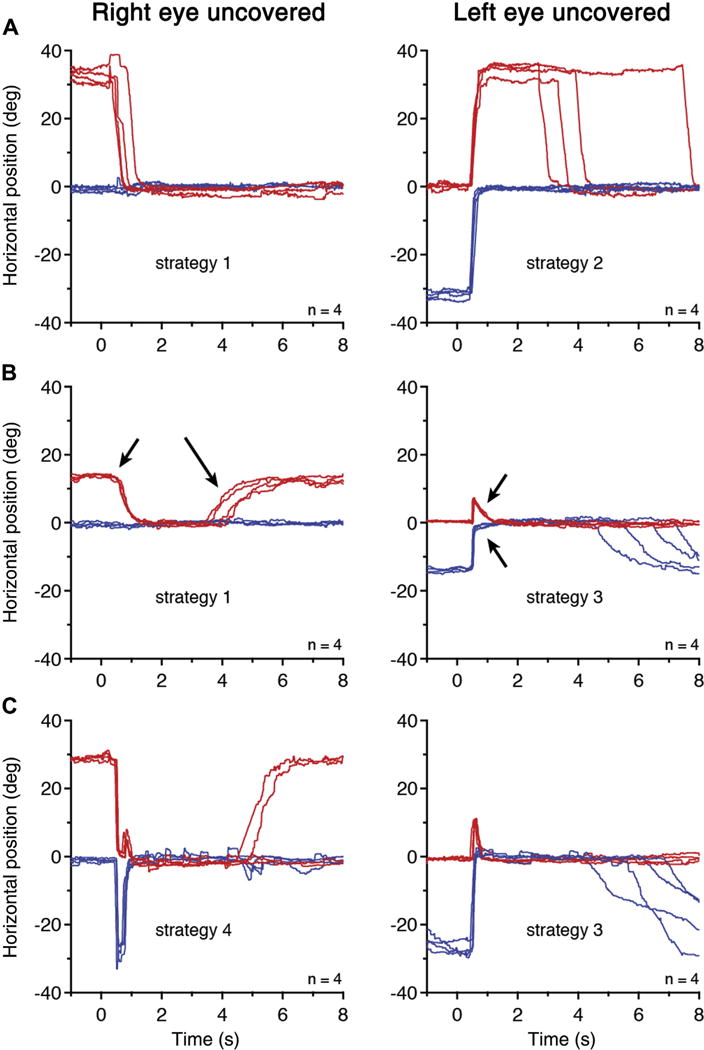
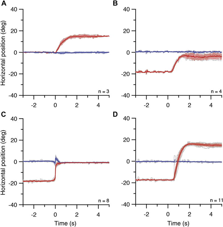
Results: In 10 of 13 subjects, the eye movement made after spontaneous loss of fusion was indistinguishable from that induced by covering 1 eye. It reached 90% of full amplitude in a mean of 1.75 seconds. Peak velocity of the deviating eye's movement was highly correlated for spontaneous and shutter-induced events. Peak velocity was also proportional to exotropia amplitude. Recovery of fusion was more rapid than loss of fusion, and often was accompanied by interjection of a disconjugate saccade.

Conclusions: Loss of fusion in intermittent exotropia is not influenced by visual feedback. Excessive divergence tone may be responsible, but breakdown of alignment occurs via a unique, pathological type of eye movement that differs from a normal, physiological divergence eye movement.


Figures

Figure 1
Figure 1
Cover-induced exotropia is equal to spontaneous exotropia. A, Recordings from a 26-year-old woman (refraction: plano both eyes [OU]) during fixation on a 0.5-degree target at 57 cm. The mean position (solid line) and standard deviation (shading) are shown for the right eye (red) and left eye (blue). At t ¼ 0 seconds, a shutter occluded either the right eye or the left eye, inducing an exodeviation. n = number of events. Positive and negative values for horizontal deviation denote right and left gaze, respectively. B, Spontaneous exotropia occurring intermittently between cover-induced episodes has an amplitude nearly equal to shutter-induced exotropia, with a difference of 0.1 degree for right exotropia and 0.4 degree for left exotropia. The shapes of the mean position traces and the variability in individual position traces are also similar. C: Recordings from a 36-year-old woman (refraction: −1.00 OU), showing a larger exotropia. D: Interspersed episodes of spontaneous fusion loss are similar to those from shutter occlusion, with an amplitude difference of 0.7 degrees for right exotropia and 1.4 degrees for left exotropia.
Figure 2
Figure 2
Cover-induced exotropia different from spontaneous exotropia. Recordings from a 35-year-old man (refraction: −1.25 +0.75 right eye; −1.25 left eye), showing that A, right exotropia induced by occlusion was smaller and more variable than B, spontaneous right exotropia. C, Covering the dominant left eye produced a highly variable outward eye movement; spontaneous left exotropia never occurred (Supplementary Video 1, available at www.aaojournal.org).
Figure 3
Figure 3
Deviation amplitude is correlated with peak velocity of the deviating eye’s movement during onset of spontaneous exotropia.
Figure 4
Figure 4
Peak velocity of the deviating eye’s movement is similar for spontaneous exotropia compared with shutter-induced exotropia.
Figure 5
Figure 5
Four strategies for ocular realignment. A, A 35-year-old man with left eye dominance. Strategy 1: After the nondominant right eye was uncovered at t = 0 seconds,it made a vergence-like adducting movement. Strategy 2: After the dominant left eye was uncovered, an alternating saccade was made, followed at variable intervals by a vergence-like movement of the right eye. Blinks have been removed (Supplementary Video 1, available at www.aaojournal.org). B, A 31-year-old man with left eye dominance. After the right eye was uncovered, strategy 1 was employed. Convergence (small arrow) was faster than divergence (large arrow), which occurred with subsequent episodes of fusion loss. Strategy 3: After the left eye was uncovered, a disconjugate saccade (much larger in the adducting, left eye) was made, followed by vergence movements (arrows). The vergence movement was larger in the nondominant right eye. C, An 11-year-old girl with right eye dominance. Strategy 4: After the dominant eye was uncovered, an alternating saccade was made to bring it onto the target (first stage of strategy 2). This was followed by a disconjugate saccade and asymmetrical vergence movement (strategy 3). After the left eye was uncovered, this child used strategy 3 to recover fusion.
Figure 6
Figure 6
Exotropia at near and distance. The eye trackers were calibrated at distance; 0 degrees refers to fixation at 305 cm. A, Task 1: Spontaneous episodes of right exotropia (amplitude 15.0 degrees, peak velocity 20.1 degrees/second) in a 46-year-old man during fixation at 305 cm. B, Task 2: Exotropia (amplitude 14.2 degrees, peak velocity 24.5 degrees/second) after shutter occlusion of the right eye, while fixating on a crosshair at ~21 cm from the left eye along its line of sight with a distant laser spot. The intent was to mimic the eye movement in (A), by moving the right eye from −15.0 degrees to 0 degrees. The crosshair may have been placed a few centimeters closer than intended, resulting in a movement of the right eye from −18.1 degrees to −3.9 degrees. C, Task 3: Gaze shift along left eye’s line of sight from a crosshair nominally at 21 cm to a laser spot at 305 cm resulted in an outward movement of the right eye (amplitude 18.1 degrees, peak velocity 142.2 degrees/second) that combined divergence with a disconjugate saccade. Replication of the right eye’s movement in (B) was intended, but was foiled by the occurrence of a saccade. D, Task 4: Same task as (C), except that when the distant laser spot appeared, a shutter simultaneously covered the right eye. The right eye moved outward (amplitude 33.0 degrees, peak velocity 43.4 degrees/second) from −17.4 degrees to 15.3 degrees, to assume the same final position as in (A).

Comment in

References

    1. Hatt SR, Mohney BG, Leske DA, Holmes JM. Variability of control in intermittent exotropia. Ophthalmology. 2008;115(2):371–376. - PMC - PubMed
    1. Chia A, Roy L, Seenyen L. Comitant horizontal strabismus: an Asian perspective. Br J Ophthalmol. 2007;91(10):1337–1340. - PMC - PubMed
    1. Kushner BJ, Morton GV. Distance/near differences in intermittent exotropia. Arch Ophthalmol. 1998;116(4):478–486. - PubMed
    1. Buck D, Powell C, Cumberland P, et al. Presenting features and early management of childhood intermittent exotropia in the UK: inception cohort study. Br J Ophthalmol. 2009;93(12):1620–1624. - PubMed
    1. Nusz KJ, Mohney BG, Diehl NN. The course of intermittent exotropia in a population-based cohort. Ophthalmology. 2006;113(7):1154–1158. - PubMed

Publication types

LinkOut - more resources