Review
. 2017 Mar;38(3):154-167.
doi: 10.1016/j.it.2016.12.002. Epub 2017 Jan 9.

Negative Regulation of Type 2 Immunity

Affiliations
Review

Negative Regulation of Type 2 Immunity

Dimitri A de Kouchkovsky et al. Trends Immunol. 2017 Mar.

Abstract

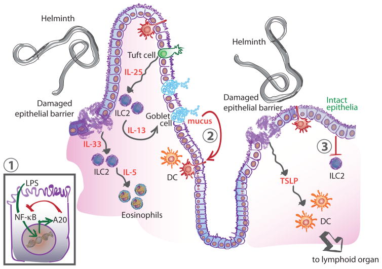
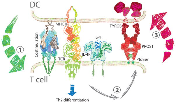
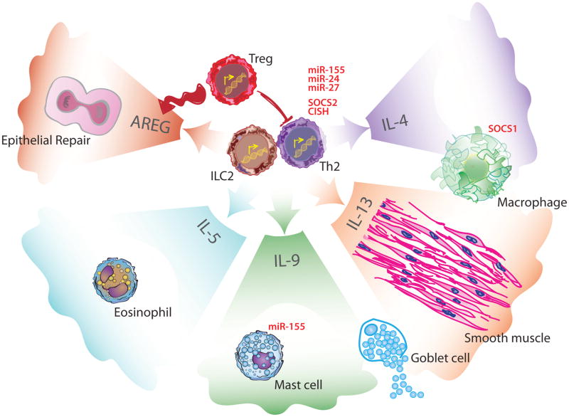
Type 2 immunity encompasses the mechanisms through which the immune system responds to helminths and an array of environmental substances such as allergens. In the developing world, billions of individuals are chronically infected with endemic parasitic helminths. In comparison, in the industrialized world, millions of individuals suffer from dysregulated type 2 immunity, referred to clinically as atopic diseases including asthma, allergic rhinitis, and atopic dermatitis. Thus, type 2 immunity must be carefully regulated to mount protective host responses yet avoid inappropriate activation and immunopathology. In this review, we describe the key players and connections at play in type 2 responses and focus on the emerging mechanisms involved in the negative regulation of type 2 immunity.


Figures

Figure 1
Figure 1. Regulation of Type 2 Immunity at Barrier Tissues
Immune stimulus (e.g. helminth) leads to tissue damage and secretion of the cytokines IL-33 and TSLP. TSLP activates DCs to adopt a Th2-promoting phenotype and migrate to lymphoid organs to activate T cells. IL-33 signals on ILC2 leading to the recruitment of eosinophils through the secretion of IL-5. ILC2s also sense the increased production of IL-25 by tuft cells and secrete IL-13 which feeds back on goblet cells leading to increased mucus production. Negative regulatory mechanisms at the barrier include inhibition of NF-κB signaling in epithelial cells through the action of A20 (1). During homeostasis, mucus limits inflammation by inducing a regulatory pathway in DCs in the context of antigen uptake (2). Similarly, an intact epithelium provides immunoregulatory signals that limit ILC2 activation (3). Abbreviations: DC, dendritic cell; IL, interleukin; ILC2, type 2 innate lymphoid cell; TSLP, thymic stromal lymphopoietin.
Figure 2
Figure 2. Regulation at the Immune Synapse
CD4+ T cells are activated and begin to differentiate in response to three signals (1). DCs present peptide antigen on the MHC class II and provide costimulation as well, to ensure productive signaling. Cytokines direct differentiation to specific CD4+ T helper subsets; here IL-4 leads to Th2 differentiation. In addition, the combination of TCR and IL-4 signaling in T cells leads to the upregulation of PROS1 on their surface (2). In turn, PROS1 interacts with TYRO3 on the DC and engages a negative feedback mechanism that limits DC activation (3). Abbreviations: DC, dendritic cell; IL-4, interleukin 4; IL-4R, interleukin 4 receptor; MCH II, class II major histocompatibility complex; PROS1, Protein S; PtdSer, phosphatidylserine; TCR, T cell receptor; Th2, type 2 helper T cell.
Figure 3
Figure 3. Regulation of Type 2 Effector Mechanisms
During the effector phase of type 2 immunity, ILC2s and Th2 cells coordinate the response through the production of effector cytokines. In Th2 cells, the SOCS proteins CISH and SOCS2 have been shown to limit production of these effector cytokines. microRNAs such as miR-24, miR-27, and miR-155 also regulate Th2 cytokine production. IL-4 is primarily produced by Th2 and, in combination with IL-13, polarizes macrophages. This polarization is negatively regulated by SOCS1. ILC2s produce AREG to trigger epithelial growth and repair. Th2 and ILC2s are both capable of producing IL-5, IL-9 and IL-13. IL-5 contributes to eosinophils recruitment to the site of injury. Mast cells are recruited by IL-9 and miR-155 limits their degranulation. IL-9 and IL-13 act on goblets cells and lead to proliferation and increased mucus secretion. Finally, IL-13 signaling on smooth muscle cells leads to increased contractility. At the tissue level, these functions act together to promote expulsion of the inciting agent. Tregs are able to suppress Th2 cell activity and also contribute to tissue repair through AREG production. Abbreviations: AREG, amphiregulin; CISH, cytokine-inducible SH2-containing protein; IL, interleukin; ILC2, type 2 innate lymphoid cell; miR, microRNA; SOCS, suppressor of cytokine signaling; Th2, type 2 helper T cell; Treg, regulatory T cell.

References

    1. Takai T. TSLP expression: cellular sources, triggers, and regulatory mechanisms. Allergol Int. 2012;61(1):3–17. - PubMed
    1. Ito T, et al. TSLP-activated dendritic cells induce an inflammatory T helper type 2 cell response through OX40 ligand. J Exp Med. 2005;202(9):1213–23. - PMC - PubMed
    1. Soumelis V, et al. Human epithelial cells trigger dendritic cell mediated allergic inflammation by producing TSLP. Nat Immunol. 2002;3(7):673–80. - PubMed
    1. Rajavelu P, et al. Airway epithelial SPDEF integrates goblet cell differentiation and pulmonary Th2 inflammation. J Clin Invest. 2015;125(5):2021–31. - PMC - PubMed
    1. Gerbe F, et al. Intestinal epithelial tuft cells initiate type 2 mucosal immunity to helminth parasites. Nature. 2016;529(7585):226–30. - PMC - PubMed

LinkOut - more resources