. 2017 Feb 20;474(5):781-795.
doi: 10.1042/BCJ20160748.

Cullin neddylation may allosterically tune polyubiquitin chain length and topology

Affiliations

Cullin neddylation may allosterically tune polyubiquitin chain length and topology

Melis Onel et al. Biochem J. .

Abstract

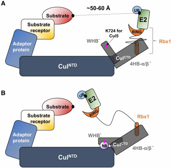
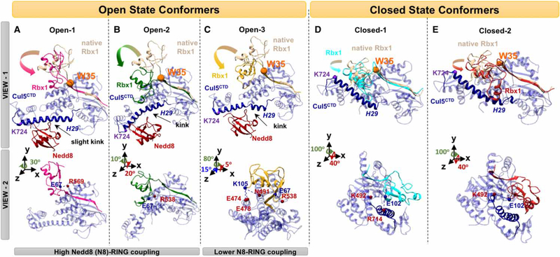
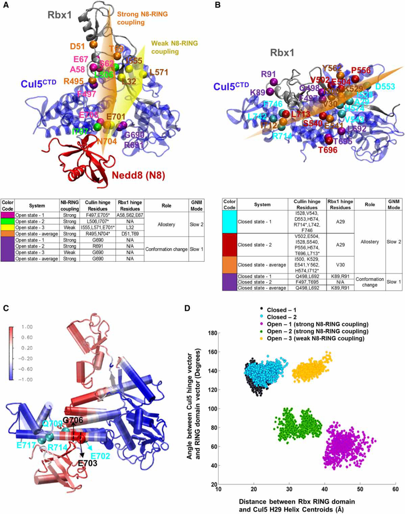
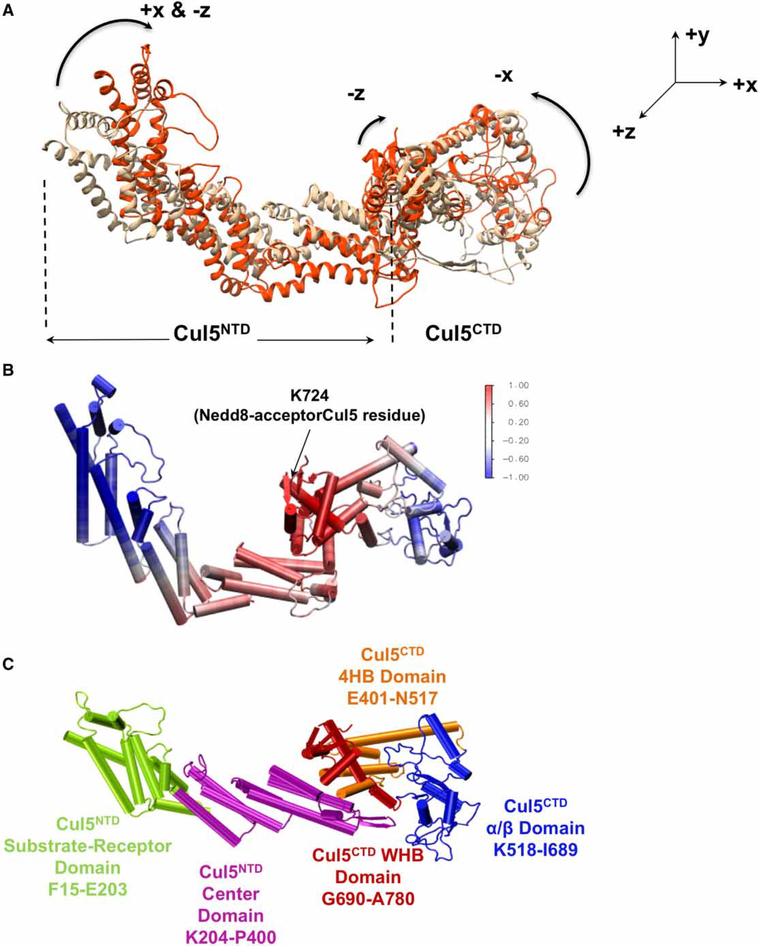
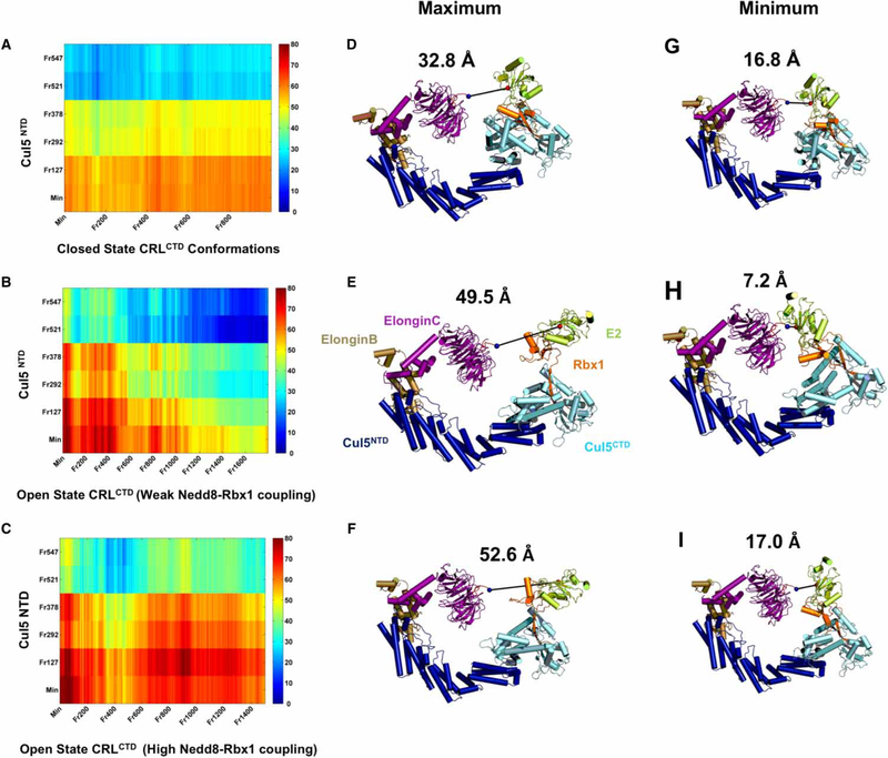
Conjugation of Nedd8 (neddylation) to Cullins (Cul) in Cul-RING E3 ligases (CRLs) stimulates ubiquitination and polyubiquitination of protein substrates. CRL is made up of two Cul-flanked arms: one consists of the substrate-binding and adaptor proteins and the other consists of E2 and Ring-box protein (Rbx). Polyubiquitin chain length and topology determine the substrate fate. Here, we ask how polyubiquitin chains are accommodated in the limited space available between the two arms and what determines the polyubiquitin linkage topology. We focus on Cul5 and Rbx1 in three states: before Cul5 neddylation (closed state), after neddylation (open state), and after deneddylation, exploiting molecular dynamics simulations and the Gaussian Network Model. We observe that regulation of substrate ubiquitination and polyubiquitination takes place through Rbx1 rotations, which are controlled by Nedd8-Rbx1 allosteric communication. Allosteric propagation proceeds from Nedd8 via Cul5 dynamic hinges and hydrogen bonds between the C-terminal domain of Cul5 (Cul5CTD) and Rbx1 (Cul5CTD residues R538/R569 and Rbx1 residue E67, or Cul5CTD E474/E478/N491 and Rbx1 K105). Importantly, at each ubiquitination step (homogeneous or heterogeneous, linear or branched), the polyubiquitin linkages fit into the distances between the two arms, and these match the inherent CRL conformational tendencies. Hinge sites may constitute drug targets.

Keywords: Nedd8; allosteric regulation; polyubiquitination; ubiquitin ligases.


Figures

Figure 1.
Figure 1.. Schematic representation of closed (A) and open (B) state Cul-RING Ligase (CRL)s.
A sketch to present the effect of Cul neddylation in CRLs. Cul have two domains: CulNTD (dark blue) and CulCTD (dark gray) where CulCTD consists of WHB domain, 4HB, and α/β-domains (shown with asterisks). Adaptor and SR proteins bind to CulNTD. Rbx1 (orange) binds to CulCTD. Ubiquitin-carrier E2 enzyme (green) binds to Rbx1 for substrate ubiquitination. Nedd8 (N8, shown in purple) conjugates to the CulCTD H29 of the WHB domain (K724 on Cul5, shown with a pink star) and leads to significant conformational change in CulCTD. Crystal structures report the distance between E2 Cys and the substrate in the closed state as ∼50–60 Å [26,57].
Figure 2.
Figure 2.. RBX RING domain rotations.
(A–C) Nedd8 (red) conjugated CRLCTD (open state) conformers that have three distinct Rbx1 RING domain rotations, and (D and E) unmodified CRLCTD (closed state) conformers are shown in two different views. The H29 of Cul (blue) are highlighted by making the rest of Cul elements transparent. Distinct Rbx1 conformations are shown with different colors (pink, green, and yellow for open; cyan and red for closed). Rotations from native — crystal structure — Rbx1 (beige) are highlighted with arrows. The Rbx1 W35 residue shown with an orange sphere has a pivotal role in RING domain rotations after (A–C) and before (D and E) Cul neddylation. Nedd8-binding site on CulCTD, K724, is shown with a purple sphere in (D and E). In view 2, the H-bonds between Cul residues (red spheres) and Rbx1 (blue spheres) stabilizing the Rbx1 RING domain conformation are shown for both states.
Figure 3.
Figure 3.. Hinge sites and coupled Cul regions on CRLCTD provide a dynamic measure for RING domain rotation.
Open-1 and closed-1 CRLCTD conformers are shown in (A and C) and (B), respectively, to illustrate the hinge sites. The open and closed state CRLCTD (Cul5CTD–Rbx1) hinge residues of the slowest mode of motion during Cul neddylation are shown in purple. Hinge residues coordinating the allosteric communication are colored differently for each conformer. Open state-average and closed state-average yield the average conformers for both states. Side tables are provided for guidelines. The Cul5 H29 hinge residues are marked with asterisk in the side tables. Approximate imaginary planes are drawn for illustration of hinge coordination. In (A), the orange and yellow hinge planes represent the hinge coordination for Nedd8–Cul5–Rbx1 allosteric communication when Nedd8–RING coupling is high and weak, respectively. In (B), the orange plane represents the hinge coordination for Cul5–Rbx1 allosteric communication in closed state conformers. (C) Open state CRLCTD conformer is colored according to the Cul5 flexible element showing highest correlation with Nedd8, Glu703–Gly706, to highlight the allosteric communication pathway from Nedd8 through CRLCTD. The normalized correlation values are in the range of [−1,1]. The Cul5 residues that H-bond with Nedd8 are shown in cyan. (D) Dynamic RING domain positions throughout each MD simulation are depicted; x-axis: distance between the RING domain (Cys42–Lys105) centroid and the Nedd8-binding Cul5 WHB domain’s H29 (Trp695–Met725) centroid, y-axis: angle between the state-specific hinge vector and the RING domain vector from Rbx1 Cys42 to the RING domain centroid.
Figure 4.
Figure 4.. Cul coupled dynamics and flexibility.
(A) Contraction motions observed in complete closed state Cul5 MD simulation are shown by aligning the most contracted Cul conformation (Frame 547 — used in Figure 5; orange) on the initial complete Cul5 model (beige). (B) Cul5 conformer is colored according to the Cul5 correlations with Nedd8-acceptor Cul5 residue K724. Color code is done based on the normalized correlation values in the range of [−1,1], where −1 reflects highest negative correlation and 1 shows the highest positive correlations in fluctuation dynamics during the MD simulation. (C) The Cul5 domains (including NTD dynamic domains determined from the network of correlated motions) are labeled.
Figure 5.
Figure 5.. Distance measurements between the CRL arms.
(A–C) show the distance maps for closed (A), and open (B and C) state CRLs. CRLCTD conformations (frames from MD simulations; x-axes) are modeled with the NTD of selected Cul5 conformations (frames from the complete closed state Cul5 MD simulation) (y-axes) to obtain complete CRL models and measure the distance between the substrate tip and active E2 Cys on CRLs. The color bar provides the measured distances in Ångstrom. (D–F) show the CRL models with the most contracted Cul5 conformation (Frame 547) having maximum distance between the arms according to the distance maps shown in (A–C), whereas (G–I) show the minimum. (D and G), (E and H), and (F and I) comparisons highlight the effect of RING domain rotations on the distance reduction between the CRL arms in each CRL functional state.
Figure 6.
Figure 6.. Schematic representation for the role of neddylated CRL dynamics during polyubiquitination.
Flexibility in Cul (A) and diverse Rbx1 conformations due to diverse RING domain rotations with varying level of Nedd8 allosteric control (B) are the key determinants of neddylated CRL dynamics. Heterogeneous (C and D) and homogeneous (E and F) polyubiquitin chains on the substrates held by neddylated CRLs are depicted with different neddylated CRL conformational tendencies (different combinations of Cul flexibility and RING domain rotations). The ubiquitin of the polyubiquitin chain which is going to be ubiquitinated and the ubiquitin on the E2 which is going to be transferred are shown by red in (C–F). It is proposed that neddylated CRL conformational tendencies may adapt/effect different polyubiquitin chain topologies.

Similar articles

Cited by

References

    1. Streich FC Jr and Lima CD (2014) Structural and functional insights to ubiquitin-like protein conjugation. Annu. Rev. Biophys. 43, 357–379 doi:10.1146/annurev-biophys-051013-022958 - DOI - PMC - PubMed
    1. Hershko A (2005) The ubiquitin system for protein degradation and some of its roles in the control of the cell division cycle. Cell Death Differ. 12, 1191–1197 doi:10.1038/sj.cdd.4401702 - DOI - PubMed
    1. Komander D and Rape M (2012) The ubiquitin code. Annu. Rev. Biochem. 81, 203–229 doi:10.1146/annurev-biochem-060310-170328 - DOI - PubMed
    1. Buetow L and Huang DT (2016) Structural insights into the catalysis and regulation of E3 ubiquitin ligases. Nat. Rev. Mol. Cell Biol. 17, 626–642 doi:10.1038/nrm.2016.91 - DOI - PMC - PubMed
    1. Ciechanover A and Stanhill A (2014) The complexity of recognition of ubiquitinated substrates by the 26S proteasome. Biochim. Biophys. Acta, Mol. Cell Res. 1843, 86–96 doi:10.1016/j.bbamcr.2013.07.007 - DOI - PubMed

Publication types

MeSH terms