Case Reports
. 2017 Jun;28(6):1723-1728.
doi: 10.1681/ASN.2016080867. Epub 2017 Jan 12.

Vancomycin-Associated Cast Nephropathy

Affiliations
Case Reports

Vancomycin-Associated Cast Nephropathy

Yosu Luque et al. J Am Soc Nephrol. 2017 Jun.

Abstract

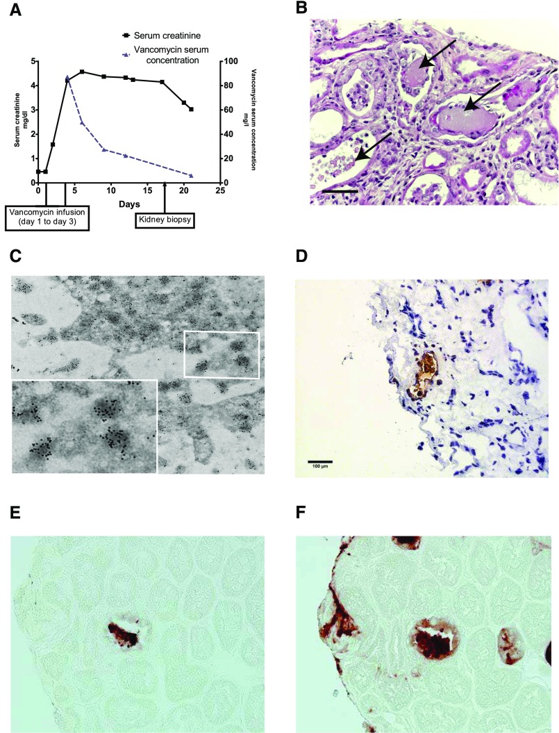
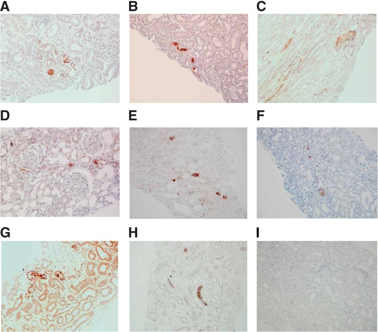
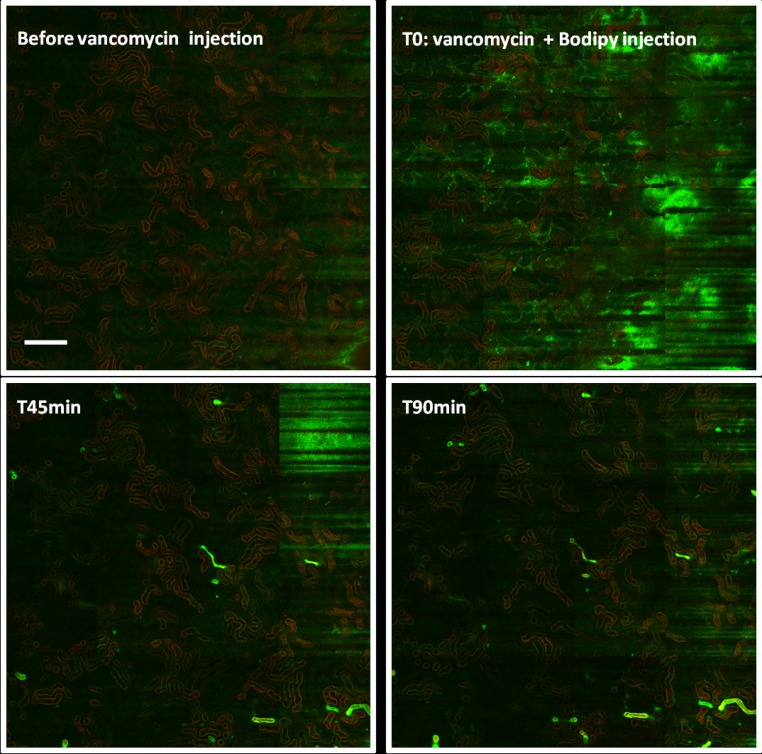
Vancomycin is a widely prescribed antibiotic, but the exact nature of vancomycin-associated nephrotoxicity is unclear, in particular when considering the frequent coadministration of aminoglycosides. We describe here the initial case of a 56-year-old woman with normal renal function developing unexplained ARF without hypovolemia after administration of vancomycin without coadministration of aminoglycosides. Studying the patient's renal biopsy specimen, we ascertained that obstructive tubular casts composed of noncrystal nanospheric vancomycin aggregates entangled with uromodulin explained the vancomycin-associated ARF. We developed in parallel a new immunohistologic staining technique to detect vancomycin in renal tissue and confirmed retrospectively that deleterious vancomycin-associated casts existed in eight additional patients with acute tubular necrosis in the absence of hypovolemia. Concomitant high vancomycin trough plasma levels had been observed in each patient. We also reproduced experimentally the toxic and obstructive nature of vancomycin-associated cast nephropathy in mice, which we detected using different in vivo imaging techniques. In conclusion, the interaction of uromodulin with nanospheric vancomycin aggregates represents a new mode of tubular cast formation, revealing the hitherto unsuspected mechanism of vancomycin-associated renal injury.

Keywords: acute renal failure; acute tubular necrosis; tubular cast; tubular epithelium; vancomycin.


Figures

Figure 1.
Figure 1.
ARF associated with nanospheric vancomycin tubular casts entangled with uromodulin. (A) Graph showing the evolution of the patient’s renal function with vancomycin plasma concentrations over time. (B) Human kidney biopsy (17 days after vancomycin injection) analysis revealed nonspecific granular tubular and proteinaceous cast formation (arrows) and ATN lesions (hematoxylin and eosin staining). Scale bar, 50 μm. (C) Transmission electron microscopy analysis with immunogold labeling detecting vancomycin nanospheres in a tubular cast formation (original, magnification, ×18,500), with detail on nanospheric vancomycin (labeled with 20-nm gold particles). (D) Staining with antivancomycin antibody (frozen section) revealed the specific accumulation of vancomycin in the tubular lumen. Note the absence of vancomycin in the surrounding tissue. Original magnification, ×400. (E) Serial biopsy (3-μm thick) showing one cast colocalizing vancomycin and (F) uromodulin showing the obstructive nature of vancomycin-associated casts. Horseradish peroxidase staining. Original magnification, ×600.
Figure 2.
Figure 2.
Vancomycin detection in a suspected case of vancomycin-associated cast nephropathy (A to H) Vancomycin staining on paraffin-embedded renal biopsy samples from 8 patients with unexplained acute renal failure occurring in the clinical context of high trough vancomycin plasma levels. Patients did not develop circulatory shock. Concomitant high trough vancomycin plasma levels were identified retrospectively from our center's database (1999-2016). Patients' clinical characteristics (A to H) are shown in Table 1. Anti-vancomycin mouse monoclonal antibody (1:1000, Abbot, 6E44-21) has been used on 4 μm dewaxed kidney sections. Nuclei are stained with hematoxylin. A strong staining has been detected in all cases in tubular lumens, with a patchy distribution among the renal cortex. Negative control has been shown in (I). Magnification × 200.
Figure 3.
Figure 3.
Vancomycin-associated cast nephropathy confirmed in mice. (A). Following vancomycin injection (n=4 per group) mice developed acute renal failure (observation made at day 2) that is not aggravated by concomitant Tazocilin injection. *P<0.05. (B) Kidney injuries have been visible as early as two days after vancomycin injection on Masson's trichrome. Acute tubular necrosis is visible with granular material found in the tubular lumen. Scale bar: 50 μm. (C) Lower magnification showing the patchy distribution of vancomycin-associated casts. Scale bar: 50 μm. (D) Intravital confocal microscopy (enlarged field reconstructed) showing the patchy distribution of vancomycin-associated tubular casts (green) magnified by a vancomycin-linked dye (boron-dipyrromethene) at 2 hours following vancomycin injection. Scale bar: 200 μm. Control mice (E) compared to vancomycin-injected mice at day 2 post-vancomycin injection (F) observed with intravital wide-field microscopy. Tubular cast formation magnified by a vancomycin-linked dye (boron-dipyrromethene) is easily visible in (F). *P<.05. Scale bar: 100 μm.
Figure 4.
Figure 4.
In vivo formation of obstructive vancomycin-associated tubular casts in mice. Intravital confocal microscopy analyses (enlarged field reconstructed) show that intratubular cast formation occurs nearly 40 minutes after vancomycin injection. The same kidney cortex area has been observed sequentially at different time points. Tubular casts are not observed before vancomycin injection (upper left panel). When vancomycin and vancomycin-linked green fluorescent dye (boron-dipyrromethene) are injected intravenously, a green fluorescence appears (upper right panel) in capillary vessels, reflecting the intravascular circulation of vancomycin. At 45 minutes (lower left panel), the first vancomycin-associated intratubular casts (green) are now visible, with their number increasing further at 90 minutes (lower right panel). Scale bar, 200 μm.

Comment in

References

    1. Rodvold KA, Blum RA, Fischer JH, Zokufa HZ, Rotschafer JC, Crossley KB, Riff LJ: Vancomycin pharmacokinetics in patients with various degrees of renal function. Antimicrob Agents Chemother 32: 848–852, 1988 - PMC - PubMed
    1. Griffith RS: Introduction to vancomycin. Rev Infect Dis 3[Suppl]: S200–S204, 1981 - PubMed
    1. Farber BF, Moellering RC Jr.: Retrospective study of the toxicity of preparations of vancomycin from 1974 to 1981. Antimicrob Agents Chemother 23: 138–141, 1983 - PMC - PubMed
    1. Levine DP: Vancomycin: A history. Clin Infect Dis 42[Suppl 1]: S5–S12, 2006 - PubMed
    1. Oktem F, Arslan MK, Ozguner F, Candir O, Yilmaz HR, Ciris M, Uz E: In vivo evidences Suggesting the role of oxidative stress in pathogenesis of vancomycin-induced nephrotoxicity: Protection by erdosteine. Toxicology 215: 227–233, 2005 - PubMed

Publication types