. 2017 Jan 13;355(6321):194-197.
doi: 10.1126/science.aal2130.

Assembly of a nucleus-like structure during viral replication in bacteria

Affiliations

Assembly of a nucleus-like structure during viral replication in bacteria

Vorrapon Chaikeeratisak et al. Science. .

Abstract

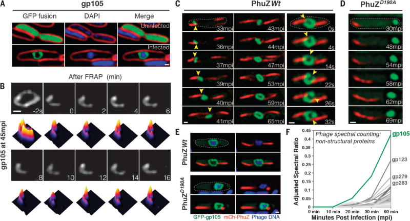
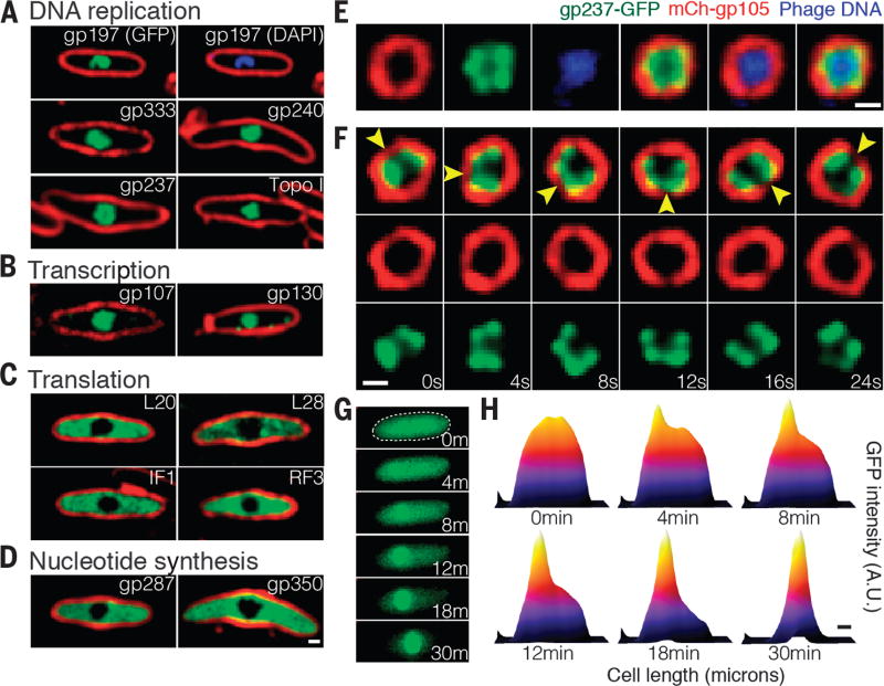
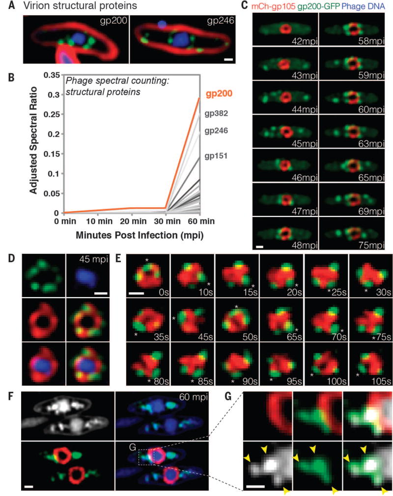
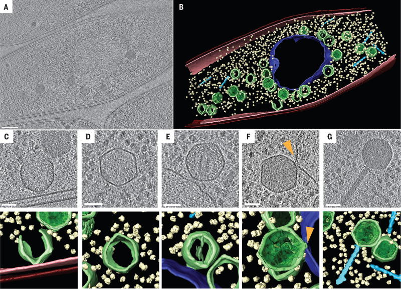
We observed the assembly of a nucleus-like structure in bacteria during viral infection. Using fluorescence microscopy and cryo-electron tomography, we showed that Pseudomonas chlororaphis phage 201φ2-1 assembled a compartment that separated viral DNA from the cytoplasm. The phage compartment was centered by a bipolar tubulin-based spindle, and it segregated phage and bacterial proteins according to function. Proteins involved in DNA replication and transcription localized inside the compartment, whereas proteins involved in translation and nucleotide synthesis localized outside. Later during infection, viral capsids assembled on the cytoplasmic membrane and moved to the surface of the compartment for DNA packaging. Ultimately, viral particles were released from the compartment and the cell lysed. These results demonstrate that phages have evolved a specialized structure to compartmentalize viral replication.


Figures

Fig. 1
Fig. 1. gp105, an early and highly expressed phage protein, forms a shell around viral DNA during phage infection
(A) N-terminal fusion of gp105 to GFP (GFP-gp105; green) is diffuse in uninfected P. chlororaphis cells (top) and assembles a shell that encloses the phage DNA at 45 min postinfection (mpi) with phage 201ϕ2-1 (bottom). Cell membranes were stained with FM 4–64 (red), and DNA was stained with DAPI (blue). (B) Fluorescence recovery after photobleaching (FRAP) of the GFP-gp105 shell at 45 mpi. The bleached area of the shell generated at time 0 min does not recover over the course of 16 min. Heat maps show GFP intensity corresponding to the images above. (C and D) Time-lapse imaging (measured in minutes postinfection or seconds) of GFP-gp105 in the presence of mCherry-tagged (red) (C) wild-type PhuZ and (D) mutant PhuZD190A. (C) As indicated by arrowheads, the shell moves to the midcell and rotates (in this case, clockwise) during phage infection in the presence of wild-type PhuZ. See also movies S1 and S3. (D) In the presence of PhuZD190A, the shell remains at the cell pole throughout the experiment and does not rotate. A lack of rotation is sometimes observed in the presence of wild-type PhuZ for larger gp105 shells such as that shown in (B). Dashed circles indicate the border of the cells. (E) Static images of infected host cells expressing GFP-gp105 together with either wild-type mCherry-PhuZ or mutant mCherry-PhuZD190A at 60 mpi. Phage DNA was stained blue with DAPI. (A to E) Scale bars, 0.5 µm. (F) Protein mass spectrometry analysis of phage-infected cells showing putative nonstructural phage proteins until 60 mpi. The gp105 protein (green line) is the most highly expressed phage protein. (See table S1.)
Fig. 2
Fig. 2. DNA replication and transcription occur inside the phage compartment, whereas translation occurs outside
(A to D) Localization profiling of GFP-tagged phage and host proteins involved in various functions at 60 mpi. All proteins were fused to GFP (green), DNA was stained with DAPI (blue), and membranes were stained with FM 4–64 (red). (A) Proteins predicted to be involved in DNA replication, including the phage proteins gp237 (RecA homolog), gp197 (helicase homolog), gp333 (ligase homolog), gp240 (RNase H homolog), and host DNA topoisomerase I. (B) Two phage-encoded proteins predicted to be involved in transcription, gp107 and gp130 (RNA polymerase β′ subunit homologs). (C) Host proteins involved in translation, including ribosomal proteins L20 and L28, translation initiation factor 1 (IF1), and peptide chain release factor 3 (RF3). (D) Phage proteins predicted to be involved in nucleotide synthesis. including gp287 (thymidylate kinase homolog) and gp350 (thymidylate synthase homolog). (E), mCherry-gp105 (red), the RecA homolog gp237-GFP (green), and DAPI-stained DNA (blue), demonstrating localization of gp237 and DNA inside the gp105 shell. (F) Time-lapse imaging (in seconds) showing rotation of the compartment and its contents. As indicated by arrowheads, the shell and gp237 rotate together (in this cell, in a counterclockwise direction), suggesting that the entire structure and its contents rotate. See also movie S5. (G) gp237 moves from the host cell cytoplasm to the compartment during phage infection in this 30-min time lapse (m, minutes). The dashed circle indicates the border of the cell. (H) Heat map of GFP intensity of gp237 in the host cell as corresponding to the fluorescent micrographs reveals that gp237 moves into and accumulates in the gp105 shell. (See also movie S4.) Scale bars in (D) to (F) and (H), 0.5 µm. A.U., arbitrary units.
Fig. 3
Fig. 3. Capsids migrate to the surface of the phage compartment for DNA encapsidation
(A) Localization of the predicted phage structural proteins gp200 (major capsid protein) and gp246 (internal head protein). The proteins were fused to GFP (green), membranes were stained with FM 4–64 (red), and DNA was stained with DAPI (blue) at 60 mpi. (B) Mass spectrometry results showing spectral counting of predicted phage structural proteins in infected host cells until 60 mpi; gp200 (orange line) is the most abundant structural protein. (C) Time-lapse microscopy of mCherry-gp105 (red) and the predicted capsid protein gp200-GFP (green), showing that gp200-GFP was initially diffuse (at 42 mpi) and assembled foci on the cell periphery (at 43 to 45 mpi) that move to the gp105 shell (at 45 to 46 mpi), remain attached for 12 min, and then are released from the shell (at 59 to 75 mpi). Foci translocate to the gp105 shell within 1 to 2 min. See also movie S6. (D) Static images showing gp200-GFP (green) on the surface of the mCherry-gp105 shell (red), with DNA (blue) inside, at 45 mpi. (E) Time lapse showing that gp200-GFP foci rotate together with mCherry-gp105 throughout this 105-s experiment. The position of a single capsid (asterisk) was tracked for the duration of the time lapse. See movie S7. (F and G) Static images of infected cells at 60 mpi to show colocalization of gp200-GFP (green) and phage DNA (blue or white) outside the mCherry-gp105 shell (red). The region indicated by the dashed box in (F) is magnified in (G) to more clearly show the colocalization of DNA (white) within the gp200 foci (green). Arrowheads indicate the colocalization. Scale bars in (A) and (C) to (G), 0.5 µm.
Fig. 4
Fig. 4. Cryo-electron tomography of the phage compartment during 201ϕ2-1 infection in P. chlororaphis
(A) Slice through a tomogram of a cryo-focused ion beam–thinned phage-infected cell at 60 mpi. Assembled capsids are docked to an apparently contiguous shell during the process of DNA encapsidation, which produces the darker, filled capsids. Scale bar, 200 nm. (B) Segmentation of the tomogram in (A), showing extracted structures, including the shell (purple), capsids (green), cytoplasmic membrane (pink), outer membrane (red), phage tails (light blue), and ribosomes (yellow). (C to G) Tomographic slices (top) and segmentation images (bottom) of (C) an assembling phage capsid, (D) an empty capsid, (E and F) two capsids docked at the compartment with (E) less or (F) more DNA, and (G) an assembled phage. Scale bars in (C) to (G), 50 nm. The arrowhead in (F) indicates a connecting collar between the capsid and the compartment shell.

References

    1. Durzyńska J, Goździcka-Józefiak A. Virol. J. 2015;12:169. - PMC - PubMed
    1. Serwer P, et al. Virology. 2004;329:412–424. - PubMed
    1. Thomas JA, et al. Virology. 2008;376:330–338. - PMC - PubMed
    1. Kraemer JA, et al. Cell. 2012;149:1488–1499. - PMC - PubMed
    1. Erb ML, et al. eLife. 2014;3:e03197.

Publication types

MeSH terms