. 2017 Jan 13;355(6321):201-206.
doi: 10.1126/science.aah7389.

Principles for designing proteins with cavities formed by curved β sheets

Affiliations

Principles for designing proteins with cavities formed by curved β sheets

Enrique Marcos et al. Science. .

Abstract

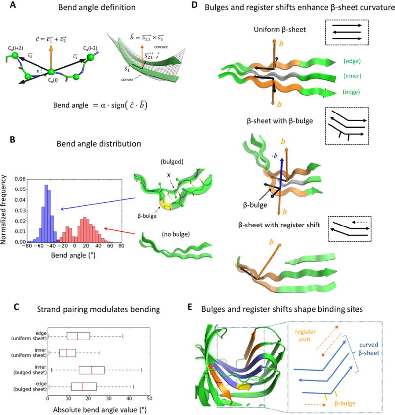
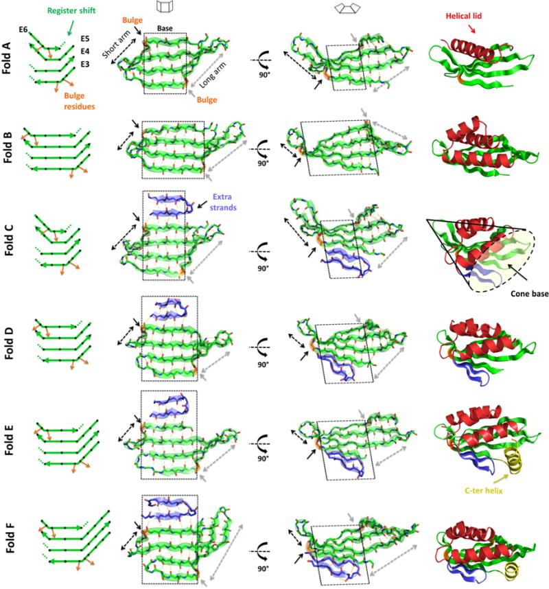
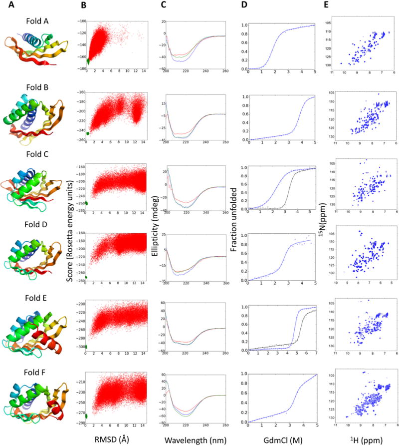
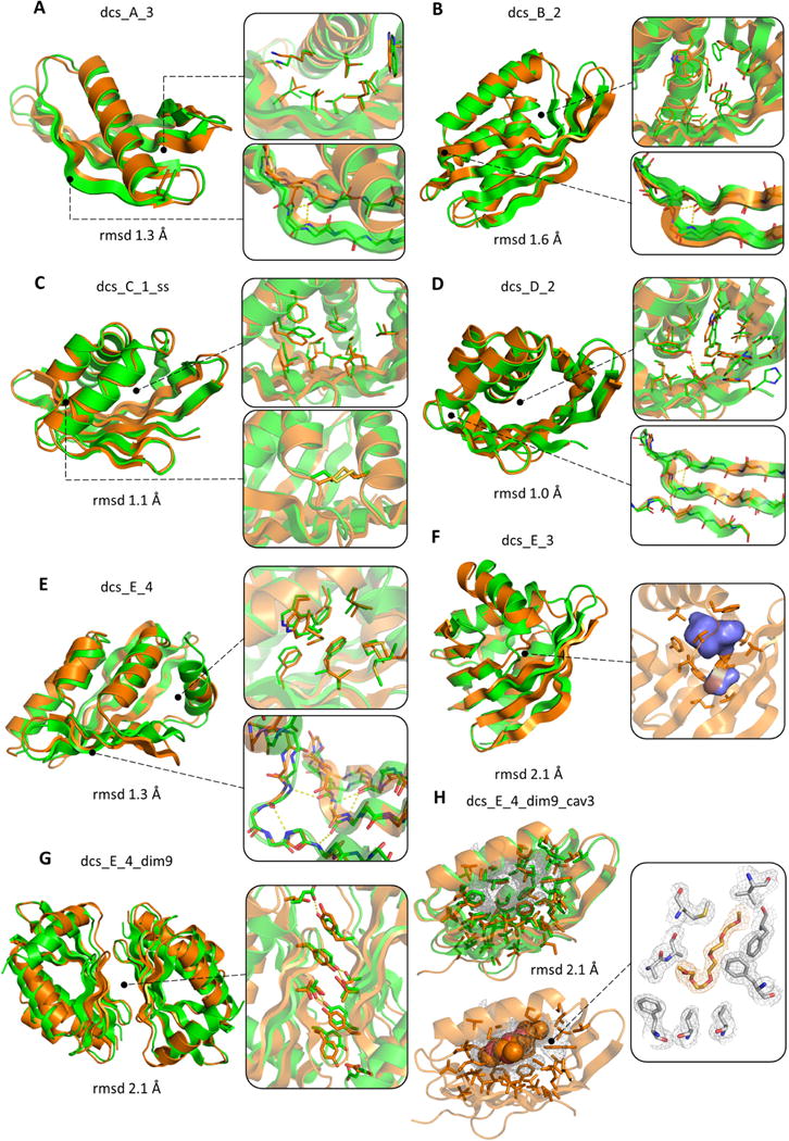
Active sites and ligand-binding cavities in native proteins are often formed by curved β sheets, and the ability to control β-sheet curvature would allow design of binding proteins with cavities customized to specific ligands. Toward this end, we investigated the mechanisms controlling β-sheet curvature by studying the geometry of β sheets in naturally occurring protein structures and folding simulations. The principles emerging from this analysis were used to design, de novo, a series of proteins with curved β sheets topped with α helices. Nuclear magnetic resonance and crystal structures of the designs closely match the computational models, showing that β-sheet curvature can be controlled with atomic-level accuracy. Our approach enables the design of proteins with cavities and provides a route to custom design ligand-binding and catalytic sites.


Figures

Fig. 1
Fig. 1. Rules for β-sheet curvature design
A) Bend angle definition. (B) Distribution of bend angles for strand pairs formed by uniform (red) and bulged (blue) strands. The local hydrogen bonding and offset in sidechain directionality at the β-bulge position are shown. The bulge and the residue following donate two backbone hydrogen bonds to the same residue X. (C) Bend angle (absolute value) box plots of strands with different pairing types in native 3-stranded β-sheets. The edge strand distribution in the bulged β-sheet case (bottom) is for the strand that does not contain the bulge. (D) Representation of the b^ vector in edge strand pairs for three types of 3-stranded β-sheets. β-sheet with β-bulge (middle) shows the - b^ vector for the bulged strand pair to indicate the natural bend direction resulting from a negative bend angle. (E) On the left, cartoon representation of the binding site formed by a curved β-sheet in a native xylanase (PDB entry 2B45). The curved 3-stranded β-sheet core is shown in blue, the β-bulge in yellow and the extra strands in orange. On the right, schematic representation of strand pairings in the curved β-sheet formed by a β-bulge and register shift.
Fig. 2
Fig. 2. Designed β-sheets and folds
On the left, diagrams of the 4-stranded antiparallel β-sheets. Black diamonds represent residues with sidechains pointing to the convex face of the β-sheet and orange arrows highlight the β-bulge offset in sidechain directionality. Dotted lines show the local termination of strand pairing due to register shift between paired strands. Second and third columns show two views of the designed β-sheets. Black and gray dashed arrows show the length of the short and long arms, respectively, that emerge from the flat central base (highlighted by a black dashed square). On the right, examples of each designed protein fold containing 4-stranded antiparallel β-sheets (green), helical lids (red), extra strands (blue) and a C-terminal helix capping the pocket entrance (yellow). The concave base of these conical folds is well suited for small molecule binding site design.
Fig. 3
Fig. 3. Experimental characterization of designed proteins for each fold
(A) Examples of design models for each fold. (B) Folding energy landscapes generated by ab initio structure prediction calculations. Each dot represents the lowest energy structure identified in an independent trajectory starting from an extended chain (red dots) or from the design model (green dots); x-axis shows the Cα-root mean squared deviation (RMSD) from the designed model; the y-axis shows the Rosetta all-atom energy. (C) Far-ultraviolet circular dichroism spectra (blue: 25 °C, red: 95 °C, green: 25 °C after cooling). (D) Chemical denaturation with GdmCl monitored with circular dichroism at 220 nm and 25 °C. For folds C and D the denaturation curves for designs stabilized by a disulfide bond or a dimer interface are shown in black lines. (E) 1H–15N HSQC spectra obtained at 25 °C.
Fig. 4
Fig. 4. Experimentally determined structures of designed proteins
In each panel the experimental structure and the design model are superimposed and colored in orange and green, respectively. Insets show comparisons of sidechain rotamers, β-bulge geometry and cavities; and designed sidechain and β-bulge hydrogen bonds are shown in yellow dashed lines. The RMSD calculated over all Ca atoms is shown in each panel. (A) dcs_A_3 and (B) dcs_B_2 were solved by NMR (comparisons utilized the lowest energy NMR model). (C) dcs_C_1_ss (3.0 Å resolution) with designed disulfide bond in inset. (D) dcs_D_2 (2.0 Å resolution). (E) dcs_E_4 (2.9 Å resolution). (F) dcs_E_3 (3.1 Å resolution); an internal hydrophobic cavity forms in both the design and the crystal structure (volume 192 Å3). (G) dcs_E_4_dim9 (2.4 Å resolution); the interface aromatic stacking and hydrogen bonding interactions are very similar in the crystal structure and design model (right inset). (H) dcs_E_4_dim9_cav3 (1.8 Å resolution). A large (520 Å3) cavity is filled with a pentaethylene glycol molecule in the crystal structure (bottom left; electron density map is on right and design model on upper left). The C-terminal helix and the dimer interface are not shown for better visualization of the cavity.

References

    1. Tinberg CE, Khare SD, Dou J, Doyle L, Nelson JW, Schena A, et al. Nature. 2013;501:212–216. - PMC - PubMed
    1. Röthlisberger D, Khersonsky O, Wollacott AM, Jiang L, Dechancie J, Betker J, et al. Nature. 2008;453:190–195. - PubMed
    1. Jiang L, Althoff EA, Clemente FR, Doyle L, Rothlisberger D, Zanghellini A, et al. Science. 2008;319:1387–1391. - PMC - PubMed
    1. Siegel JB, Zanghellini A, Lovick HM, Kiss G, Lambert AR, St Clair JL, et al. Science. 2010;329:309–313. - PMC - PubMed
    1. Rajagopalan S, Wang C, Yu K, Kuzin AP, Richter F, Lew S, et al. Nat Chem Biol. 2014;10:386–391. - PMC - PubMed

Publication types