. 2017 Mar 1;156(1):96-108.
doi: 10.1093/toxsci/kfw248.

Prenatal Exposure to DEHP Induces Premature Reproductive Senescence in Male Mice

Affiliations

Prenatal Exposure to DEHP Induces Premature Reproductive Senescence in Male Mice

Radwa Barakat et al. Toxicol Sci. .

Abstract

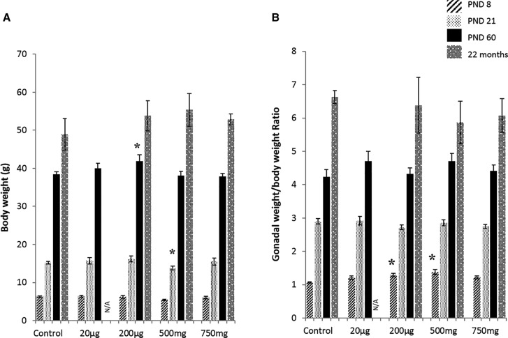
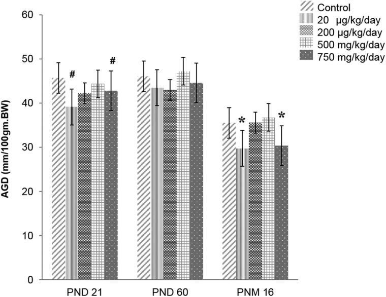
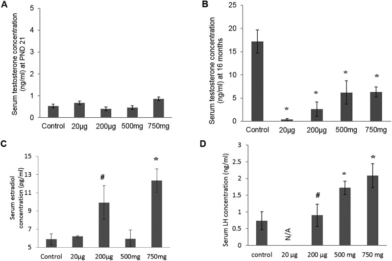
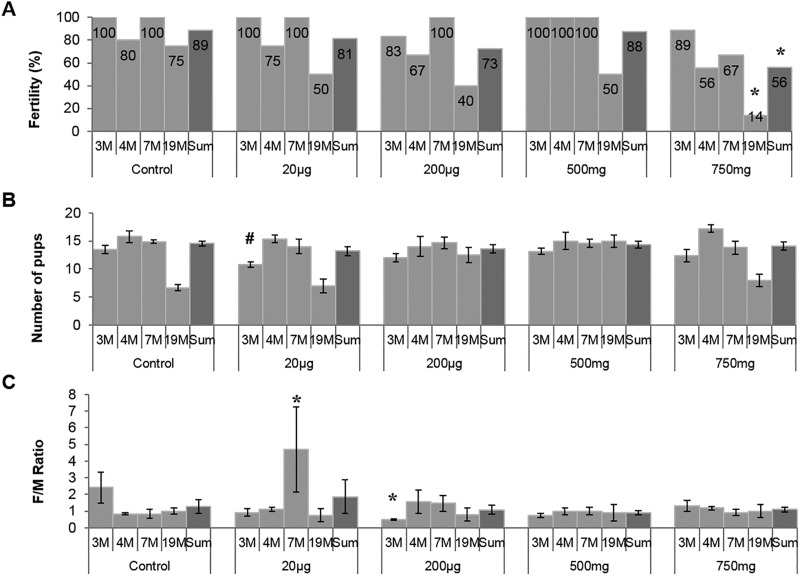
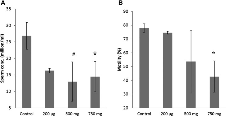
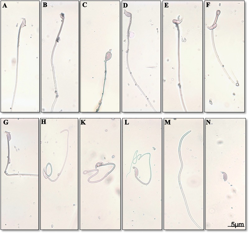
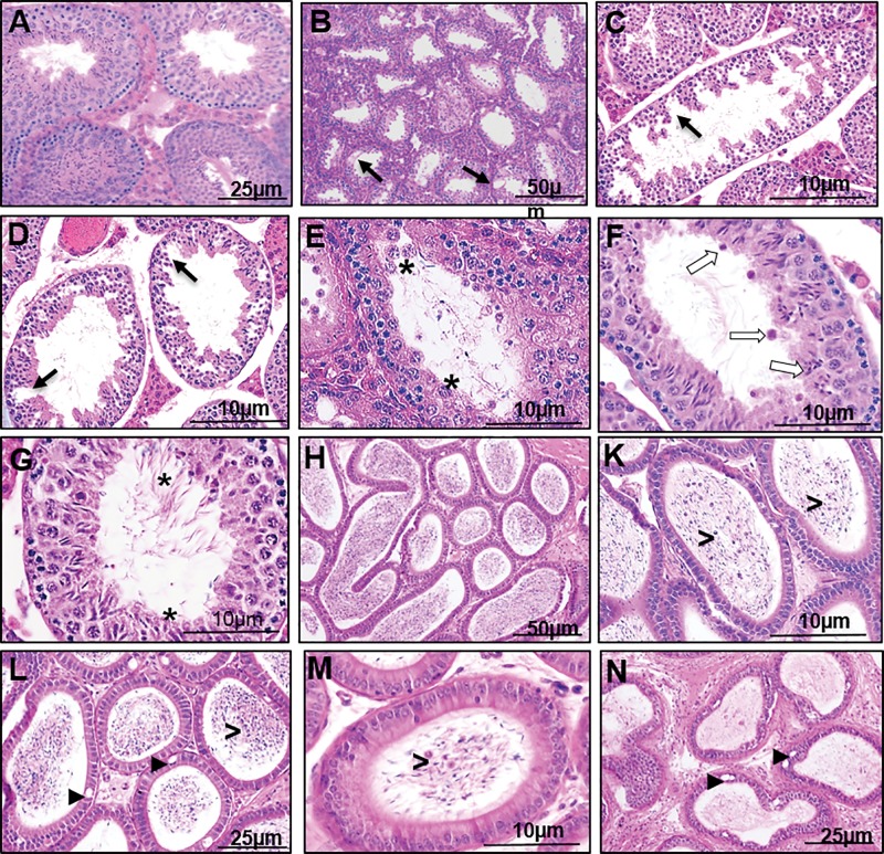
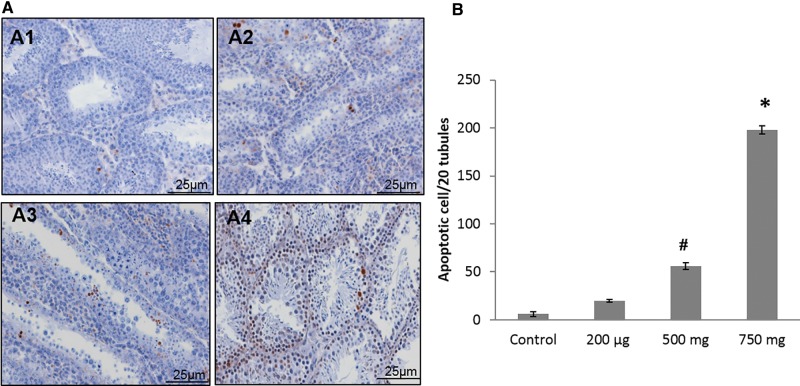
Di-(2-ethylhexyl) phthalate (DEHP) is the most commonly used phthalate, and it is an endocrine-disrupting chemical. This study tested a hypothesis that prenatal exposure to DEHP lays the foundation for premature gonadal dysfunction and subsequent reproductive senescence in male mice. Pregnant female CD-1 mice were orally dosed with vehicle control (tocopherol-stripped corn oil) or with 20 μg/kg/day, 200 μg/kg/day, 500 mg/kg/day, or 750 mg/kg/day of DEHP from gestational day 11 to birth. Overall, the prenatal DEHP exposure did not cause any overt physical health problems in male offspring, as no significant differences in their body nor gonadal weight were seen up to the age of 23 months. However, an age- and dose-dependent gonadal dysfunction was observed. As early as 7 months of age, the 750 mg/kg/day group of mice exhibited significantly reduced fertility. At 19 months of age, 86% of the 750 mg/kg/day mice became infertile, whereas only 25% of the control mice were infertile. At this age, all of the DEHP-exposed mice had lower serum testosterone levels, higher serum estradiol levels, and higher LH levels compared with control mice. Histological evaluations showed that mice prenatally exposed to DEHP displayed a wide array of gonadal and epididymal abnormalities such as increased germ cell apoptosis, degenerative seminiferous tubules, oligozoospermia, asthenozoospermia, and teratozoospermia in comparison to age-matching control mice. In summary, this study shows that prenatal exposure to DEHP induces premature reproductive senescence in male mice.

Keywords: DEHP; endocrine disruptor; fertility; semen.; testes.


Figures

FIG. 1
FIG. 1
Effects of prenatal DEHP exposure on the body and gonadal weights. Body weight growth (A), and gonadal (testis and epididymis) weights to body weight ratio (B) were measured at the ages of postnatal day (PND) 8, 21, 60, and 22 months of age. Graphs show mean ± SEM. Asterisks indicate P ≤ .05, (n = 4–7 per treatment group). N/A: all of mice of 20 µg/kg/day group dead at age of 22 months.
FIG. 2
FIG. 2
Effect of prenatal DEHP exposure on anogenital distance (AGD) at PND 21, PND 60 and 16 months of age. The AGD was normalized to body weight as mm/100gm BW (n = 4–7 per treatment group). Asterisks indicates P ≤ .05 when compared with control group, # indicate P ≤ .09. Note that AGD was also measured at 22 months, it was the same as AGD at 16 months of old.
FIG. 3
FIG. 3
Effect of prenatal DEHP exposure on hormonal level. Serum testosterone concentrations was measured at the ages of PND 21 (A) and at 16 months of age (B), serum estradiol concentrations was measured at 16 months of age (C), serum LH concentrations was measured at 22 months of age (D). Graphs show mean ± SEM, asterisks indicates P ≤ .05 when compared with control group, # indicate P ≤ .09. (n = 4–7 per treatment group), N/A: all of mice of 20 µg/kg/day group dead at 22 months of age.
FIG. 4
FIG. 4
Effect of prenatal DEHP exposure on fertility outcomes. Four fertility tests were done at 3, 4, 7, and 19 months of age, this graph shows the fertility % (percent of males that produced a litter at each trial, A), litter size (numbers of pups per litter, B), and sex ratio (number of female to male pups produced in each litter, C). The numbers of mice used for this experiment (n = 4–7) and the ages of mice that were used at each fertility trial are indicated in months (M). Graphs show mean ± SEM, asterisks indicate P ≤ .05 when compared with control group, # indicate P ≤ .09. Fertility data was compared using Fisher’s exact test for each treatment group against control group.
FIG. 5
FIG. 5
Effect of prenatal DEHP exposure on sperm quality at 22 months of age. Sperm concentrations (millions/mL, A), sperm motility % (percent of motile sperm, B) are shown. Graphs show mean ± SEM, asterisks indicates P ≤ .05 when compared with control group, # indicate P ≤ .09 (n = 4–7 per treatment group). All of mice of the 20 µg/kg/day group were dead at this time point.
FIG. 6
FIG. 6
Effect of prenatal DEHP exposure on sperm morphology at 22 months of age. Normal sperm (A), knobbed hook sperm (B), sperm with amorphous head (C), bent head sperm (D), sperm with incorrect head neck connection (E), folded midpiece (F), bent tail (G), looping midpiece (H), folded tail (K), coiled tail (L), headless sperm (M), detached head (N).
FIG. 7
FIG. 7
Effect of DEHP exposure on the histology of the testes and epididymis at 22 months of age. Testis of a control group (A). Testes of DEHP treated groups (B–G). Epididymis of a control group (H). Epididymis of DEHP treated groups (K–N). Note hypospermatogenesis with degenerative changes in the seminiferous tubules and germ cell degeneration (black arrows), desquamation of large multinucleated cell into the lumen (asterisks), failure of spermiation with abnormal residual bodies found in the lumen (white arrows), abnormal stages of spermatogenesis (stars), vacuoles in the epididymis epithelium (arrow heads), desquamated germ cell the in lumen of epididymis (>), ductal atrophy of the epididymis (N).
FIG. 8
FIG. 8
Effect of DEHP exposure on cell death at 22 months of age. Shown are representative images of each treatment group (A) examined by high power (20×), testis of control group (A1), testis of 200 µg/kg/day DEHP treated group (A2), testis of 500mg/kg/day DEHP treated group (A3), testis of 750mg/kg/day DEHP treated group (A4). Number of apoptotic cell/20 seminiferous tubules for all treated groups (B). Graphs show mean ± SEM, asterisks indicates P ≤ .05 when compared with control group, # indicate P ≤ .09. (n = 4–7 per treatment group). All of mice of 20 µg/kg/day group were dead at this time point.

References

    1. Adibi J. J., Hauser R., Williams P. L., Whyatt R. M., Calafat A. M., Nelson H., Robert H., Swan S. H. (2009). Maternal urinary metabolites of Di-(2-ethylhexyl) phthalate in relation to the timing of labor in a US multicenter pregnancy cohort study. Am. J. Epidemiol. 169, 1015–1024. - PMC - PubMed
    1. Agency for Toxic Substances and Disease Registry ( 2002). Toxicological profile: di(2-ethylhexyl)phthalate (DEHP). ATSDR, Atlanta, GA. Available from: http://www.atsdr.cdc.gov/toxprofiles/tp9.html. - PubMed
    1. Andrade A. J., Grande S. W., Talsness C. E., Grote K., Golombiewski A., Sterner-Kock A., Chahoud I. (2006). A dose-response study following in utero and lactational exposure to di-(2-ethylhexyl) phthalate (DEHP): Effects on androgenic status, developmental landmarks and testicular histology in male offspring rats. Toxicology 225, 64–74. - PubMed
    1. Atanassova N., McKinnell C., Fisher J., Sharpe R. M. (2005). Neonatal treatment of rats with diethylstilboestrol (DES) induces stromal-epithelial abnormalities of the vas deferens and cauda epididymis in adulthood following delayed basal cell development. Reproduction 129, 589–601. - PubMed
    1. Barlow N. J., Foster P. M. D. (2003). Pathogenesis of male reproductive tract lesions from gestation through adulthood following in utero exposure to Di(n-butyl) phthalate. Toxicol. Pathol. 31, 397–410. - PubMed

Publication types

MeSH terms