. 2016 Dec;9(4):496-508.
doi: 10.1007/s12195-016-0448-5. Epub 2016 May 26.

Biorheology of platelet activation in the bloodstream distal to thrombus formation

Affiliations

Biorheology of platelet activation in the bloodstream distal to thrombus formation

Jevgenia Zilberman-Rudenko et al. Cell Mol Bioeng. 2016 Dec.

Abstract

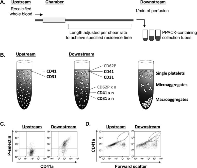
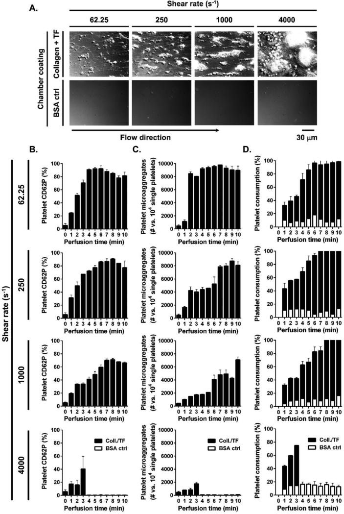
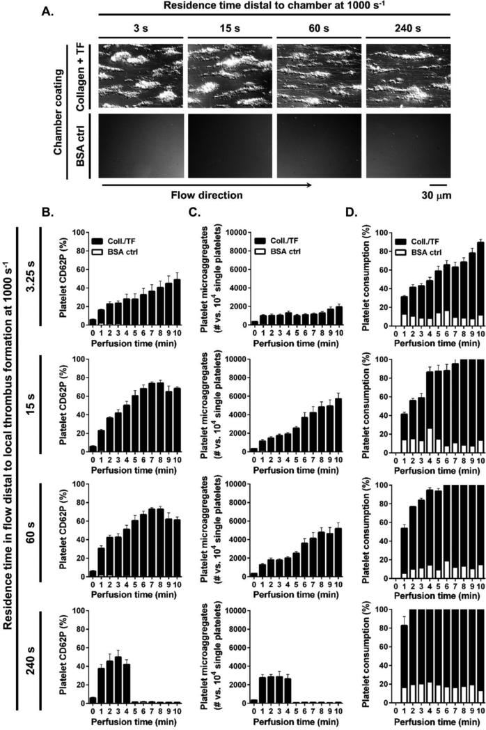
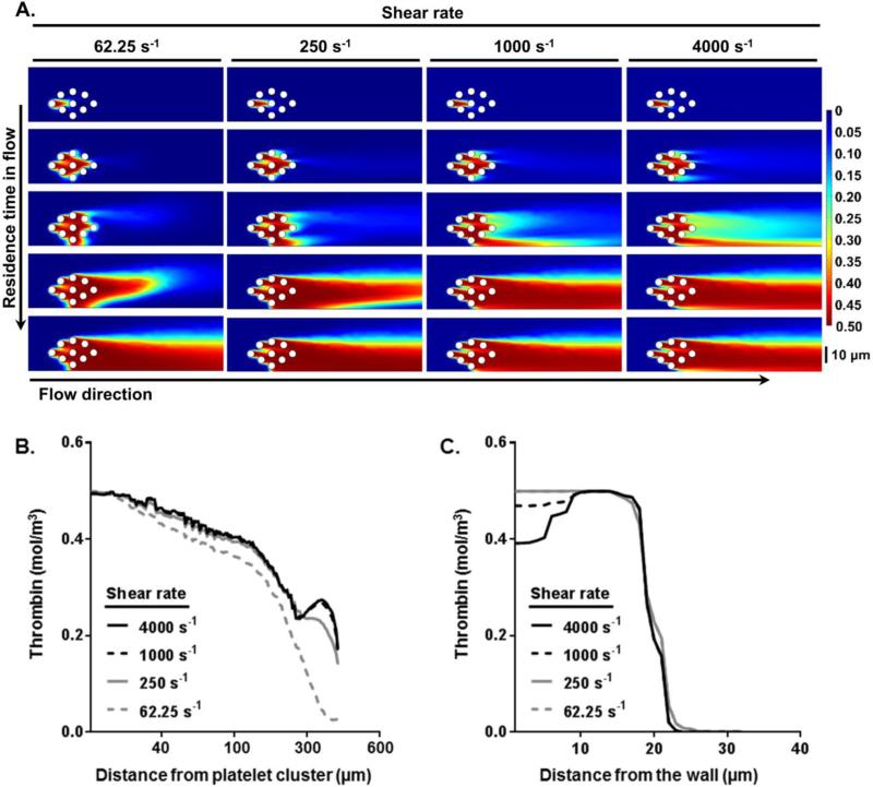
Thrombus growth at the site of vascular injury is mediated by the sequential events of platelet recruitment, activation and aggregation concomitant with the initiation of the coagulation cascade, resulting in local thrombin generation and fibrin formation. While the biorheology of a localized thrombus formation has been well studied, it is unclear whether local sites of thrombin generation propagate platelet activation within the bloodstream. In order to study the physical biology of platelet activation downstream of sites of thrombus formation, we developed a platform to measure platelet activation and microaggregate formation in the bloodstream. Our results show that thrombi formed on collagen and tissue factor promote activation and aggregation of platelets in the bloodstream in a convection-dependent manner. Pharmacological inhibition of the coagulation factors (F) X, XI or thrombin dramatically reduced the degree of distal platelet activation and microaggregate formation in the bloodstream without affecting the degree of local platelet deposition and aggregation on a surface of immobilized collagen. Herein we describe the development and an example of the utility of a platform to study platelet activation and microaggregate formation in the bloodstream (convection-limited regime) relative to the local site of thrombus formation.

Keywords: biophysics; factor X; factor XI; physical biology; platelets; shear; thrombin.


Figures

Figure 1
Figure 1. The schematic of the flow chamber and FACS analysis
Whole blood samples were taken before the blood perfusion (upstream) or at distal site to a collagen and tissue factor-coated flow chamber (downstream) and analyzed by FACS. (B) Platelet activation and microaggregate formation as well as total single platelet consumption (single platelet integration into forming microaggregates and precipitating out of blood macroaggregates) were assessed. Samples were labeled for constitutively expressed platelet CD41a or CD31 and P-selectin (CD62P) expressed on activated platelets. (C) Percent platelet activation was determined by a dot plot with PE-CD41a and APC-CD62P fluorescence. (D) Platelet microaggregate formation was defined by CD41a mean fluorescence intensity and size (forward scatter) shift, as indicated by the region marked with the circle. Gated platelets before (upstream; left) and after (downstream; right) perfusion over a collagen-coated surface are shown.
Figure 2
Figure 2. Effect of shear on local platelet aggregate formation and distal platelet activation
Recalcified whole blood was perfused over collagen/TF or BSA-coated control surfaces for 10 min at 62.25, 250 or 1000 s−1 shear rate and for 3 min (collagen/TF-coated chambers occluded by 3 min of perfusion) at 4000 s−1 shear rate. (A) Final local platelet aggregates formed were evaluated by differential interference contrast (DIC) microscopy. Samples were collected distally at each minute of perfusion after 15 s of residence time in flow and (B) distal platelet activation, (C) microaggregate formation and (D) single platelet consumption were quantified by FACS. Images and histograms are representative from each 3 fields of view from two different blood donors.
Figure 3
Figure 3. Effect of distal residence time in flow on platelet activation in the bloodstream
Recalcified whole blood was perfused over collagen/TF or BSA-coated control surfaces for 10 min at 1000 s−1 shear rate. (A) Local platelet aggregates formed after 10 min of blood perfusion were evaluated by differential interference contrast (DIC) microscopy. Samples were collected distally to local thrombus formation after 3.25, 15, 60 or 240 s of residence time in flow and (B) distal platelet activation, (C) microaggregate formation and (D) single platelet consumption over perfusion time (min) were assessed by FACS. Images and histograms are representative from each 3 fields of view from four different blood donors.
Figure 4
Figure 4. Thrombin mass transfer in flow distal to thrombus formation
Thrombin distribution (mol/m3) in the bloodstream distal to local thrombus formation at increasing shear rates was simulated using COMSOL (A) over different residence time in flow; final residence time shown is 15 s. Predictions of (B) axial thrombin distribution from a platelet cluster in flow and (C) radial thrombin distribution from the wall to the channel center at different set shear rates after 15 s of residence time in flow.
Figure 5
Figure 5. 14E11, 1A6, and rivaroxaban prolonged the clotting time of human plasma
Pooled human plasma was incubated with increasing concentrations of (A) 14E11, (B) 1A6 or (C) rivaroxaban for 10 min and aPTT was recorded, as described in Methods.
Figure 6
Figure 6. 14E11, 1A6, and rivaroxaban did not affect local platelet aggregate formation under flow
Recalcified whole blood was perfused over collagen-coated surfaces at 1000 s−1 shear rate. (A) Platelet aggregates formed on collagen surfaces in the presence of 14E11 (50 μg/ml), 1A6 (50 μg/ml) or rivaroxaban (100 nM). (B) Aggregate volume (left) and surface area (right) in the presence of 14E11 (top), 1A6 (middle) or rivaroxaban (bottom) at indicated concentrations. Images and histograms are representative from each 3 fields of view from two different blood donors. Scale bar = 20 μm.
Figure 7
Figure 7. FXI and FXa inhibitors abrogated distal platelet CD62P expression and microaggregate formation under shear
Whole blood, treated with 14E11, 1A6, rivaroxaban or melagatran at indicated concentrations, was perfused over a collagen-coated chambers immediately after recalcification; PBS or DMSO was used as vehicle. In parallel, whole blood samples were collected after treatment with vehicle or inhibitor, and incubated with TRAP-6 (10 μg/ml) or vehicle for 5 min. Data are reported as (A) mean ± SEM percentage of CD62P-positive platelets, or (B) mean ± SEM platelet microaggregate count versus 104 gated single platelets in gated population of at least three experiments. The maximal CD62P expression levels or microaggregate counts after 5 mins of perfusion are shown in the graphs for each treatment.

References

    1. Abulencia JP, Tien N, McCarty OJ, Plymire D, Mousa SA, Konstantopoulos K. Comparative antiplatelet efficacy of a novel, nonpeptide GPIIb/IIIa antagonist (XV454) and abciximab (c7E3) in flow models of thrombosis. Arterioscler. Thromb. Vasc. Biol. 2001;21:149–156. - PubMed
    1. Andrews RK, Berndt MC. Platelet physiology and thrombosis. Thromb. Res. 2004;114:447–453. - PubMed
    1. Baker-Groberg SM, Cianchetti FA, Phillips KG, McCarty OJ. Development of a method to quantify platelet adhesion and aggregation under static conditions. Cell. Mol. Bioeng. 2014;7:285–290. - PMC - PubMed
    1. Baker SM, Phillips KG, McCarty OJT. Development of a label-free imaging technique for the quantification of thrombus formation. Cell. Mol. Bioeng. 2012;5:488–492. - PMC - PubMed
    1. Chang JY. Thrombin specificity. Requirement for apolar amino acids adjacent to the thrombin cleavage site of polypeptide substrate. Eur. J. Biochem. FEBS. 1985;151:217–224. - PubMed

LinkOut - more resources