. 2016 Dec 15;4(4):044003.
doi: 10.1063/1.4972069. eCollection 2017 Jul.

Structural enzymology using X-ray free electron lasers

Affiliations

Structural enzymology using X-ray free electron lasers

Christopher Kupitz et al. Struct Dyn. .

Abstract

Mix-and-inject serial crystallography (MISC) is a technique designed to image enzyme catalyzed reactions in which small protein crystals are mixed with a substrate just prior to being probed by an X-ray pulse. This approach offers several advantages over flow cell studies. It provides (i) room temperature structures at near atomic resolution, (ii) time resolution ranging from microseconds to seconds, and (iii) convenient reaction initiation. It outruns radiation damage by using femtosecond X-ray pulses allowing damage and chemistry to be separated. Here, we demonstrate that MISC is feasible at an X-ray free electron laser by studying the reaction of M. tuberculosis ß-lactamase microcrystals with ceftriaxone antibiotic solution. Electron density maps of the apo-ß-lactamase and of the ceftriaxone bound form were obtained at 2.8 Å and 2.4 Å resolution, respectively. These results pave the way to study cyclic and non-cyclic reactions and represent a new field of time-resolved structural dynamics for numerous substrate-triggered biological reactions.


Figures

FIG. 1.
FIG. 1.
Data collection schematic showing the T-junction set-up used for a mixing time of about 2 s. The T-junction was placed outside of the nozzle rod in our experiment but could also be engineered to fit inside closer to the interaction region for shorter mixing times.
FIG. 2.
FIG. 2.
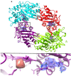
Electron density in the catalytic cleft of BlaC. (a) Refined model of the entire tetramer (σ = 1.1) in the asymmetric unit after mixing. The mixed electron density (2Fo-Fc) is shown in blue in the binding pockets. Subunits A and C contain phosphate while subunits B and D have a bound ceftriaxone, with the electron density of D being slightly stronger. (b) Enlarged section of subunit D showing the unmixed ED, which corresponds to a bound phosphate. (c) Enlarged section of subunit D showing the mixed ED (blue electron density) with ceftriaxone modelled in. Slightly different views of the same subunit binding pocket are shown in (b) and (c); however, there are minimal changes to the ligand binding sphere (see supplementary material, Table S2).

References

    1. Lewis K., “ Platforms for antibiotic discovery,” Nat. Rev. Drug Discovery , 371–387.10.1038/nrd3975 - DOI - PubMed
    1. Singh G. S., “ Beta-lactams in the new millennium. Part-I: monobactams and carbapenems,” Mini Rev. Med. Chem. , 69–92 (2004).10.2174/1389557043487501 - DOI - PubMed
    1. Page M. G., “ Cephalosporins in clinical development,” Expert Opin. Invest. Drugs , 973–985 (2004).10.1517/13543784.13.8.973 - DOI - PubMed
    1. Meini M. R., Llarrull L. I., and Vila A. J., “ Overcoming differences: The catalytic mechanism of metallo-beta-lactamases,” FEBS Lett. , 3419–3432 (2015).10.1016/j.febslet.2015.08.015 - DOI - PMC - PubMed
    1. Palzkill T., “ Metallo-beta-lactamase structure and function,” Ann. N.Y. Acad. Sci. , 91–104 (2013).10.1111/j.1749-6632.2012.06796.x - DOI - PMC - PubMed