Review
. 2017 Jun;74(11):1999-2017.
doi: 10.1007/s00018-016-2451-3. Epub 2017 Jan 12.

Mitochondrial dynamics as regulators of cancer biology

Affiliations
Review

Mitochondrial dynamics as regulators of cancer biology

Andrew Paul Trotta et al. Cell Mol Life Sci. 2017 Jun.

Abstract

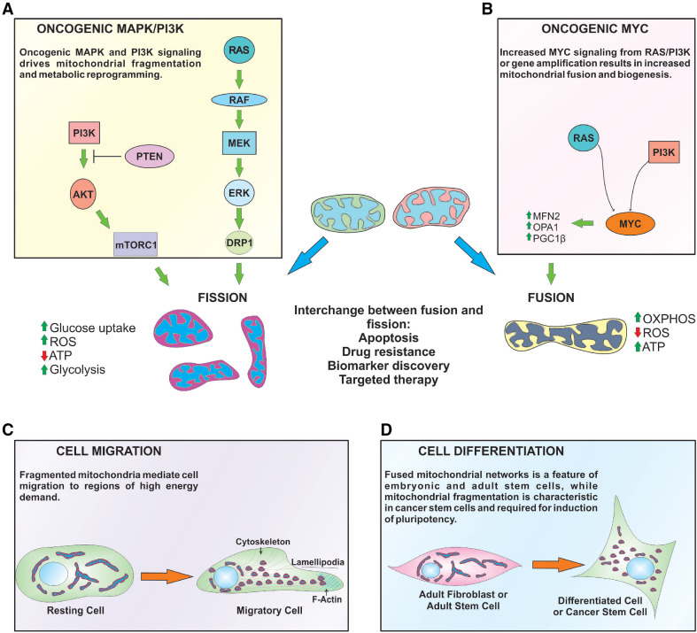
Mitochondria are dynamic organelles that supply energy required to drive key cellular processes, such as survival, proliferation, and migration. Critical to all of these processes are changes in mitochondrial architecture, a mechanical mechanism encompassing both fusion and fragmentation (fission) of the mitochondrial network. Changes to mitochondrial shape, size, and localization occur in a regulated manner to maintain energy and metabolic homeostasis, while deregulation of mitochondrial dynamics is associated with the onset of metabolic dysfunction and disease. In cancers, oncogenic signals that drive excessive proliferation, increase intracellular stress, and limit nutrient supply are all able to alter the bioenergetic and biosynthetic requirements of cancer cells. Consequently, mitochondrial function and shape rapidly adapt to these hostile conditions to support cancer cell proliferation and evade activation of cell death programs. In this review, we will discuss the molecular mechanisms governing mitochondrial dynamics and integrate recent insights into how changes in mitochondrial shape affect cellular migration, differentiation, apoptosis, and opportunities for the development of novel targeted cancer therapies.

Keywords: Apoptosis; Cancer; Differentiation; Migration; Mitochondrial dynamics; Oncogenic signaling.


Conflict of interest statement

The authors declare no conflict of interest.

Figures

Fig. 1
Fig. 1
Dynamins are large GTPases that regulate mitochondrial fusion and fission. Schematic of Mfn1, Mfn2, OPA1, and DRP1 protein structures. a Amino-terminal region of Mfn1 and Mfn2 contain a GTPase domain. A centralized transmembrane (TM) domain enables insertion of the protein into the OMM. Flanking the TM region are two coiled–coiled (CC1, CC2) domains that permit homo- or hetero-dimerization of Mfn1 and Mfn2 proteins, and allow for fusion between adjacent mitochondria. b OPA1 consists of an amino-terminal mitochondrial localization signal, followed by two transmembrane domains, and a CC1 domain. The GTPase region is centrally located and is followed by a second CC domain (CC2), and GTPase effector domain (GED) at the carboxyl-terminus. OPA1 is proteolytically processed to produce long (L-OPA1) and short (S-OPA1) isoforms. c DRP1 consists of an amino-terminally located GTPase domain; followed by a middle domain that is involved in self-assembly and a variable CC region called “Insert B”. At the carboxyl-terminus is a GED, which is involved in intra-molecular interactions with the GTPase domain
Fig. 2
Fig. 2
Mechanisms of mitochondrial fusion and fission. a Fusion of the OMM is mediated by Mfn1 and Mfn2. The orientation of Mfn1 and Mfn2 domains suggests that the amino and carboxyl termini face the cytosol to facilitate interactions with other mitofusins on adjacent mitochondria, while the TM domain is embedded within the OMM and IMS. The mitofusin GTPase domain is required to pull the two opposing OMMs together resulting in bilayer fusion. Fusion of the OMM requires homo- or heterotypic interactions between Mfn1 and Mfn2, although heterotypic dimers (i.e., Mfn1:Mfn2) are more efficient at fusion compared to homotypic complexes. IMM fusion is coordinated by OPA1. L-OPA1 isoforms are anchored within the IMM and have their GTPase and GED exposed to the IMS. Proteolytic cleavage of L-OPA1 results in the generation of S-OPA1, allowing both to coordinate IMM fusion. OPA1 interacts with both Mfn1 and Mfn2 to form a bridge between the IMM and OMM, and is required for lipid mixing during fusion. b DRP1 regulates mitochondrial fission. Soluble, cytosolic DRP1 exists in the cytosol as dimers or trimers. Following activation via phosphorylation at Ser616, DRP1 translocates to the OMM where it binds to adaptor proteins (e.g., MFF, MiD49, MiD51, and FIS1). At the OMM, DRP1 undergoes conformational change, so that the middle domain and GED form a stalk-like structure. The DRP1 GTPase domain faces away from the OMM and connects with other DRP1 proteins on mitochondria. Once a DRP1 helix has completely spiralled around mitochondrion, GTP hydrolysis causes constriction of the helix and scission of the OMM and IMM. Actin polymerization also occurs at ER-mitochondrial junctions and facilitates migration of individual mitochondrion away from each other during fission
Fig. 3
Fig. 3
Role of mitochondrial dynamics in cancer processes. a, b Oncogenic signaling results in DRP1-dependent mitochondrial fragmentation. RAS and PI3K signaling up-regulates MYC, which subsequently promotes expression of pro-fusion and mitochondria biogenesis proteins. MYC gene amplification can also phenocopy these events in the absence of upstream stimuli. Mitochondrial shape also plays distinctive roles in regulating cellular metabolism. Fused mitochondria have increased oxidative metabolism, ATP production, and decreased ROS. Oncogenic signaling that fragments mitochondria increases glucose uptake, ROS, and decreases OXPHOS, which leads to a metabolic switch to glycolysis. Mitochondrial morphology is interchangeable between fused and fission states, and has implications for apoptosis, drug resistance, and clinical applications, such as biomarker discovery and targeted therapies. c Cell migration requires mitochondrial fission to enable movement of mitochondria to regions of the cell that have higher ATP requirements (i.e., lamellipodia). d Fused mitochondria are common in adult fibroblasts and stem cells, but mitochondrial network fragmentation is an initiating event following induction of pluripotency (i.e., iPCs) and cancer stem cells maintenance

References

    1. Wai T, Langer T. Mitochondrial dynamics and metabolic regulation. TEM. 2016;27(2):105–117. - PubMed
    1. Archer SL. Mitochondrial dynamics–mitochondrial fission and fusion in human diseases. N Engl J Med. 2013;369(23):2236–2251. doi: 10.1056/NEJMra1215233. - DOI - PubMed
    1. Lewis MR, Lewis WH. Mitochondria (and other cytoplasmic structures) in tissue cultures. Am J Anat. 1915;17(3):339–401. doi: 10.1002/aja.1000170304. - DOI
    1. Jakobs S. High resolution imaging of live mitochondria. Biochim Biophys Acta. 2006;1763(5–6):561–575. doi: 10.1016/j.bbamcr.2006.04.004. - DOI - PubMed
    1. Karbowski M, Lee YJ, Gaume B, Jeong SY, Frank S, Nechushtan A, Santel A, Fuller M, Smith CL, Youle RJ. Spatial and temporal association of Bax with mitochondrial fission sites, Drp1, and Mfn2 during apoptosis. J Cell Biol. 2002;159(6):931–938. doi: 10.1083/jcb.200209124. - DOI - PMC - PubMed

Publication types