. 2017 Mar;11(3):266-279.
doi: 10.1002/1878-0261.12033. Epub 2017 Feb 7.

Sorafenib analogue SC-60 induces apoptosis through the SHP-1/STAT3 pathway and enhances docetaxel cytotoxicity in triple-negative breast cancer cells

Affiliations

Sorafenib analogue SC-60 induces apoptosis through the SHP-1/STAT3 pathway and enhances docetaxel cytotoxicity in triple-negative breast cancer cells

Chun-Yu Liu et al. Mol Oncol. 2017 Mar.

Abstract

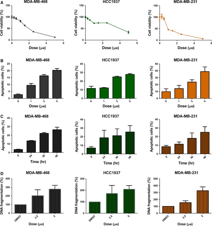
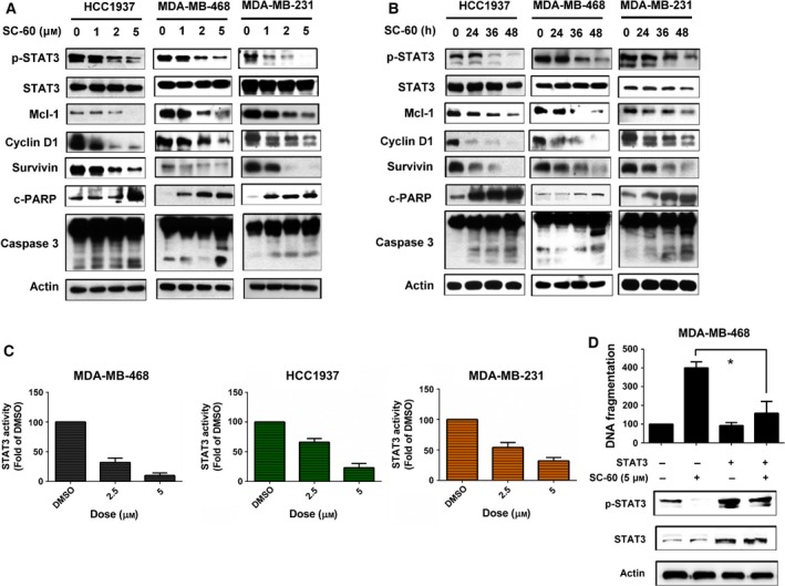
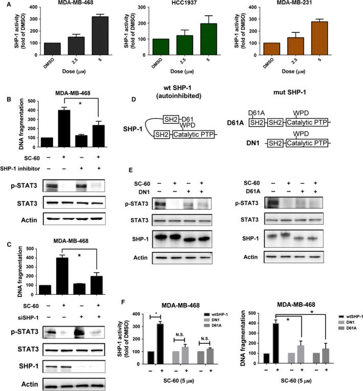
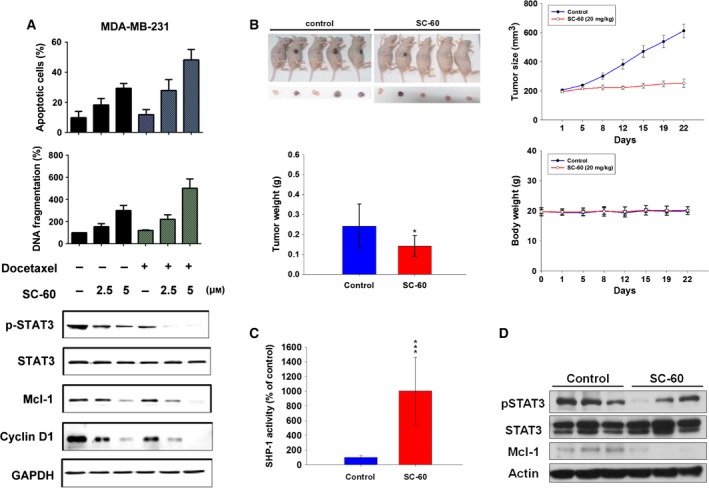
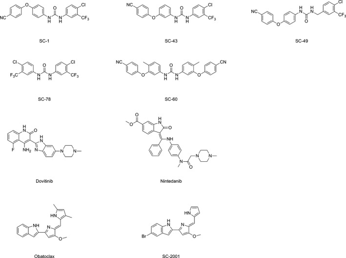
Recurrent triple-negative breast cancer (TNBC) needs new therapeutic targets. Src homology region 2 domain-containing phosphatase-1 (SHP-1) can act as a tumor suppressor by dephosphorylating oncogenic kinases. One major target of SHP-1 is STAT3, which is highly activated in TNBC. In this study, we tested a sorafenib analogue SC-60, which lacks angiokinase inhibition activity, but acts as a SHP-1 agonist, in TNBC cells. SC-60 inhibited proliferation and induced apoptosis by dephosphorylating STAT3 in both a dose- and time-dependent manner in TNBC cells (MDA-MB-231, MDA-MB-468, and HCC1937). By contrast, ectopic expression of STAT3 rescued the anticancer effect induced by SC-60. SC-60 also increased the SHP-1 activity, but this effect was inhibited when the N-SH2 domain (DN1) was deleted or with SHP-1 point mutation (D61A), implying that SHP-1 is the major target of SC-60 in TNBC. The use of SC-60 in combination with docetaxel synergized the anticancer effect induced by SC-60 through the SHP-1/STAT3 pathway in TNBC cells. Importantly, SC-60 also displayed a significant antitumor effect in an MDA-MB-468 xenograft model by modulating the SHP-1/STAT3 axis, indicating the anticancer potential of SC-60 in TNBC treatment. Targeting SHP-1/p-STAT3 and the potential combination of SHP-1 agonist with chemotherapeutic docetaxel is a feasible therapeutic strategy for TNBC.

Keywords: SHP-1 agonist; STAT3; triple-negative breast cancer.


Figures

Figure 1
Figure 1
SC‐60 shows antiproliferative effects in human triple‐negative breast cancer cells. (A) Cells were exposed to SC‐60 at the indicated doses for 72 h, and cell viability was assessed by MTT assay. (B,C) Flow cytometry assay revealed that SC‐60 treatment resulted in increased percentage of apoptotic cells in the aforementioned cell lines in a dose (B)‐ and time (C)‐dependent manner. (D) SC‐60 treatment led to increased DNA fragmentation in TNBC cells in a dose‐dependent manner. Means of at least three independent experiments performed in triplicate are shown. Data are shown as mean ± SD.
Figure 2
Figure 2
SC‐60 enhances apoptosis and reduces p‐STAT3 signaling in TNBC cells. (A,B) The effects of SC‐60 on p‐STAT3 and its downstream molecules (Mcl‐1, cyclin D1, and survivin) were analyzed by western blot. Cells were treated with SC‐60 at the (A) indicated doses for 48 h or (B) treated with SC‐60 (5 μm) at the indicated times. (C) Cells were transfected with inducible STAT3‐responsive firefly luciferase construct and constitutively expressing Renilla luciferase construct as internal control for 48 h; then, the cells were treated with SC‐60 at indicated dose for 6 h. The activities of STAT3 were measured by dual luciferase assay. The activities of STAT3 of tested samples were normalized with cells treated with DMSO. (D) Overexpression of STAT3 reversed the apoptotic effect of SC‐60. MDAMB‐468 cells were transfected with STAT3‐expressing vector with myc‐tag for 24 h and then treated with DMSO or SC‐60 at 5 μm for another 24 h. DNA fragmentation was measured, and the effect on p‐STAT3 was analyzed by western blot. Means of at least three independent experiments performed in triplicate are shown. *< 0.05. Data are shown as mean ± SD.
Figure 3
Figure 3
SC‐60 induces cell apoptosis by SHP‐1/p‐STAT3 signaling in TNBC cells. (A) The activities of SHP‐1 were measured at the indicated doses for 48 h in MDAMB‐468, HCC1937, and MDAMB‐231 TNBC cell lines. (B) The protective effects of SHP‐1 inhibitor on SC‐60‐induced apoptosis in MDAMB‐468 cells. Cells were pretreated with 50 μm SHP‐1 inhibitor (PTP inhibitor III) for 1 h and then treated with SC‐60 at 5 μm for 36 h. DNA fragmentation was determined by the Cell Death Detection ELISAPLUS Kit. (C) Knockdown of SHP‐1 reversed the biological effects of SC‐60 on apoptosis (left) and p‐STAT3 inhibition (right). MDAMB‐468 cells were transfected with control siRNA (scrambled) or SHP‐1 siRNA for 24 h and then treated with SC‐60 at 5 μm for another 24 h. The protein levels of p‐STAT3, STAT3, SHP‐1, and actin (as loading control) were analyzed by western blot, and DNA fragmentation was measured by Cell Death Detection ELISAPLUS Kit. (D) Schematic representation of wild‐type SHP‐1 (autoinhibited), deletion and single mutants of SHP‐1 (mimic activated SHP‐1). (E,F) MDAMB‐468 cells were transfected with mutant SHP‐1 (DN1 or D61A) for 24 h and then treated with SC‐60 at 5 μm for another 24 h. (E) The protein levels of p‐STAT3, STAT3, SHP‐1, and actin (as loading control) were analyzed by western blot. (F) The SHP‐1 activity was assessed by SHP‐1 phosphatase activity (left) and DNA fragmentation (right) was measured by Cell Death Detection ELISAPLUS Kit as described in 2. Means of at least three independent experiments performed in triplicate are shown. *< 0.05. Data are shown as mean ± SD.
Figure 4
Figure 4
Combination of SC‐60 and docetaxel increases cell apoptosis by reducing p‐STAT3, and SC‐60 diminishes xenograft tumor growth of TNBC cells. (A) Cells were treated with docetaxel (0.1 μm) for 24 h, then treated with SC‐60 at the indicated doses (0, 2.5, and 5 μm) for another 24 h. Effect of docetaxel/SC‐60 combination on cell apoptosis (upper), DNA fragmentation (middle), p‐STAT3 and its downstream signaling (lower) were measured. Means of at least three independent experiments performed in triplicate are shown. Data are shown as mean ± SD. (B–D) MDAMB‐468‐bearing mice were treated with vehicle or SC‐60 orally at 20 mg·kg−1 three times a week. (B) Mice images (upper left), growth curves (lower left), tumor weight (upper right), body weight (lower right), (C) SHP‐1 activity, and (D) western blot analysis of p‐STAT3, STAT3, and Mcl‐1 were measured. Data of growth curve (n = 5) are shown as mean ± SE. Data of tumor weight, body weight, and SHP‐1 activity (n = 5) are shown as mean ± SD. *< 0.05. ***< 0.001.
Figure 5
Figure 5
Chemical structures of compounds or drugs that have been reported to enhance SHP‐1 activity.

References

    1. Arabaci G, Guo XC, Beebe KD, Coggeshall KM and Pei D (1999) alpha‐Haloacetophenone derivatives as photoreversible covalent inhibitors of protein tyrosine phosphatases. J Am Chem Soc 121, 5085–5086.
    1. Balmanno K and Cook SJ (1999) Sustained MAP kinase activation is required for the expression of cyclin D1, p21Cip1 and a subset of AP‐1 proteins in CCL39 cells. Oncogene 18, 3085–3097. - PubMed
    1. Banerjee K and Resat H (2015) Constitutive activation of STAT3 in breast cancer cells: a review. Int J Cancer 138, 2570–2578. - PMC - PubMed
    1. Berishaj M, Gao SP, Ahmed S, Leslie K, Al‐Ahmadie H, Gerald WL, Bornmann W and Bromberg JF (2007) Stat3 is tyrosine‐phosphorylated through the interleukin‐6/glycoprotein 130/Janus kinase pathway in breast cancer. Breast Cancer Res 9, R32. - PMC - PubMed
    1. Binai NA, Damert A, Carra G, Steckelbroeck S, Lower J, Lower R and Wessler S (2010) Expression of estrogen receptor alpha increases leptin‐induced STAT3 activity in breast cancer cells. Int J Cancer 127, 55–66. - PubMed

MeSH terms

LinkOut - more resources