. 2017 Jan 13:7:40304.
doi: 10.1038/srep40304.

Regulation of sleep plasticity by a thermo-sensitive circuit in Drosophila

Affiliations

Regulation of sleep plasticity by a thermo-sensitive circuit in Drosophila

Angelique Lamaze et al. Sci Rep. .

Abstract

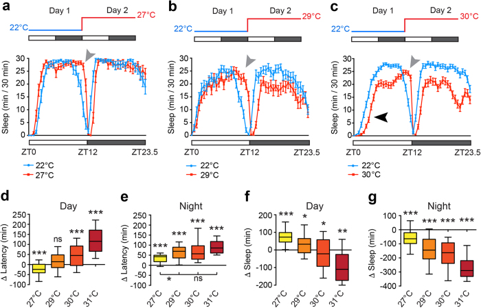
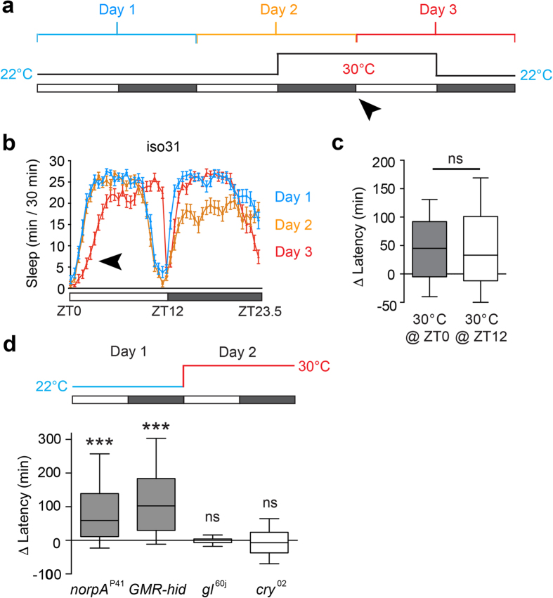
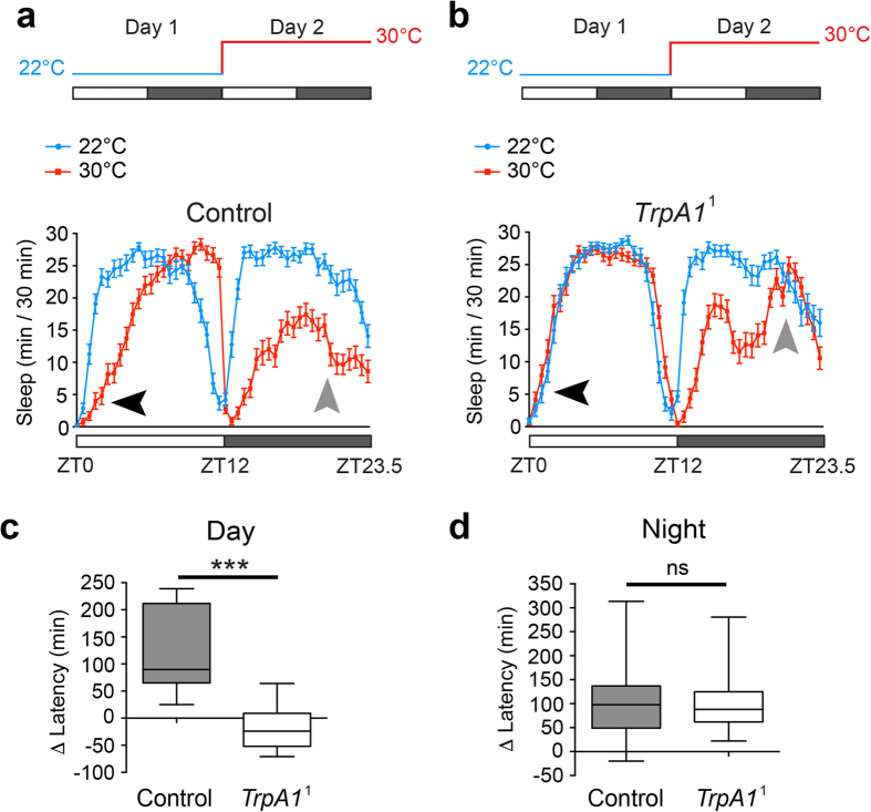
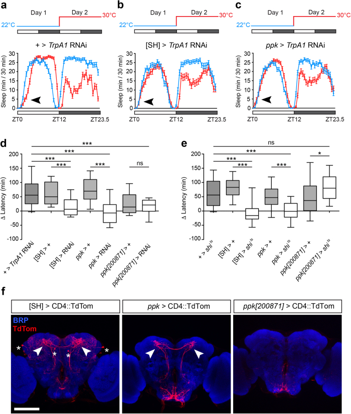
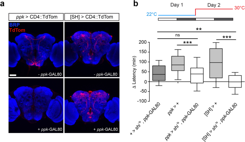
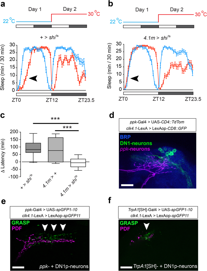
Sleep is a highly conserved and essential behaviour in many species, including the fruit fly Drosophila melanogaster. In the wild, sensory signalling encoding environmental information must be integrated with sleep drive to ensure that sleep is not initiated during detrimental conditions. However, the molecular and circuit mechanisms by which sleep timing is modulated by the environment are unclear. Here we introduce a novel behavioural paradigm to study this issue. We show that in male fruit flies, onset of the daytime siesta is delayed by ambient temperatures above 29 °C. We term this effect Prolonged Morning Wakefulness (PMW). We show that signalling through the TrpA1 thermo-sensor is required for PMW, and that TrpA1 specifically impacts siesta onset, but not night sleep onset, in response to elevated temperatures. We identify two critical TrpA1-expressing circuits and show that both contact DN1p clock neurons, the output of which is also required for PMW. Finally, we identify the circadian blue-light photoreceptor CRYPTOCHROME as a molecular regulator of PMW, and propose a model in which the Drosophila nervous system integrates information encoding temperature, light, and time to dynamically control when sleep is initiated. Our results provide a platform to investigate how environmental inputs co-ordinately regulate sleep plasticity.


Figures

Figure 1
Figure 1. Warm temperatures prolong morning wakefulness in male Drosophila.
(a–c) Average sleep patterns of adult male flies shifted from 22 °C to either 27 °C (n = 20) (a), 29 °C (n = 32) (b) or 30 °C (n = 69) (c). Sleep traces are presented as mean ± SEM for each time point in these and all subsequent figures. Temperature-shift paradigms are indicated above. Sleep was measured under 12 h light: 12 h dark conditions (white/grey bars) with Zeitgeber Times (ZT) shown below. Black arrowhead indicates the delay of the sleep onset observed at 30 °C (PMW). Grey arrowheads indicate the delay of sleep offset induced at 27 °C or above. (d,e) Change in time taken to the first day sleep episode (d) or night sleep episode (e) (Δ Latency) between consecutive 24 h periods at 22 °C and 27–31 °C. (f,g) Difference in total sleep during the day (f) or night (g) between consecutive 24 h periods at 22 °C and 27–31 °C. n-values: 27 °C, n = 39; 29 °C, n = 32; 30 °C, n = 79; 31 °C, n = 18. In this and all subsequent figures, box plots show the 10th, 25th, median, 75th and 90th percentiles, and p-values are indicated as follows: *p < 0.05, **p < 0.005, ***p < 0.0005, ns – p > 0.05. Statistical comparisons: (d–g) Wilcoxon signed rank test compared to a theoretical median of zero and (e) Kruskal-Wallis test with Dunn’s post-hoc test.
Figure 2
Figure 2. PMW is CRYPTOCHROME- and GLASS-dependent.
(a) Three-day temperature-shift paradigm to test whether PMW is an acute avoidance response. Ambient temperature is raised from 22 °C to 30 °C at ZT12 on Day 2 for 24 h. Sleep latency was subsequently measured on Day 3 (black arrowhead). (b) Average sleep patterns of control adult male flies during the above temperature-shift paradigm. Subsequent days are juxtaposed to allow direct comparison. Day 1: blue, Day 2: orange, Day 3: red. Black arrowhead indicates PMW during Day 3. n = 43. (c) Comparison of the PMW when ambient temperature is increased at either ZT0 on the experimental day, or at ZT12 – the beginning of the previous night. ns – p > 0.05, Mann-Whitney U-test. ZT0: n = 79 (data also presented in Fig. 1); ZT12: n = 45. (d) PMW in light-pathway mutants. Statistical comparison: Wilcoxon signed rank test compared to a theoretical median of zero. norpAP41: n = 31, GMR-hid: n = 38, gl60j: n = 44, cry02: n = 58.
Figure 3
Figure 3. The TrpA1 thermo-sensor is required for PMW.
(a,b) Average sleep patterns of adult male control or TrpA11 homozygotes shifted from 22 °C to 30 °C at ZT0. Temperature-shift paradigms are indicated above. Black arrowheads: presence/absence of PMW. Grey arrowheads: presence/absence of enhanced wakefulness prior to lights-on during a warm night. (c,d) Comparison of change in latency to the first sleep episode between control and TrpA11 homozygote males during the day (c) and night (d) following a shift from 22 °C to 30 °C. Statistical comparison: Mann-Whitney U-test. Control: n = 43; TrpA11: n = 62.
Figure 4
Figure 4. TrpA1-expressing TrpA1[SH]- and ppk-neurons are necessary for PMW.
(a–c) Average sleep patterns of adult males containing a UAS-TrpA1 RNAi transgene but lacking a promoter-GAL4 driver (a), or expressing UAS-TrpA1 RNAi under control of the TrpA1[SH]- and ppk-GAL4 drivers (b,c respectively). Temperature-shift paradigms are indicated above. Black arrowheads: presence/absence of PMW. (d) Comparison of PMW in males expressing UAS-TrpA1 RNAi under GAL4 drivers and their corresponding controls. n = 32–120. Statistical comparison: Kruskal-Wallis test with Dunn’s post-hoc test. All controls are ***p < 0.0001. [SH] > RNAi: p = 0.0481; ppk > RNAi: p = 0.2420; ppk[200871] > RNAi: p = 0.0054, Wilcoxon signed rank test compared to a theoretical median of zero. (e) Effect of acute inhibition of synaptic output (using UAS-shits) from TrpA1[SH]-, ppk-, and ppk[200871]-neurons on PMW. Statistical comparison: Kruskal-Wallis test with Dunn’s post-hoc test. n = 24–77. All controls except ppk[200871] > + (**p = 0.0008) are ***p < 0.0001. [SH] > shits: ns (p = 0.06); ppk > shits: ns (p = 0.89); ppk[200871] > shits: ***p < 0.0001, Wilcoxon signed rank test compared to a theoretical median of zero. (f) Expression patterns of TrpA1[SH]-, ppk- and ppk[200871]-GAL4 in the adult Drosophila brain. CD4::TdTom is labeled with anti DsRed. Synaptic neuropil (BRP) is labeled using an anti-nc82 antibody. Stars: cell bodies labeled by TrpA1[SH]-GAL4. Arrows: projections to the dorsal posterior protocerebrum from subsets of TrpA1[SH]- and ppk-neurons. Similar projection were not observed in ppk[200871]-positive neurons. Scale bar: 100 μm.
Figure 5
Figure 5. TrpA1[SH]- and ppk-neurons are distinct cellular populations.
(a) Expression patterns of the ppk- and TrpA1[SH]-GAL4 drivers in adult male brains in the absence (top) or presence (bottom) of ppk-GAL80 - a GAL4-inhibitory protein under control of the ppk-promoter. Scale bar: 50 μm. (b) Effect of acute inhibition of synaptic output from ppk-and TrpA1[SH]-neurons on PMW using UAS-shits in the presence of ppk-GAL80. n = 34–62, Kruskal-Wallis test with Dunn’s post-hoc test. All controls p < 0.0001; ppk > shits, ppk- GAL80: p = 0.0005; [SH] > shits, ppk- GAL80: p = 0.91. Statistical comparison: Wilcoxon signed rank test compared to a theoretical median of zero.
Figure 6
Figure 6. DN1p clock-neurons are necessary for PMW.
(a,b) Average sleep patterns of adult males with synaptic output of DN1p neurons inhibited using UAS-shits (4.1 M > shits, a), and + > shits control (b). Temperature-shift paradigms are indicated above. Black arrowheads: presence/absence of PMW. (c) Comparison of PMW in 4.1 M > shits males and associated controls. n = 37–53, Kruskal-Wallis test with Dunn’s post-hoc test. All controls p < 0.0001; 4.1 M > shits: p = 0.24, using Wilcoxon signed rank test compared to a theoretical median of zero. (d) Co-localization of projections from ppk-neurons (magenta) and DN1p neurons (green) in the dorsal posterior protocerebrum of the adult Drosophila brain. BRP-positive neuropil is labeled with an anti-nc82 antibody. (e,f) GRASP between DN1p neurons and ppk-neurons (e) or TrpA1[SH]-neurons (f). Arrows indicate regions of punctate GRASP signal. Scale bars: 20 μm.

References

    1. Borbély A. A. A two process model of sleep regulation. Hum Neurobiol 1, 195–204 (1982). - PubMed
    1. Sehgal A. & Mignot E. Genetics of sleep and sleep disorders. Cell 146, 194–207, doi: 10.1016/j.cell.2011.07.004 (2011). - DOI - PMC - PubMed
    1. Bushey D., Tononi G. & Cirelli C. Sleep and synaptic homeostasis: structural evidence in Drosophila. Science 332, 1576–1581, doi: 10.1126/science.1202839 (2011). - DOI - PMC - PubMed
    1. Gilestro G. F., Tononi G. & Cirelli C. Widespread changes in synaptic markers as a function of sleep and wakefulness in Drosophila. Science 324, 109–112, doi: 10.1126/science.1166673 (2009). - DOI - PMC - PubMed
    1. Kayser M. S., Yue Z. & Sehgal A. A critical period of sleep for development of courtship circuitry and behavior in Drosophila. Science 344, 269–274, doi: 10.1126/science.1250553 (2014). - DOI - PMC - PubMed

Publication types

LinkOut - more resources