. 2017 Jan 13:7:40800.
doi: 10.1038/srep40800.

Proteoliposomal formulations of an HIV-1 gp41-based miniprotein elicit a lipid-dependent immunodominant response overlapping the 2F5 binding motif

Affiliations

Proteoliposomal formulations of an HIV-1 gp41-based miniprotein elicit a lipid-dependent immunodominant response overlapping the 2F5 binding motif

Luis M Molinos-Albert et al. Sci Rep. .

Abstract

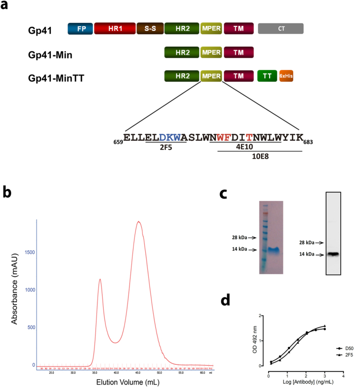
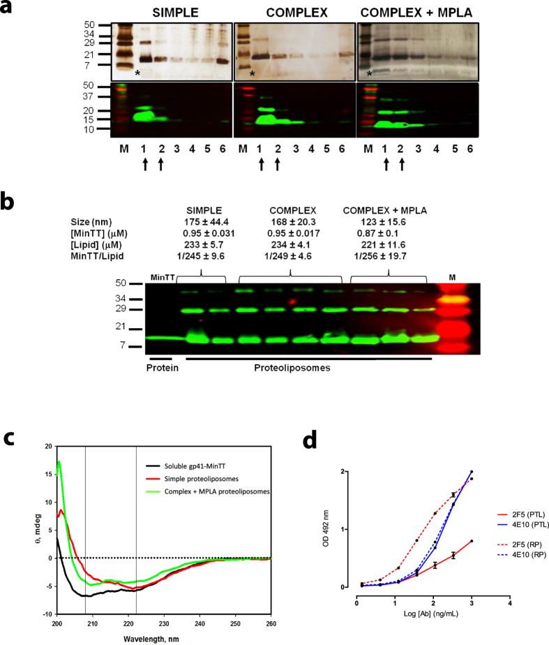
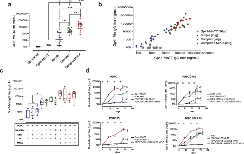
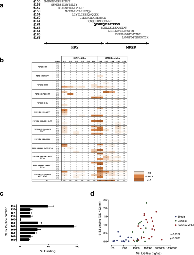
The HIV-1 gp41 Membrane Proximal External Region (MPER) is recognized by broadly neutralizing antibodies and represents a promising vaccine target. However, MPER immunogenicity and antibody activity are influenced by membrane lipids. To evaluate lipid modulation of MPER immunogenicity, we generated a 1-Palmitoyl-2-oleoylphosphatidylcholine (POPC)-based proteoliposome collection containing combinations of phosphatidylserine (PS), GM3 ganglioside, cholesterol (CHOL), sphingomyelin (SM) and the TLR4 agonist monophosphoryl lipid A (MPLA). A recombinant gp41-derived miniprotein (gp41-MinTT) exposing the MPER and a tetanus toxoid (TT) peptide that favors MHC-II presentation, was successfully incorporated into lipid mixtures (>85%). Immunization of mice with soluble gp41-MinTT exclusively induced responses against the TT peptide, while POPC proteoliposomes generated potent anti-gp41 IgG responses using lower protein doses. The combined addition of PS and GM3 or CHOL/SM to POPC liposomes greatly increased gp41 immunogenicity, which was further enhanced by the addition of MPLA. Responses generated by all proteoliposomes targeted the N-terminal moiety of MPER overlapping the 2F5 neutralizing epitope. Our data show that lipids impact both, the epitope targeted and the magnitude of the response to membrane-dependent antigens, helping to improve MPER-based lipid carriers. Moreover, the identification of immunodominant epitopes allows for the redesign of immunogens targeting MPER neutralizing determinants.


Figures

Figure 1
Figure 1. Gp41-MinTT expression and purification.
(a) Schematic representation of gp41, gp41-Min and gp41-MinTT proteins is shown. FP, fusion peptide (blue); HR1, N-terminal heptad repeat (red); S-S, disulfide loop (brown); HR2, C-terminal heptad repeat (green); MPER, membrane proximal external region (yellow); TM, transmembrane domain (purple); CT, cytoplasmic tail (gray); TT, tetanus toxoid epitope (light green); 6xHis, 6-histidine tag (orange). MPER-spanning sequence (residues 659–683, HXB2 numbering) is depicted. MPER sequences containing the 2F5, 4E10 and 10E8 epitopes are underlined. 2F5 neutralizing core and residues equally recognized by 4E10 and 10E8 are highlighted in blue and red respectively. (b) Gel filtration chromatography. Elution profile of the latter purification step is shown. (c) A highly pure 15KDa protein was recovered and concentrated from central fractions of the largest peak shown in panel B (44–49 mL fractions), as confirmed by SDS-PAGE and coomassie staining (left) and by Western blot using the 2F5 antibody (right). Molecular markers are indicated. (d) Antigenicity of purified gp41-MinTT protein determined by ELISA using serial dilutions of D50 (anti-HR2) and 2F5 (anti-MPER) antibodies.
Figure 2
Figure 2. Proteoliposomes characterization.
(a) Silver stained gels (top) and Western blots (bottom) of each sucrose gradient fractions corresponding to representative proteoliposomes of different composition (simple, complex and complex + MPLA). After proteoliposomes floatation, a sample of each recovered fraction was used for silver staining and Western blot analysis. Proteoliposomes could be detected in the first two fractions (arrows) by the signals of the protein and lipids (asterisks). M: Molecular Weight Marker and numbering 1 to 6 corresponds to the collected fractions from the top to the bottom of the ultracentrifuge tube. (b) Mean values and standard deviations of vesicle size, quantified protein and lipid amounts of each proteoliposome group are indicated. Ranges of protein/lipid ratios are also shown. Western blot analysis of the selected proteoliposome fractions for immunization is shown below. Purified gp41-MinTT protein was used as reference (left lane). (c) Secondary structure determination by circular dichroism. Near UV Spectra of gp41-MinTT in solution (black), simple (red) and complex + MPLA (green) proteoliposome formulations. Data are representative of two independent preparations. (d) Comparison of the antigenicity of gp41-MinTT-containing proteoliposomes (PTL, solid lines) and gp41-MinTT recombinant protein (RP, dotted lines) by ELISA using 2F5 (red) and 4E10 (blue) antibodies. Graph shows specific signal (OD 492 nm) of serial dilutions of monoclonal antibodies against both antigens captured by D50 antibody. Samples were assayed in duplicate and values are expressed as the mean and the interquartile range. Data are representative of two independent experiments.
Figure 3
Figure 3. Effect of lipid composition on anti-gp41 IgG response.
(a) Summary of anti-gp41-Min specific IgG titers, at sacrifice day, of C57 BL/6 mice immunized with gp41-MinTT proteoliposomes of simple (blue), complex (green) and complex incorporating MPLA (red) compositions. Data from animals immunized with control liposomes (black) and soluble gp41-MinTT protein (grey) are included. (b) Correlation of IgG titers, at sacrifice, against gp41-MinTT and gp41-Min antigens of mice immunized with gp41-MinTT as recombinant protein or formulated in proteoliposomes. (c) Influence of lipid mixtures over gp41-MinTT immunogenicity. Gp41-IgG titer at sacrifice day is shown. (d) Evolution of the gp41-Min IgG response of animals immunized with gp41-MinTT proteoliposomes based on POPC (upper left); POPC and GM3 (upper right); POPC and PS (down left); POPC combined with GM3 and PS (down right) in a simple, complex or complex with MPLA composition (blue, green and red lines respectively). Asterisks indicate immunization time points. In all panels, IgG titer is displayed as ng/mL referred to the D50 antibody, which was used as standard. Data show the median and interquartile range of at least two independent determinations. In panels (a) and (c), ***, ** and * denote p < 0.0001, p < 0.001 and p < 0.01 respectively. ns, not significant.
Figure 4
Figure 4. Mapping of humoral response against gp41-overlapping peptides.
(a) 15-mer overlapping peptide collection covering the HR2 (OLP peptides #155–161) and the MPER (OLP peptides #162–166) used for mapping. (b) OD values of the indicated immunized mice sera (60-fold dilution) against OLP peptides in ELISA. 2F5 binding is also indicated. Color code is shown. (c) For each animal the peptide yielding the strongest signal was assigned 100%. Bar diagram shows the percentage of signal of each peptide in all animals (mean+/− SD), highlighting the immunodominance of the #162 peptide sequence. (d) Spearman’s correlation of #162 binding signals and gp41-Min titer of immunized mice sera. Data are representative of two independent experiments. Spearman’s correlation coefficient and p value are indicated.
Figure 5
Figure 5. Alanine-scanning analysis of the immunodominance against #OLP-162.
(a) 29 mice sera displaying the highest signals against OLP#162 peptide were tested for binding against a collection of OLP#162-alanine mutants. Values indicate the binding percentage relative to the wild type peptide signal. 2F5 profile is also included. Color code is indicated. (b) Percentage of residual binding (mean+/− SD) for each alanine mutant peptide is shown. Immunodominant residues (<50% residual binding) are highlighted in red in both panels. HXB2 numbering of Q653 and W666 residues is indicated in grey.
Figure 6
Figure 6. In silico structural model of gp41-Min.
(a) Structural model embedded in a POPC bilayer. (b,c) Detail of the helix turns containing relevant residues for #162 binding in POPC (b) and complex (c) bilayers. Gp41-Min protein is shown in a ribbon representation. The helix turn containing L660 and L661 is shown in black and the turn containing W666 is shown in grey. In addition, all tryptophans present in gp41-Min and some residues specifically named in the text are labeled with arrows and represented as sticks. Lipid bilayers are represented as solvent accessible surface, truncated along the main protein axis to allow its visualization. (d,e) Number of water molecules within a sphere of 3 Å around lateral chains of residues D664 (left), K665 (middle) and W666 (right) in the gp41-Min model embedded in a POPC (d) and complex (e) bilayers throughout the molecular dynamics. Mean values are represented by a continuous horizontal line. The respective mean values obtained from the molecular dynamics performed in the absence of the lipid bilayer (water-only solvated model) are also shown for comparison (discontinuous line).

References

    1. Ward A. B. & Wilson I. A. Insights into the trimeric HIV-1 envelope glycoprotein structure. Trends Biochem. Sci. 40, 101–107 (2015). - PMC - PubMed
    1. Walker L. M. et al. A limited number of antibody specificities mediate broad and potent serum neutralization in selected HIV-1 infected individuals. PLoS Pathog. 6, e1001028 (2010). - PMC - PubMed
    1. Binley J. M. et al. Profiling the specificity of neutralizing antibodies in a large panel of plasmas from patients chronically infected with human immunodeficiency virus type 1 subtypes B and C. J. Virol. 82, 11651–11668 (2008). - PMC - PubMed
    1. Binley J. M. et al. Comprehensive Cross-Clade Neutralization Analysis of a Panel of Anti-Human Immunodeficiency Virus Type 1 Monoclonal Antibodies Comprehensive Cross-Clade Neutralization Analysis of a Panel of Anti-Human Immunodeficiency Virus Type 1 Monoclonal Antibodies. J. Virol. 78, 13232–13252 (2004). - PMC - PubMed
    1. Simek M. D. et al. Human immunodeficiency virus type 1 elite neutralizers: individuals with broad and potent neutralizing activity identified by using a high-throughput neutralization assay together with an analytical selection algorithm. J. Virol. 83, 7337–7348 (2009). - PMC - PubMed

Publication types