. 2017 Jan 13:7:40462.
doi: 10.1038/srep40462.

Improvement of Uveal and Capsular Biocompatibility of Hydrophobic Acrylic Intraocular Lens by Surface Grafting with 2-Methacryloyloxyethyl Phosphorylcholine-Methacrylic Acid Copolymer

Affiliations

Improvement of Uveal and Capsular Biocompatibility of Hydrophobic Acrylic Intraocular Lens by Surface Grafting with 2-Methacryloyloxyethyl Phosphorylcholine-Methacrylic Acid Copolymer

Xuhua Tan et al. Sci Rep. .

Abstract

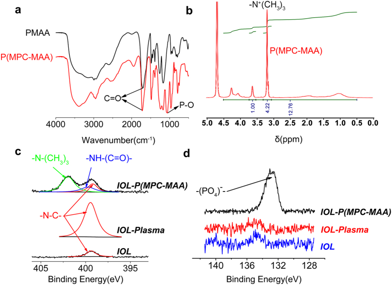
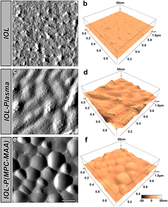
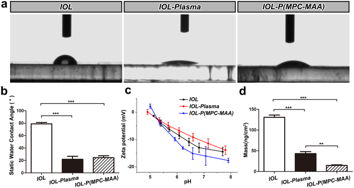
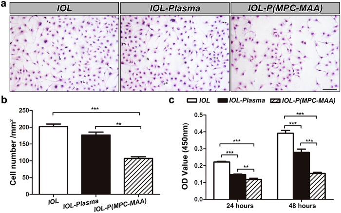
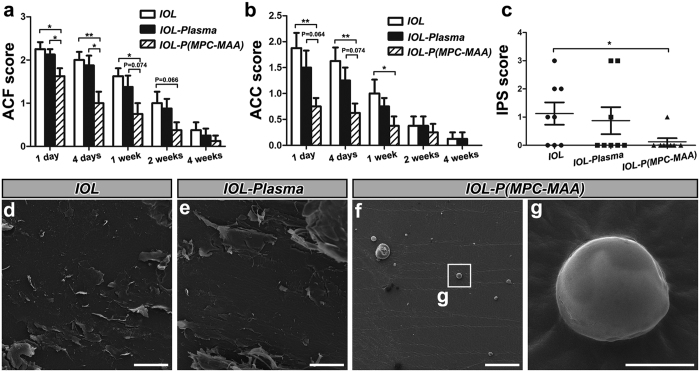
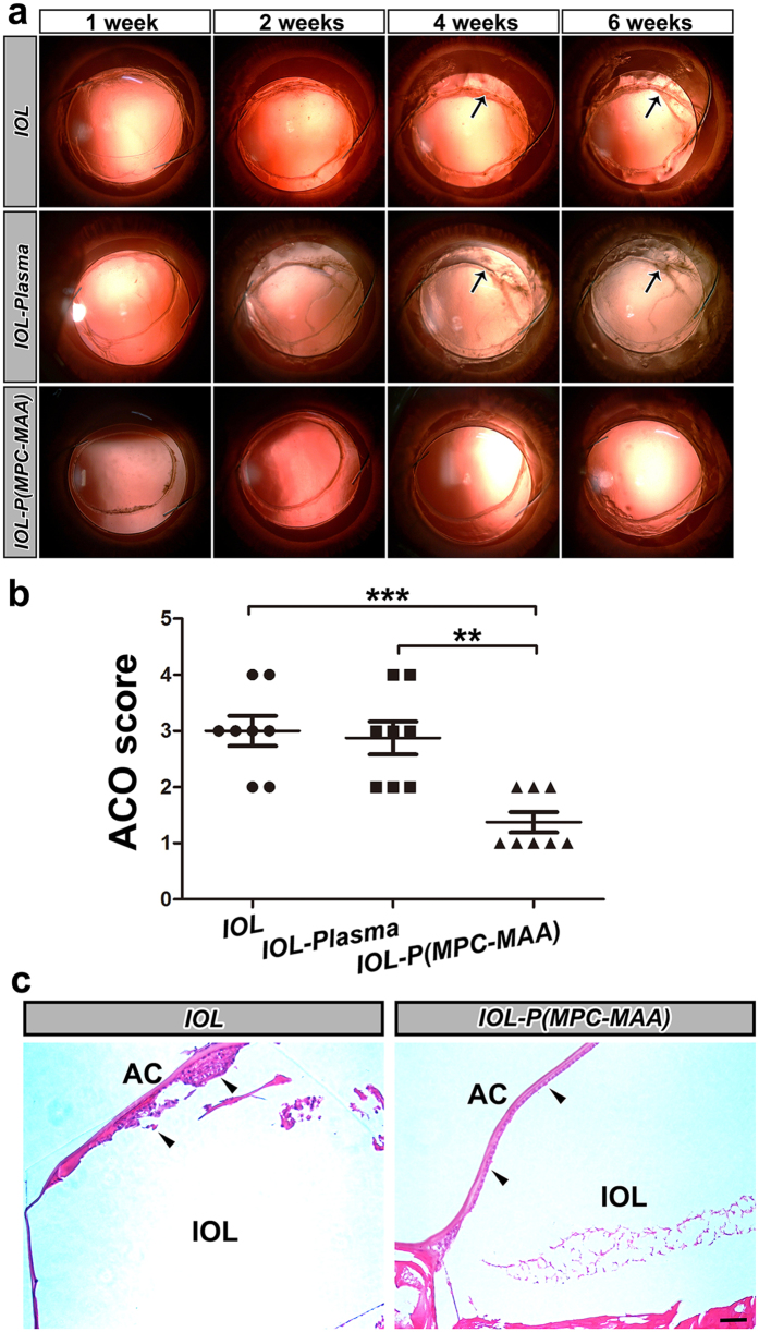
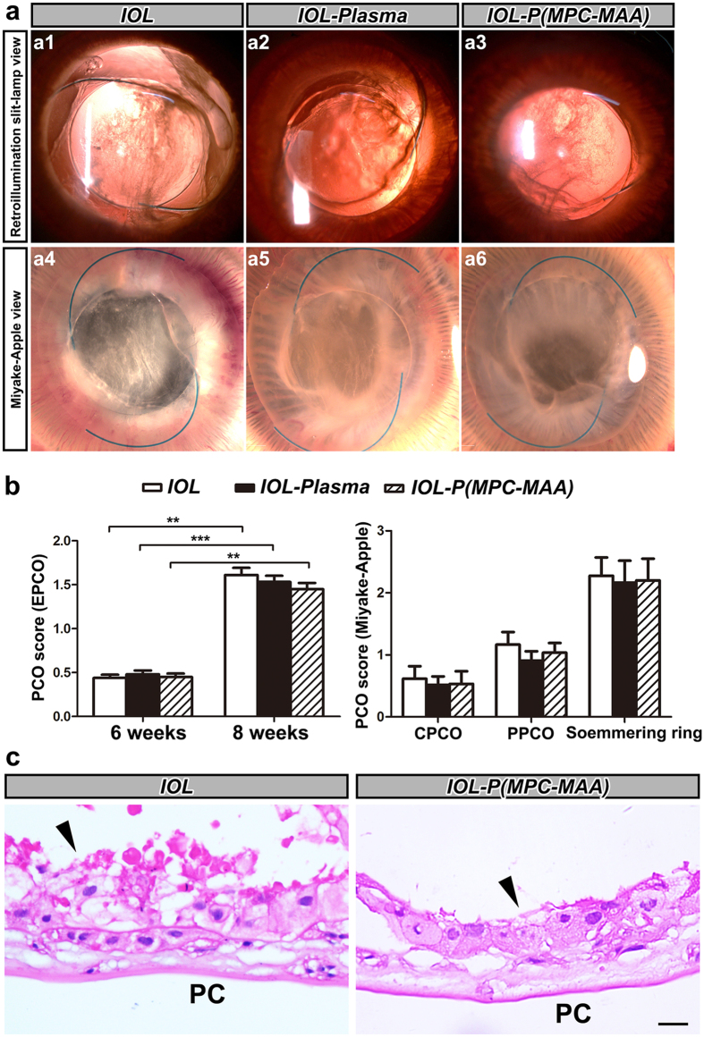
Biocompatibility of intraocular lens (IOL) is critical to vision reconstruction after cataract surgery. Foldable hydrophobic acrylic IOL is vulnerable to the adhesion of extracellular matrix proteins and cells, leading to increased incidence of postoperative inflammation and capsule opacification. To increase IOL biocompatibility, we synthesized a hydrophilic copolymer P(MPC-MAA) and grafted the copolymer onto the surface of IOL through air plasma treatment. X-ray photoelectron spectroscopy, atomic force microscopy and static water contact angle were used to characterize chemical changes, topography and hydrophilicity of the IOL surface, respectively. Quartz crystal microbalance with dissipation (QCM-D) showed that P(MPC-MAA) modified IOLs were resistant to protein adsorption. Moreover, P(MPC-MAA) modification inhibited adhesion and proliferation of lens epithelial cells (LECs) in vitro. To analyze uveal and capsular biocompatibility in vivo, we implanted the P(MPC-MAA) modified IOLs into rabbits after phacoemulsification. P(MPC-MAA) modification significantly reduced postoperative inflammation and anterior capsule opacification (ACO), and did not affect posterior capsule opacification (PCO). Collectively, our study suggests that surface modification by P(MPC-MAA) can significantly improve uveal and capsular biocompatibility of hydrophobic acrylic IOL, which could potentially benefit patients with blood-aqueous barrier damage.


Figures

Figure 1
Figure 1. P(MPC-MAA) was successfully synthesized and grafted onto the IOL surface.
(a) FT-IR spectra of PMAA and P(MPC-MAA) detecting with the potassium bromide pressed-disk technique for 32 scans over the 500–4,000 cm−1 range at a resolution of 4.0 cm−1. (b) H-NMR spectra of P(MPC-MAA) determined at 400 MHz. (c,d) N1s high-resolution spectra and P1s high-resolution spectra of IOL, IOL-Plasma, and IOL-P(MPC-MAA), respectively.
Figure 2
Figure 2. Surface characterization of IOL by AFM.
Representative AFM images of the IOL, IOL-Plasma, and IOL-P(MPC-MAA) surfaces. Area size for each scan: 1.0 × 1.0 μm2. Scale bar = 200 nm.
Figure 3
Figure 3. P(MPC-MAA) modification increases surface hydrophilicity and decreases protein adsorption.
(a) Representative images of static WCA measurement in each group at 25 °C. (b) Quantification of static WCA in each group. n = 5. **P < 0.01, ***P < 0.001. (c) Quantification of zeta potential in each group. n = 4. (d) Measurement of BSA adsorption by QCM. n = 3. **P < 0.01, ***P < 0.001.
Figure 4
Figure 4. IOL-P(MPC-MAA) inhibits the adhesion and proliferation of LECs in vitro.
(a) Representative inverted phase contrast microscope images of LECs attached to the surface of IOL, IOL-Plasma, and IOL-P(MPC-MAA), respectively. Scale bar = 100 μm. (b) Quantification of the number of cells attached to the surfaces of IOLs in (a). In each group, 5 IOLs were chosen, and in each IOL, five fields were selected with one in the central and four in peripheral quadrants at random. (c) Cell viability assay shows the proliferation of LECs on the surfaces of IOLs after incubation for 24 and 48 hours, respectively. n = 3. **P < 0.01, ***P < 0.001.
Figure 5
Figure 5. IOL-P(MPC-MAA) reduces postoperative inflammation after cataract surgery.
(a,b) Quantification of ACF and ACC scores in each group 1 day, 4 days, 1 week, 2 weeks, and 4 weeks after surgery, respectively. n = 8. *P < 0.05, **P < 0.01. (c) Quantification of IPS scores in each group 8 weeks after surgery. n = 8. *P < 0.05, **P < 0.01. (dg) Representative SEM images of IOL, IOL-Plasma, and IOL-P(MPC-MAA) surfaces 8 weeks after surgery. (df) Scale bar = 100 μm. (g) Scale bar = 5 μm.
Figure 6
Figure 6. IOL-P(MPC-MAA) suppresses ACO formation after cataract surgery.
(a) Representative retroillumination slit lamp photos of anterior capsule opacification in each group 1 week, 2 weeks, 4 weeks, and 6 weeks after surgery, respectively. Black arrows indicate anterior capsule fibrosis and shrinkage. (b) Quantification of ACO scores in each group 6 weeks after surgery. n = 8. **P < 0.01, ***P < 0.001. (c) Representative HE staining images of anterior capsule in each group 8 weeks after surgery. Black arrowheads indicate LECs under anterior capsule. AC: anterior capsule. Scale bar = 50 μm.
Figure 7
Figure 7. IOL-P(MPC-MAA) does not affect PCO formation after cataract surgery.
(a) Representative photos of posterior capsule opacification in each group 8 weeks after surgery. a1–a3: retroillumination slit-lamp view, a4–a6: Miyake-Apple view. (b) Quantification of PCO scores in each group by EPCO 2000 software or Miyake-Apple view analysis, respectively. n = 8. **P < 0.01, ***P < 0.001. CPCO: central posterior capsule opacification, PPCO: periphery posterior capsule opacification. (c) Representative HE staining images of posterior capsule in each group 8 weeks after surgery. Black arrowheads indicate migration and proliferation of LECs on the posterior capsule. PC: posterior capsule. Scale bar = 50 μm.

References

    1. Khairallah M. et al. Number of People Blind or Visually Impaired by Cataract Worldwide and in World Regions, 1990 to 2010. Invest Ophthalmol Vis Sci 56, 6762–6769, doi: 10.1167/iovs.15-17201 (2015). - DOI - PubMed
    1. Wang G. et al. In vivo implantation of hydrophobic acrylic intraocular lenses with surface modification. Eye Sci 28, 176–179 (2013). - PubMed
    1. Abela-Formanek C., Amon M., Kahraman G., Schauersberger J. & Dunavoelgyi R. Biocompatibility of hydrophilic acrylic, hydrophobic acrylic, and silicone intraocular lenses in eyes with uveitis having cataract surgery: Long-term follow-up. J Cataract Refract Surg 37, 104–112, doi: 10.1016/j.jcrs.2010.07.038 (2011). - DOI - PubMed
    1. Richter-Mueksch S. et al. Uveal and capsular biocompatibility after implantation of sharp-edged hydrophilic acrylic, hydrophobic acrylic, and silicone intraocular lenses in eyes with pseudoexfoliation syndrome. J Cataract Refract Surg 33, 1414–1418, doi: 10.1016/j.jcrs.2007.05.009 (2007). - DOI - PubMed
    1. Saika S., Miyamoto T. & Ohnishi Y. Histology of anterior capsule opacification with a polyHEMA/HOHEXMA hydrophilic hydrogel intraocular lens compared to poly(methyl methacrylate), silicone, and acrylic lenses. J Cataract Refract Surg 29, 1198–1203 (2003). - PubMed

Publication types

MeSH terms