. 2017 Oct;35(10):2243-2250.
doi: 10.1002/jor.23519. Epub 2017 Mar 23.

Predicting early symptomatic osteoarthritis in the human knee using machine learning classification of magnetic resonance images from the osteoarthritis initiative

Affiliations

Predicting early symptomatic osteoarthritis in the human knee using machine learning classification of magnetic resonance images from the osteoarthritis initiative

Beth G Ashinsky et al. J Orthop Res. 2017 Oct.

Abstract

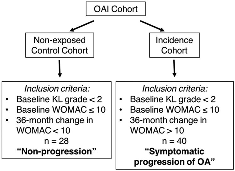
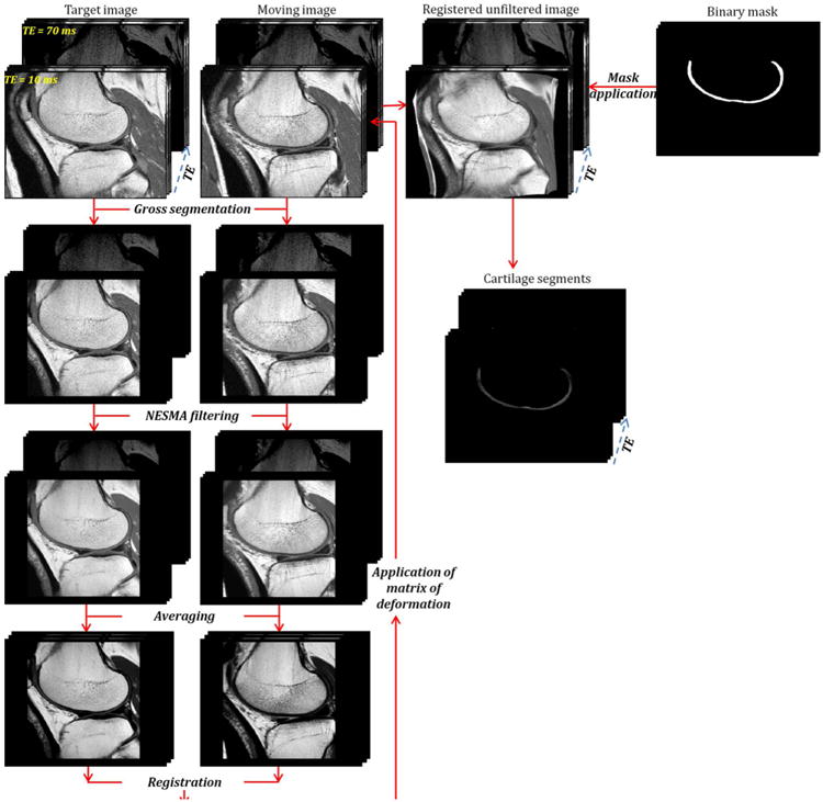
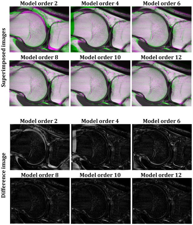
The purpose of this study is to evaluate the ability of a machine learning algorithm to classify in vivo magnetic resonance images (MRI) of human articular cartilage for development of osteoarthritis (OA). Sixty-eight subjects were selected from the osteoarthritis initiative (OAI) control and incidence cohorts. Progression to clinical OA was defined by the development of symptoms as quantified by the Western Ontario and McMaster Universities Arthritis (WOMAC) questionnaire 3 years after baseline evaluation. Multi-slice T2 -weighted knee images, obtained through the OAI, of these subjects were registered using a nonlinear image registration algorithm. T2 maps of cartilage from the central weight bearing slices of the medial femoral condyle were derived from the registered images using the multiple available echo times and were classified for "progression to symptomatic OA" using the machine learning tool, weighted neighbor distance using compound hierarchy of algorithms representing morphology (WND-CHRM). WND-CHRM classified the isolated T2 maps for the progression to symptomatic OA with 75% accuracy.

Clinical significance: Machine learning algorithms applied to T2 maps have the potential to provide important prognostic information for the development of OA. © 2017 Orthopaedic Research Society. Published by Wiley Periodicals, Inc. J Orthop Res 35:2243-2250, 2017.

Keywords: MRI; classification; osteoarthritis; pattern recognition; registration; segmentation.


Conflict of interest statement

Conflict of interest: The authors have no conflicts of interest to declare. The study sponsors had no involvement in the study design, collection and interpretation of data, writing of the manuscript or manuscript submission.

Figures

Figure 1
Figure 1
Outline of study design. The non-progression group was randomly selected from the OAI Control cohort based on criteria of baseline KL grade < 2, baseline WOMAC ≤ 10 and a 36-month change in WOMAC < 10 (n = 28). The symptomatic progression of OA group was randomly selected from the OAI Incidence cohort based on the criteria of baseline KL grade < 2, baseline WOMAC ≤ 10 and a 36-month change in WOMAC > 10 (n = 40). MRIs from these 68 patients were then processed and used for classification analysis.
Figure 2
Figure 2
Flow chart of processing steps leading from the unfiltered and unregistered moving image to the cartilage segments. The target and unregistered images were cropped to reduce the field of view and to suppress regions of fat and muscle. Then, a multispectral non-local filter was applied to these images. After filtering, both the multi-spectral target and unregistered filtered weighted images were each averaged across TE. The averaged filtered unregistered image was then registered to the averaged filtered target image. The calculated matrix of deformation derived from this registration was directly applied to the original unfiltered and unregistered T2W image at each TE. A 3D cartilage mask was generated through manual segmentation of each slice from the target image, which was then systematically applied to each of the registered images to isolate cartilage segments from all subjects.
Figure 3
Figure 3
Examples of the quality of registration for six different orders of the nonlinear polynomial transformation model. For each model order, performance of the registration was evaluated through superimposed and difference images between target and registered images. The green and purple colors in the superimposed images indicate misregistered regions. Both the overlap and difference between target and registered images decrease as the order of the model increases from second to eighth order, with no improvement seen for higher order corrections.
Figure 4
Figure 4
Representative T2 maps calculated before and after registration in knee cartilage regions. T2 maps were superimposed on their corresponding T2W images before and after registration. The T2 values are shown to be minimally changed before and after registration, indicating that the registration methods did not introduce bias to the T2 maps.

References

    1. Swedberg JA, Steinbauer JR. Osteoarthritis. Am Fam Physician. 1992;45:557–568. - PubMed
    1. Davis MA. Epidemiology of osteoarthritis. Clin Geriatr Med. 1988;4:241–255. - PubMed
    1. Braun HJ, Gold GE. Advanced MRI of articular cartilage. Imaging Med. 2011;3:541–555. - PMC - PubMed
    1. Braun HJ, Gold GE. Diagnosis of osteoarthritis: imaging. Bone. 2012;51:278–288. - PMC - PubMed
    1. Ashinsky B, Bouhrara M, Urish KL, Coletta CE, Goldberg IG, Spencer RG. The Orthopaedic Research Society. Las Vegas, NV: 2015. Predicting Early Osteoarthritis in the Human Knee: Pattern Recognition and Machine Learning Classification of Magnetic Resonance Images. - PMC - PubMed

Publication types