

The Rotator Cuff Organ: Integrating Developmental Biology, Tissue Engineering, and Surgical Considerations to Treat Chronic Massive Rotator Cuff Tears
- PMID: 28084902
- PMCID: PMC5567418
- DOI: 10.1089/ten.TEB.2016.0446
The Rotator Cuff Organ: Integrating Developmental Biology, Tissue Engineering, and Surgical Considerations to Treat Chronic Massive Rotator Cuff Tears
Abstract
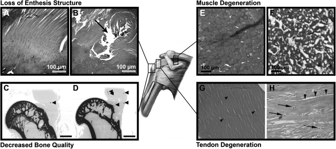
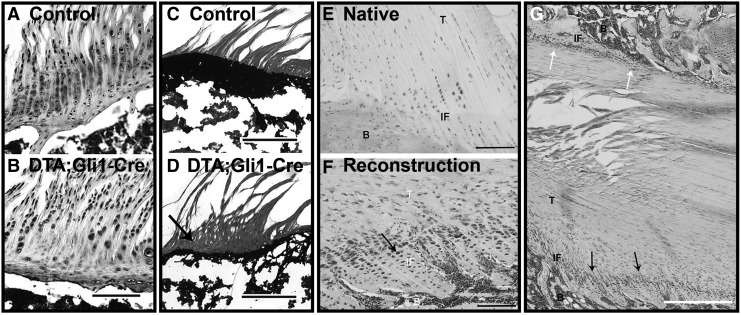
The torn rotator cuff remains a persistent orthopedic challenge, with poor outcomes disproportionately associated with chronic, massive tears. Degenerative changes in the tissues that comprise the rotator cuff organ, including muscle, tendon, and bone, contribute to the poor healing capacity of chronic tears, resulting in poor function and an increased risk for repair failure. Tissue engineering strategies to augment rotator cuff repair have been developed in an effort to improve rotator cuff healing and have focused on three principal aims: (1) immediate mechanical augmentation of the surgical repair, (2) restoration of muscle quality and contractility, and (3) regeneration of native enthesis structure. Work in these areas will be reviewed in sequence, highlighting the relevant pathophysiology, developmental biology, and biomechanics, which must be considered when designing therapeutic applications. While the independent use of these strategies has shown promise, synergistic benefits may emerge from their combined application given the interdependence of the tissues that constitute the rotator cuff organ. Furthermore, controlled mobilization of augmented rotator cuff repairs during postoperative rehabilitation may provide mechanotransductive cues capable of guiding tissue regeneration and restoration of rotator cuff function. Present challenges and future possibilities will be identified, which if realized, may provide solutions to the vexing condition of chronic massive rotator cuff tears.
Keywords: developmental engineering; enthesis; mechanobiology; rotator cuff.
Conflict of interest statement
No competing financial interests exist.
Figures
References
-
- Tashjian R.Z. Epidemiology, natural history, and indications for treatment of rotator cuff tears. Clin Sports Med 31, 589, 2012 - PubMed
-
- Miller R.M., Popchak A., Vyas D., Tashman S., Irrgang J.J., Musahl V., and Debski R.E. Effects of exercise therapy for the treatment of symptomatic full-thickness supraspinatus tears on in vivo glenohumeral kinematics. J Shoulder Elbow Surg 25, 641, 2016 - PubMed
-
- Moosmayer S., Tariq R., Stiris M., and Smith H.J. The natural history of asymptomatic rotator cuff tears a three-year follow-up of fifty cases. J Bone Joint Surg Am 95A, 1249, 2013 - PubMed
-
- Galatz L.M., Ball C.M., Teefey S.A., Middleton W.D., and Yamaguchi K. The outcome and repair integrity of completely arthroscopically repaired large and massive rotator cuff tears. J Bone Joint Surg Am 86A, 219, 2004 - PubMed
MeSH terms
Grants and funding
LinkOut - more resources
Full Text Sources
Other Literature Sources
Medical
