

Nailfold capillary morphology in exfoliation syndrome
- PMID: 28085140
- PMCID: PMC5437324
- DOI: 10.1038/eye.2016.312
Nailfold capillary morphology in exfoliation syndrome
Abstract
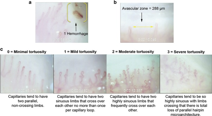
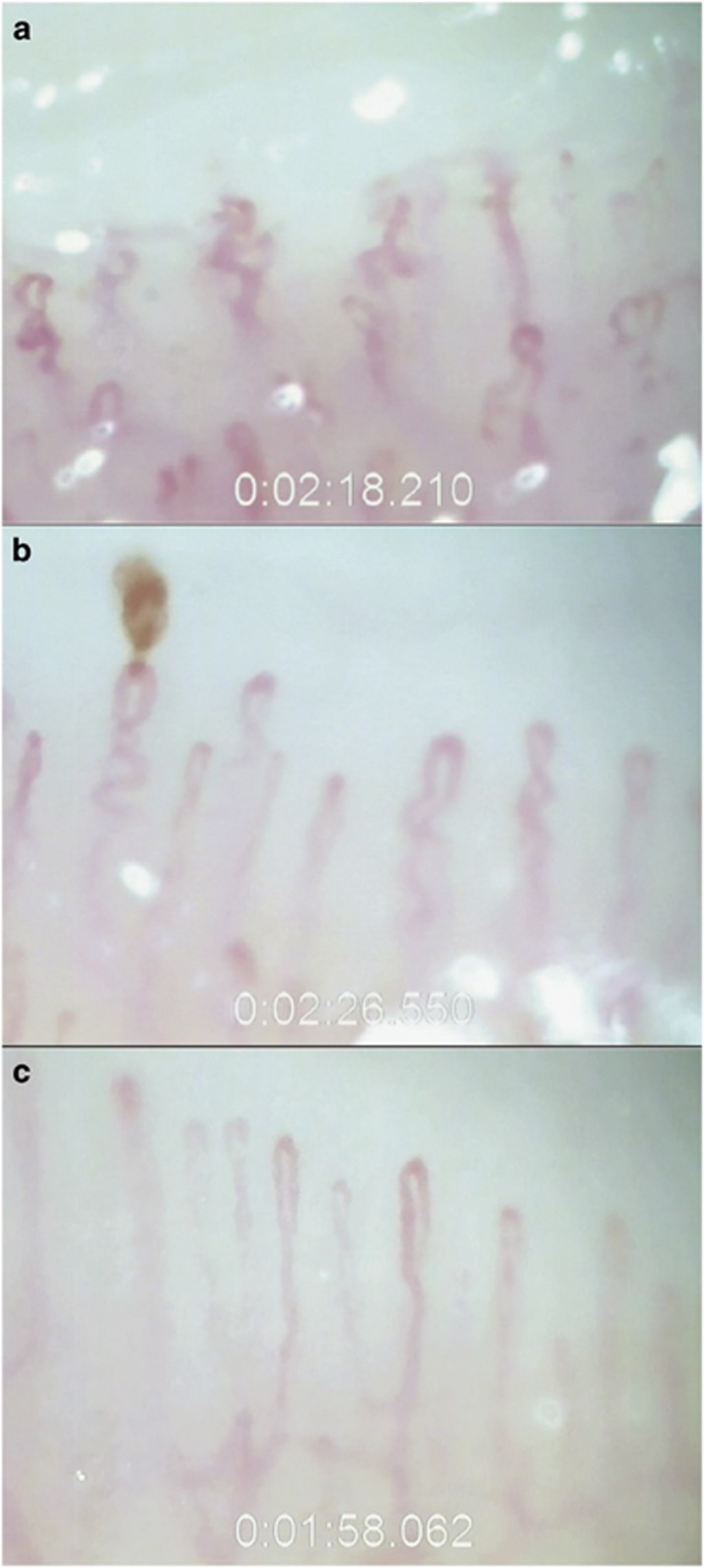
PurposeThe purpose of the study was to investigate nailfold microvascular morphology in exfoliation syndrome with or without glaucoma (XFS/XFG) compared with primary open-angle glaucoma (POAG) and control subjects using nailfold capillary videomicroscopy.Patients and methodsWe used a JH-1004 capillaroscope to perform nailfold capillary videomicroscopy on the fourth and fifth digit of the non-dominant hand. We enrolled 56 XFS/XFG patients, 87 POAG patients, and 75 control subjects. Masked observers graded the videos for hemorrhages, avascular zones ≥200 microns (μm), and degree of microvascular tortuosity on a four-point subjective scale. Multivariable odds ratios, 95% confidence intervals and P-for trends for assessing the relation between morphological changes and POAG or XFS/XFG were obtained from logistic regression analyses. We also assessed this relation with XFS/XFG compared with POAG in multivariable models.ResultsAfter adjusting for multiple covariates, nailfold hemorrhages, avascular zones ≥200 μm, and higher degree of vascular tortuosity were more common in XFS/XFG vs controls (P-for trend ≤0.0001) and in POAG vs controls (P-for trend ≤0.01). For each 100 capillaries, the number of hemorrhages was similar (P-for trend=0.91) between XFS/XFG and POAG patients; however, there were more avascular zones per 100 capillaries with borderline significance (P-for trend=0.04) in the XFS/XFG group. XFS/XFG patients had more tortuosity than POAG patients; specifically, having a tortuosity score ≥1.5 was associated with a 4.4-fold increased odds of XFS/XFG (95% confidence interval: 1.5-13.3) relative to a tortuosity score <1.0 (P-for trend=0.005).ConclusionA high degree of nailfold capillary tortuosity is a distinct non-ocular feature associated with XFS/XFG compared with either POAG or controls.
Conflict of interest statement
The authors declare no conflict of interest.
Figures
References
-
- Prince AM, Ritch R. Clinical signs of the pseudoexfoliation syndrome. Ophthalmology 1986; 93: 803–807. - PubMed
-
- Vesti E, Kivelä T. Exfoliation syndrome and exfoliation glaucoma. Prog Retin Eye Res 2000; 19: 345–368. - PubMed
-
- Ritch R, Schlötzer-Schrehardt U. Exfoliation syndrome. Surv Ophthalmol 2001; 45: 265–315. - PubMed
-
- Schlötzer-Schrehardt UM, Koca MR, Naumann GO, Volkholz H. Pseudoexfoliation syndrome. Ocular manifestation of a systemic disorder? Arch Ophthalmol 1992; 110: 1752–1756. - PubMed
-
- Streeten BW, Li ZY, Wallace RN, Eagle RC Jr., Keshgegian AA. Pseudoexfoliative fibrillopathy in visceral organs of a patient with pseudoexfoliation syndrome. Arch Ophthalmol 1992; 110: 1757–1762. - PubMed
Publication types
MeSH terms
Grants and funding
LinkOut - more resources
Full Text Sources
Other Literature Sources
