. 2017 Mar;24(3):523-533.
doi: 10.1038/cdd.2016.155. Epub 2017 Jan 13.

The BCL-2 pro-survival protein A1 is dispensable for T cell homeostasis on viral infection

Affiliations

The BCL-2 pro-survival protein A1 is dispensable for T cell homeostasis on viral infection

Selma Tuzlak et al. Cell Death Differ. 2017 Mar.

Abstract

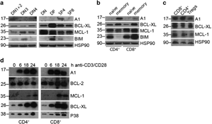
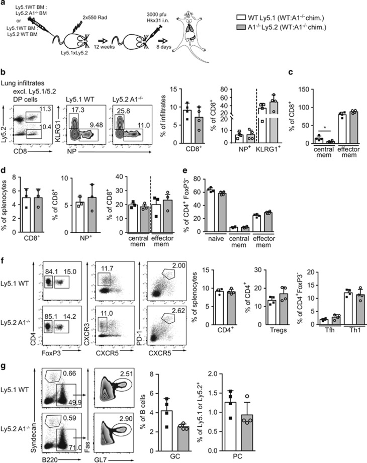
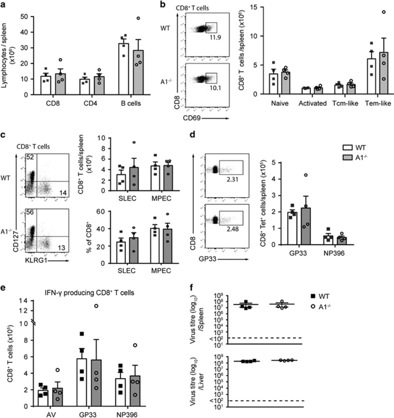
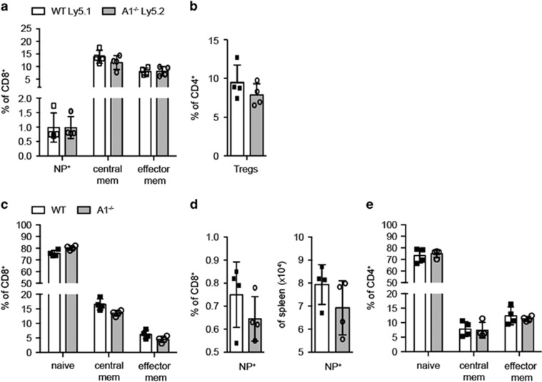
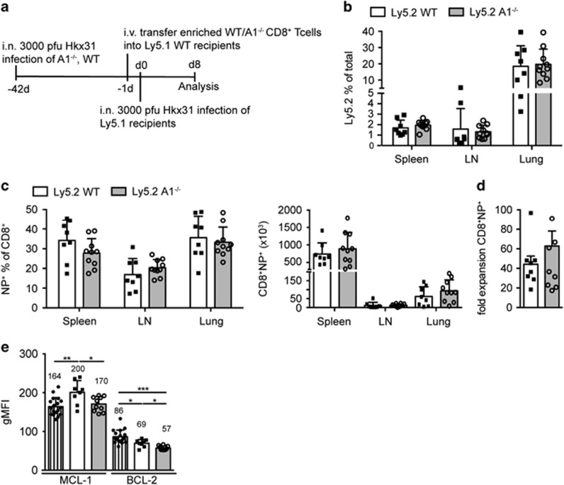
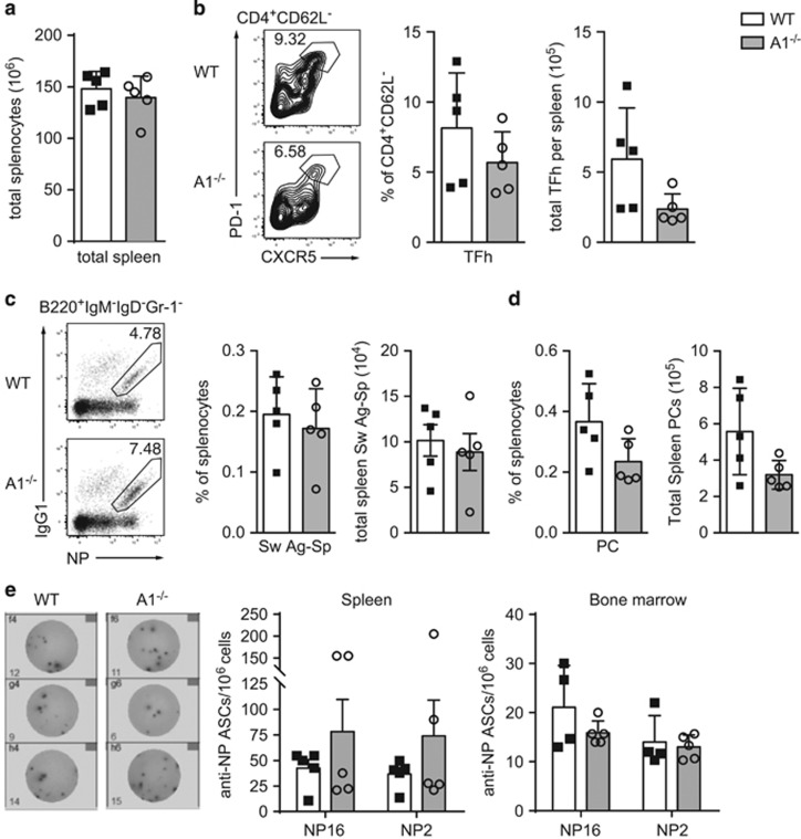
The physiological role of the pro-survival BCL-2 family member A1 has been debated for a long time. Strong mRNA induction in T cells on T cell receptor (TCR)-engagement suggested a major role of A1 in the survival of activated T cells. However, the investigation of the physiological roles of A1 was complicated by the quadruplication of the A1 gene locus in mice, making A1 gene targeting very difficult. Here, we used the recently generated A1-/- mouse model to examine the role of A1 in T cell immunity. We confirmed rapid and strong induction of A1 protein in response to TCR/CD3 stimulation in CD4+ as well as CD8+ T cells. Surprisingly, on infection with the acute influenza HKx31 or the lymphocytic choriomeningitis virus docile strains mice lacking A1 did not show any impairment in the expansion, survival, or effector function of cytotoxic T cells. Furthermore, the ability of A1-/- mice to generate antigen-specific memory T cells or to provide adequate CD4-dependent help to B cells was not impaired. These results suggest functional redundancy of A1 with other pro-survival BCL-2 family members in the control of T cell-dependent immune responses.


Conflict of interest statement

The authors declare no conflict of interest.

Figures

Figure 1
Figure 1
A1 is mainly expressed in SP4 thymocytes and memory T cells and is rapidly upregulated in T cells on TCR/CD3 stimulation. (a) CD4CD8CD44+ double-negative stage (DN)1 and DN2, CD4CD8CD25+CD44 DN3, CD4CD8CD44CD25 DN4, CD4CD8 total DN, CD4+CD8+ DP, CD4+CD8 single CD4 positive (SP4) and CD4CD8+ (SP8) wild-type thymocytes were FACS-sorted and lysed in NP-40 containing lysis buffer. Western blots were probed for the proteins indicated. Probing for HSP90 served as loading control. (b) Naive (CD62L+CD44) and memory-like (CD62LCD44+) CD4+ or CD8+ T cells were sorted from wild-type splenocytes and treated as described in (a). (c) 8.5 × 10⁵ CD8+, CD4+FOXP3 conventional T cells and CD4+FOXP3+ regulatory T cells (Tregs), respectively, were FACS-sorted from Foxp3YFP-cre mice according to their expression of CD8, CD4 and YFP. Cells were lysed directly in 1 × Laemmli buffer for western blot analysis. (d) CD4+ (left graph) or CD8+ (right graph) T cells were sorted from spleens of wild-type mice and activated in vitro with 5 μg/ml plate-bound anti-CD3 and 1 μg/ml anti-CD28 antibodies for the indicated times. Cells were lysed and western blot analysis performed for the indicated proteins. Membranes were probed with an antibody specific for p38 MAPK to control for equal protein loading. Western blots shown are representative of two independent experiments
Figure 2
Figure 2
A1−/− cells in chimaeric mice exhibit normal T cell immune responses in an acute influenza infection model. (a) Schematic overview of the influenza infection workflow: C57BL/6-Ly5.1xLy5.2 F1 mice were lethally irradiated and reconstituted with 1:1 mixed bone-marrow from C57BL/6-Ly5.1 wild-type and C57BL/6-Ly5.2 A1−/− mice. Reconstituted mice were infected intra-nasally (i.n.) with 3000 pfu HKx31 influenza virus and lungs and spleens were analysed 8 days post infection by flow cytometry. (b) Leukocyte infiltrates were isolated from lungs of chimaeric mice by gradient centrifugation. The recovered cells were analysed by flow cytometry for tetramer-positive antigen-specific (NP+) CD8+ T cells, KLRG1+ short-lived CD8+ effector T cells and (c) central (CD44+CD62L+) as well as effector (CD44+CD62L) memory-like CD8+ T cells. (d) Spleen cells from infected bone-marrow chimaeric mice were isolated and analysed for CD8+ NP+ cells, or central as well as effector memory-like CD8+ T cells. (e) CD4+ splenic T cells were further analysed for CD4+FoxP3 naive (CD62L+CD44-), central or effector memory-like subsets. (f) CD4+ splenic T cells were analysed for FoxP3+ Treg cells, FoxP3CXCR3+ Th1 cells, and FoxP3CXCR5+PD-1+ Tfh cells. (g) Spleen cells from infected mice were analysed for Syndecan1+ plasma cells (PC) and B220+FAS+GL7+ germinal centre B cells (GC). Bars represent means±S.E.M. (n=4), irradiation-resistant Ly5.1/5.2 DP cells were excluded by electronic gating
Figure 3
Figure 3
A1−/− mice exhibit normal T cell immune responses to chronic infection with LCMV docile. Wild-type and A1−/− mice were infected i.v. with 2 × 10⁶ pfu LCMV docile and spleens were analysed 8 days post infection by flow cytometry. (a) Total numbers of CD8+ as well as CD4+ T cells and B cells were assessed by cell counting and flow cytometric analysis. (b) CD8+ T cells were further analysed for CD62L+CD44 naive, CD69+ activated, CD44+CD62L+ central memory (Tcm) like, CD44+CD62L effector memory (Tem) like T cells. (c) CD8+ T cells were also analysed for CD127-KLRG1+ short-lived effector cells (SLEC) and CD127+KLRG1 memory precursor effector cells (MPEC). (d) The numbers of GP33+ low-affinity antigen-specific, or NP396+ high-affinity antigen-specific CD8+ T cells were assessed. (e) Spleen cells from infected mice were cultured in the presence of adenovirus, GP33, or NP396 antigen overnight and intracellular staining for IFN-γ was performed and cells analysed by flow cytometry. (f) LCMV titres were calculated from spleens and livers of infected mice 8 days post infection. Bars represent means±S.E.M. (n=4)
Figure 4
Figure 4
A1−/− T cells show normal in vivo memory formation. (a) 1 : 1 C57BL/6-Ly5.1 wild-type and C57BL/6-Ly5.2 A1−/− bone-marrow chimaeric mice were infected i.n. with 3000 pfu HKx31 influenza virus and spleen cells were analysed 41 days post infection by flow cytometry. CD8+ T cells were analysed for NP+ antigen-specific T cells as well as for central memory and effector memory-like phenotypes. (b) The frequencies of Treg cells within the CD4+ T cell population were analysed by intracellular flow cytometry for FoxP3 expression. (c) Wild-type and A1−/− mice were infected i.n. with 3000 pfu HKx31 influenza virus and spleen cells were analysed 41 days post infection by flow cytometry. CD8+ T cell populations were analysed for naive, central memory, and effector memory T cells. (d) The frequencies and total numbers of NP+ antigen-specific CD8+ T cells were assessed by flow cytometry. (e) CD4+ T cells in the spleens of infected mice were analysed for naive, central memory, and effector memory T cells. Bars represent means ±S.E.M. (n=4)
Figure 5
Figure 5
A1−/− T cells expand normally in an influenza rechallenging model. (a) Schematic overview of the influenza recall immune response workflow: wild-type and A1−/− mice were infected i.n. with 3000 pfu HKx31 influenza virus and after 41 days mice were killed and CD8+ T cells were enriched by negative magnetic bead sorting. A total of 2 × 10⁴ NP-specific CD8+ T cells were transferred i.v. into naive C57BL/6-Ly5.1 wild-type recipient mice. One day later mice were infected i.n. with 3000 pfu HKx31 influenza and 8 days later lungs, mLN and spleens were isolated and analysed by flow cytometry. (b) The frequencies of Ly5.2+ donor cells were analysed in the spleen, mLN and lung infiltrates. (c) The frequencies and total numbers of NP+ antigen-specific Ly5.2+CD8+ T cells were analysed in the spleens, mLN, and lung infiltrates of infected mice. (d) The fold expansion of total Ly5.2+CD8+NP+ T cells was calculated by comparing the numbers of injected Ly5.2+CD8+NP+ T cells to the number of recovered Ly5.2+CD8+NP+ T cells from spleen, mLN and lung. (e) Intracellular staining for MCL-1 and BCL-2 was performed on total spleen cells of infected mice and the geometric mean fluorescent intensity (gMFI) for MCL-1 and BCL-2 protein in Ly5.2+CD8+ donor T cells was compared with that from similarly stained Ly5.1+CD8+ host T cells. Bars represent means±S.E.M. (n=8–10). A Mann Whitney statistical test was performed, *P<0.05, **P<0.01, ***P<0.0001
Figure 6
Figure 6
CD4 T-helper cell function is not impaired in A1−/− mice immunised with NP-KLH/Alum. (a) The numbers of total spleen cells of wild-type and A1−/− mice that had been immunised i.p. with 100 μg/20 g body weight NP-KLH/Alum 14 days earlier were determined. (b) The frequencies and total numbers of CD4+CXCR5+PD-1+ Tfh cells in immunised wild-type and A1−/− mice were analysed by flow cytometry. (c) The numbers of IgG1+NP+B220+ class-switched antigen-specific B cells and (d) Syndecan1+ plasma cells in the spleen were determined by flow cytometry. (e) Overall, 1 × 10⁶ spleen cells or bone-marrow cells were cultured overnight on NP16- or NP2-coated ELISA plates and ELISpot assays were performed. Antigen-specific plasma cells producing low (NP16) or high-affinity (NP2) antibodies against NP per 106 cells were quantified. Pictures show representative ELISpots from wild-type or A1−/− bone-marrow cells cultured overnight on NP2-coated ELISA plates. Bars represent means±S.E.M. (n=5)

References

    1. Tan JT, Dudl E, LeRoy E, Murray R, Sprent J, Weinberg KI et al. IL-7 is critical for homeostatic proliferation and survival of naive T cells. Proc Natl Acad Sci USA 2001; 98: 8732–8737. - PMC - PubMed
    1. Opferman JT, Letai A, Beard C, Sorcinelli MD, Ong CC, Korsmeyer SJ. Development and maintenance of B and T lymphocytes requires antiapoptotic MCL-1. Nature 2003; 426: 671–676. - PubMed
    1. Koenen P, Heinzel S, Carrington EM, Happo L, Alexander WS, Zhang JG et al. Mutually exclusive regulation of T cell survival by IL-7 R and antigen receptor-induced signals. Nat Commun 2013; 4: 1735. - PMC - PubMed
    1. Sandalova E, Wei CH, Masucci MG, Levitsky V. Regulation of expression of Bcl-2 protein family member Bim by T cell receptor triggering. Proc Natl Acad Sci USA 2004; 101: 3011–3016. - PMC - PubMed
    1. Wensveen FM, van Gisbergen KP, Derks IA, Gerlach C, Schumacher TN, van Lier RA et al. Apoptosis threshold set by Noxa and Mcl-1 after T cell activation regulates competitive selection of high-affinity clones. Immunity 2010; 32: 754–765. - PubMed

Publication types

MeSH terms

Substances

LinkOut - more resources