. 2017 Jan 13:2:16253.
doi: 10.1038/nmicrobiol.2016.253.

RodA as the missing glycosyltransferase in Bacillus subtilis and antibiotic discovery for the peptidoglycan polymerase pathway

Affiliations

RodA as the missing glycosyltransferase in Bacillus subtilis and antibiotic discovery for the peptidoglycan polymerase pathway

Kaveh Emami et al. Nat Microbiol. .

Erratum in

Abstract

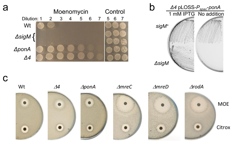
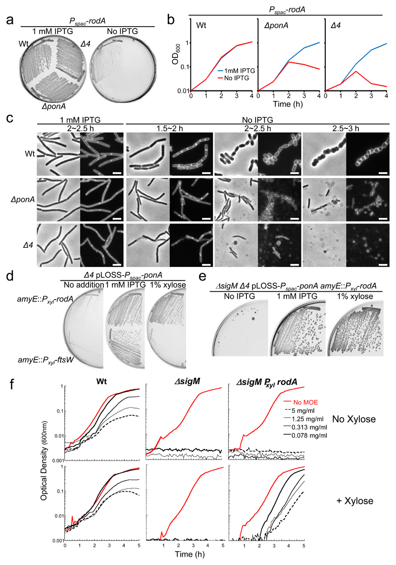
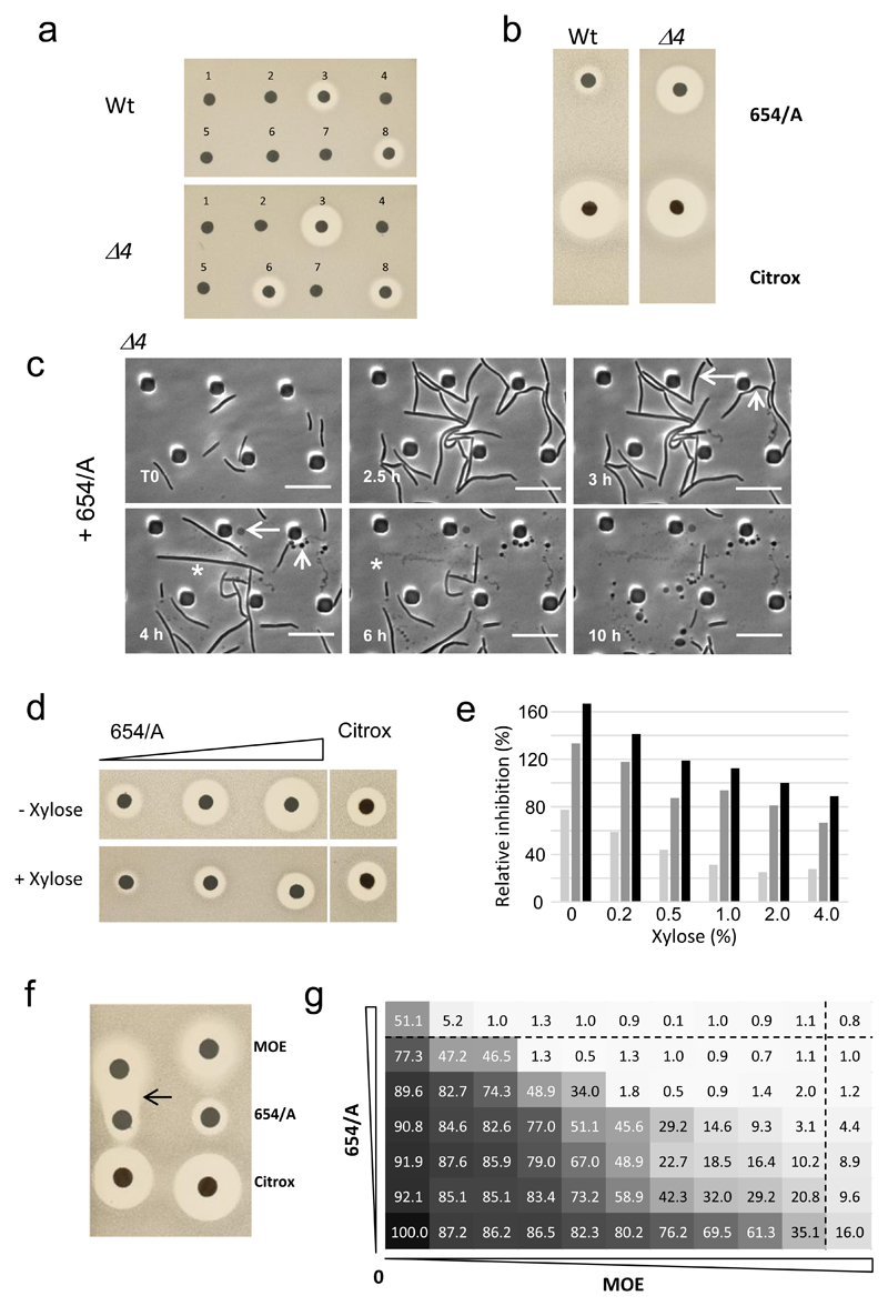
The bacterial cell wall is a highly conserved essential component of most bacterial groups. It is the target for our most frequently used antibiotics and provides important small molecules that trigger powerful innate immune responses. The wall is composed of glycan strands crosslinked by short peptides. For many years, the penicillin-binding proteins were thought to be the key enzymes required for wall synthesis. RodA and possibly other proteins in the wider SEDS (shape, elongation, division and sporulation) family have now emerged as a previously unknown class of essential glycosyltranferase enzymes, which play key morphogenetic roles in bacterial cell wall synthesis. We provide evidence in support of this role and the discovery of small natural product molecules that probably target these enzymes. The SEDS proteins have exceptional potential as targets for new antibacterial therapeutic agents.


Conflict of interest statement

Author conflicts

JE is a Director and shareholder at Demuris Ltd. NA and JD are employees at Demuris Ltd.

Figures

Figure 1
Figure 1. Use of MOE sensitivity to screen for missing GTase candidate genes.
a, Relative MOE sensitivity of the sigM mutant and resistance of the ponA and Δ4 mutant compared to the wild type. Overnight cultures of each strain were diluted (10-fold series; left to right) and 10 µl spots were placed on NA containing MOE at 10 µg/ml. Right hand spots show the last three dilutions plated on NA with no MOE. sigM spottings were done in duplicate. b, Synthetic lethality of a Δ4 mutant in a ΔsigM background. Growth on NA plates with or without 1 mM IPTG of strains AG221 (Δ4 pLOSS-Pspac-ponA) and AG831 (Δ4 pLOSS-Pspac-ponA ΔsigM::kan) at 30°C. c, Hypersensistivity of rodA, mreC and mreD mutants compared to wild type, ponA and Δ4 strains. Disc diffusion assays on MSM plates using 10 µg MOE per disc. The experiments have been repeated several times during the course of the project, producing consistent results. The figures are representative of at least two independent experiments.
Figure 2
Figure 2. Evidence for RodA as a candidate for the missing GTase.
a, Synthetic lethality of a rodA mutation in a ΔponA and Δ4 mutants. Growth on NA/MSM plates with or without 1 mM IPTG of strains YK2245 (Pspac-rodA), YK2246 (ΔponA::spc Pspac-rodA) and YK2242 (Δ4 Pspac-rodA) at 30°C. b, Growth profiles of YK2245 (Pspac-rodA), YK2246 (ΔponA::spc Pspac-rodA) and YK2242 (Δ4 Pspac-rodA) strains in NB/MSM with or without 1 mM IPTG at 37°C. c, Morphological effects of RodA depletion in ΔponA and Δ4 mutants. Phase-contrast and the corresponding cell membrane-stained images of typical fields of cells of YK2245 (Pspac-rodA), YK2246 (ΔponA::spc Pspac-rodA) and YK2242 (Δ4 Pspac-rodA) cultured in NB/MSM medium with or without 1 mM IPTG at 37°C. Time (h) of cultivation of cells after removal of IPTG is indicated. Scale bar represents 3 μm. d, Rescue of the growth defect of a Δ4 mutant by overexpression of rodA, but not ftsW. Growth on PAB plates with or without 1 mM IPTG, or 1% xylose, of strains YK2256 (Δ4 pLOSS-Pspac-ponA amyE::Pxyl-rodA) and YK2257 (Δ4 pLOSS-Pspac-ponA amyE::Pxyl-ftsW) at 30°C. e, Rescue of the synthetic lethality of Δ4 and ΔsigM strains by the overexpression of rodA. Growth on NA plates with or without 1 mM IPTG, or 1% xylose of strain YK2259 (Δ4 pLOSS-Pspac-ponA amyE::Pxyl-rodA ΔsigM::kan) at 30°C. f, Restoration of resistance to MOE in a sigM mutant overproducing RodA. Wild type (168CA), AG494 (ΔsigM) or YK2268 (ΔsigM amyE::Pxyl-rodA) were grown in the presence (bottom) or absence (top) of xylose, and treated with MOE as indicated. All the results in the figure are representative of at least two independent experiments.
Figure 3
Figure 3. Isolation and characterization of a putative inhibitor of the missing GTase.
a, Validation of a selection of hits from rescreening for antimicrobial activity using disc diffusion test. Fresh culture media extracts of different Actinomycete strains were prepared and tested against B. subtilis Wt and the Δ4 mutant. Discs 3 (Strain DEM20654) and 6 showed reproducible differential activity (larger zones of inhibition) on the Δ4 vs the Wt strain. Other extracts showed either no significant activity or large zones on both strains (disc 8). b, Confirmation of the differential activity of purified 654/A on Δ4 relative to a control by disc diffusion assay. c, Effects of purified 654/A on the Δ4 mutant. Individual frames are extracted from the video in Supplementary Video 1. Numbers in the bottom left corner of each frame represent time elapsed in the video. Examples of different lytic events are shown by asterisks (complete cell destruction), arrows (formation of phase pale bulbous cells), and arrowheads (formation of chains of small phase dark “minicells”).. Scale bar represents 10 µm. d,e, Increased 654/A resistance in ponA mutant cells containing an extra xylose-inducible copy of rodA (Pxyl-rodA). d, Disk diffusion tests were done with three different concentrations of 654/A (corresponding to 2, 5 or 10 µl amounts of purified material, left to right). Citrox was used as a control. Duplicate plates without or with 4% xylose (to induce overexpression of rodA), as indicated, were inoculated with strain YK2251 and incubated overnight. e, Dose response results for three different concentrations of 654/A, and at a range of concentrations of xylose. Average zones of inhibition were measured for duplicate disc diffusion tests and expressed as a percentage of the inhibition zone for the control antibiotic (citrox) measured on the same plate. f,g, Synergy between 654/A and MOE as demonstrated by disc diffusion (f) or checkerboard analysis (g) with the wild type strain (168CA). f, MOE, 654/A and a control antibiotic (citrox) were placed on a lawn of bacteria on an NA plate. The arrow points to an enhanced zone of clearing at the interface of the MOE and 654/A sub-inhibitory regions. g, Serial dilutions (2-fold) of MOE and 654/A were transferred to a 96 well plate, with MOE along the horizontal axis and 654/A in the vertical as indicated below and to the left. A dilute culture of wild type bacteria was used to inoculate each well and the plate was incubated for 24 h with continuous optical density monitoring. Dotted lines identify the minimum inhibitory concentrations derived by reference to the wells containing only one antibiotic (top row for MOE and far right column for 654/A). OD600 readings at the 18 h time point were used to calculate percentage inhibition values (shown in each rectangular segment) relative to the no antibiotic control. All the results in the figure are representative of at least two independent experiments.

References

    1. Errington J. L-form bacteria, cell walls and the origins of life. Open biology. 2013;3:120143. doi: 10.1098/rsob.120143. - DOI - PMC - PubMed
    1. Typas A, Banzhaf M, Gross CA, Vollmer W. From the regulation of peptidoglycan synthesis to bacterial growth and morphology. Nat Rev Microbiol. 2012;10:123–136. doi: 10.1038/nrmicro2677. - DOI - PMC - PubMed
    1. Lovering AL, Safadi SS, Strynadka NC. Structural perspective of peptidoglycan biosynthesis and assembly. Annu Rev Biochem. 2012;81:451–478. doi: 10.1146/annurev-biochem-061809-112742. - DOI - PubMed
    1. Spratt BG. Distinct penicillin-binding proteins involved in the division, elongation and shape of Escherichia coli K-12. Proc Natl Acad Sci U S A. 1975;72:2999–3003. - PMC - PubMed
    1. Wei Y, Havasy T, McPherson DC, Popham DL. Rod shape determination by the Bacillus subtilis class B penicillin-binding proteins encoded by pbpA and pbpH . J Bacteriol. 2003;185:4717–4726. - PMC - PubMed

MeSH terms