Review
. 2017 Jan;11(1):5-27.
doi: 10.1002/1878-0261.12019. Epub 2016 Dec 9.

Modes of invasion during tumour dissemination

Affiliations
Review

Modes of invasion during tumour dissemination

Pahini Pandya et al. Mol Oncol. 2017 Jan.

Abstract

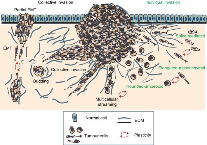
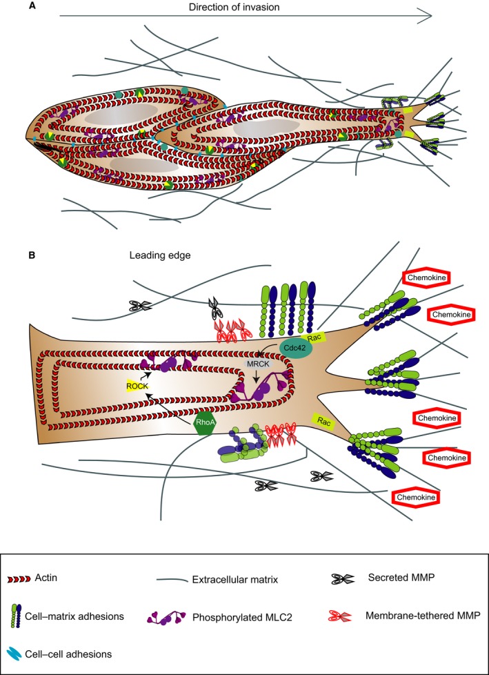
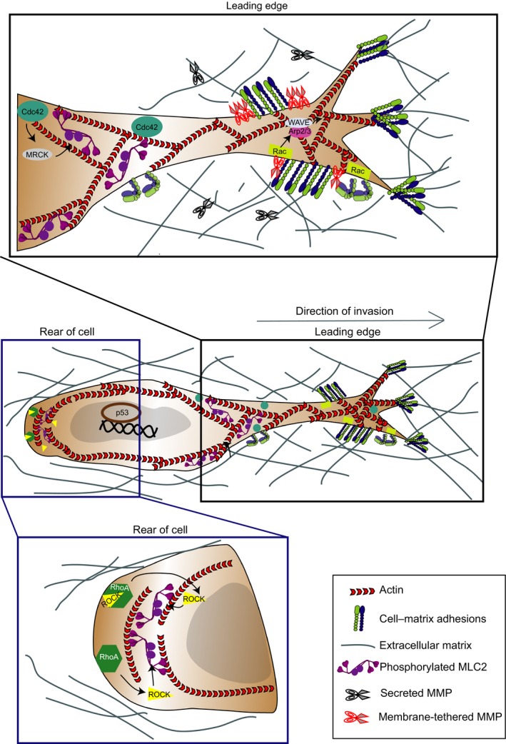
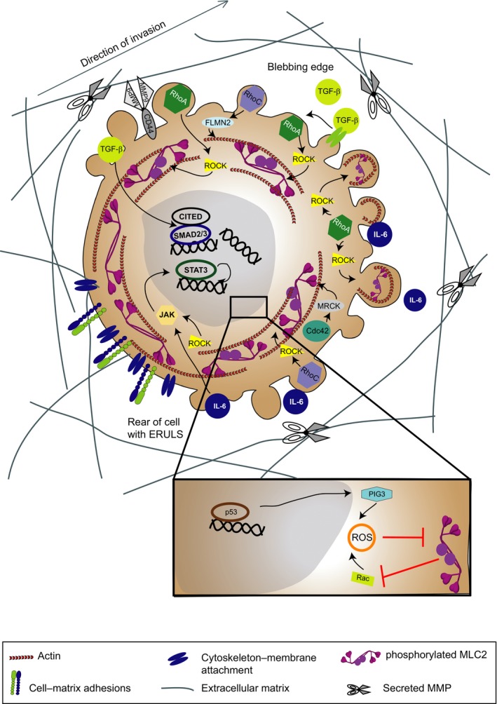
Cancer cell migration and invasion underlie metastatic dissemination, one of the major problems in cancer. Tumour cells exhibit a striking variety of invasion strategies. Importantly, cancer cells can switch between invasion modes in order to cope with challenging environments. This ability to switch migratory modes or plasticity highlights the challenges behind antimetastasis therapy design. In this Review, we present current knowledge on different tumour invasion strategies, the determinants controlling plasticity and arising therapeutic opportunities. We propose that targeting master regulators controlling plasticity is needed to hinder tumour dissemination and metastasis.

Keywords: Rho GTPases; actomyosin contractility; cancer metastasis; invasion; plasticity.


Figures

Figure 1
Figure 1
Modes of invasion during tumour dissemination. Diagram showing the main individual and collective modes of tumour invasion and plasticity that allows interconversion between modes. Cells invading individually can use protrusion‐based elongated‐mesenchymal, bleb‐ and contractility‐driven rounded‐amoeboid and filopodial spike‐mediated strategies. When cell–cell junctions are maintained, cells can move collectively as multicellular streams, budding or larger clusters (collective invasion). Migratory plasticity drives interconversion between the different modes.
Figure 2
Figure 2
Signalling pathways controlling collective modes of invasion. (A) Diagram showing the key regulators of collective migration. The leading edge of the multicellular group comprises one (or several) leader cells with mesenchymal characteristics. Leader cells extend actomyosin‐mediated actin‐rich protrusions that generate integrin‐mediated forward traction and pericellular proteolysis yielding a re‐aligned ECM that guides the group. Following cells are passively dragged behind along the established migration track by cell–cell adhesion. (B) Diagram showing the intracellular pathways activated in response to external stimuli and proteolysis of ECM. Membrane receptors such as β1 integrins control migration of individual elongated‐mesenchymal cells. Rac activation at the leading edge allows for protrusion formation that is linked to a ‘supracellular’ cytoskeleton. Activation of myosin II‐based contractile forces by Rho‐ROCK and Cdc42‐MRCK signalling allows for contraction of cell body and retraction of the rear.
Figure 3
Figure 3
Signalling pathways controlling elongated‐mesenchymal mode of invasion. Diagram showing key regulators of elongated‐mesenchymal mode of migration in cells. During this mode, cells adopt an elongated morphology that is characterized by actin‐rich protrusions, focal adhesion formation, matrix metalloproteinase (MMP) activity and actomyosin contractility localized at the rear of the cells. Top inset: signalling activity at the leading edge of cells exhibiting elongated‐mesenchymal migration. Polarized signalling of GTPase Rac1 directs Arp2/3 via WAVE2 to drive actin polymerization in branched filaments against the plasma membrane. Bottom inset: signalling activity at the rear of cells exhibiting elongated‐mesenchymal migration. Rho‐ROCK signalling is required for the contractile activity of actomyosin scaffold to retract the cell rear. Transcription driven by p53 promotes elongated‐mesenchymal strategies.
Figure 4
Figure 4
Signalling pathways controlling rounded‐amoeboid mode of invasion. Diagram showing key regulators of rounded‐amoeboid mode of migration. Rounded‐amoeboid cells squeeze through the matrix using small, unstable blebs present throughout the surface of the cells except at the rear, due to the presence of ezrin‐rich uropod‐like structures (ERULS) that determine polarity. Blebs are a consequence of low membrane–cortex attachment, increased intracellular pressure, high actomyosin contractility, low degree of β1 integrin‐mediated adhesion, reduced focal adhesion size and force generation. Rounded‐amoeboid motility is supported by high levels of actomyosin contractility downstream of Rho‐ROCK. While there is significant overlap in the RhoA‐ and RhoC‐mediated activation of actomyosin contractility, the assembly of cortical actin as a consequence of formin FLMN2 activation seems to be specific to RhoC. Maintenance of rounded‐amoeboid movement is driven by IL‐6 family of cytokines and the transcription factor STAT3. Conversely, ROCK can activate JAK/STAT3 signalling generating a positive feedback loop. TGF‐β promotes rounded‐amoeboid migration, which is perpetuated via SMAD2/CITED1‐mediated transcription. In addition, Rho/ROCK suppresses p53/PIG3‐mediated ROS production. On the other hand, Rac suppresses actomyosin contractility via ROS generation.

References

    1. Aceto N, Bardia A, Miyamoto DT, Donaldson MC, Wittner BS, Spencer JA, Yu M, Pely A, Engstrom A, Zhu H et al (2014) Circulating tumor cell clusters are oligoclonal precursors of breast cancer metastasis. Cell 158, 1110–1122. - PMC - PubMed
    1. Ackerman AB and Ragaz A (1984) The Lives of Lesions. Masson Publishers, New York, NY.
    1. Ahn J, Sanz‐Moreno V and Marshall CJ (2012) The metastasis gene NEDD9 product acts through integrin beta3 and Src to promote mesenchymal motility and inhibit amoeboid motility. J Cell Sci 125, 1814–1826. - PubMed
    1. Alexander S, Koehl GE, Hirschberg M, Geissler EK and Friedl P (2008) Dynamic imaging of cancer growth and invasion: a modified skin‐fold chamber model. Histochem Cell Biol 130, 1147–1154. - PubMed
    1. Amann KJ and Pollard TD (2001) The Arp2/3 complex nucleates actin filament branches from the sides of pre‐existing filaments. Nat Cell Biol 3, 306–310. - PubMed

Publication types

LinkOut - more resources