

Host-Microbial Interactions in Idiopathic Pulmonary Fibrosis
- PMID: 28085486
- PMCID: PMC5476909
- DOI: 10.1164/rccm.201607-1408OC
Host-Microbial Interactions in Idiopathic Pulmonary Fibrosis
Abstract
Rationale: Changes in the respiratory microbiome are associated with disease progression in idiopathic pulmonary fibrosis (IPF). The role of the host response to the respiratory microbiome remains unknown.
Objectives: To explore the host-microbial interactions in IPF.
Methods: Sixty patients diagnosed with IPF were prospectively enrolled together with 20 matched control subjects. Subjects underwent bronchoalveolar lavage (BAL), and peripheral whole blood was collected into PAXgene tubes for all subjects at baseline. For subjects with IPF, additional samples were taken at 1, 3, and 6 months and (if alive) 1 year. Gene expression profiles were generated using Affymetrix Human Gene 1.1 ST arrays.
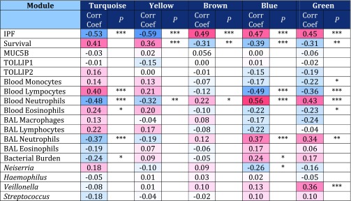
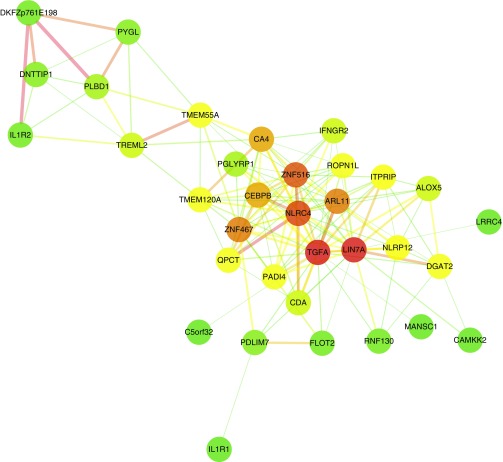
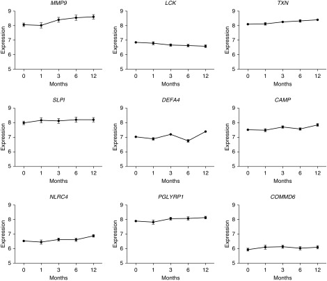
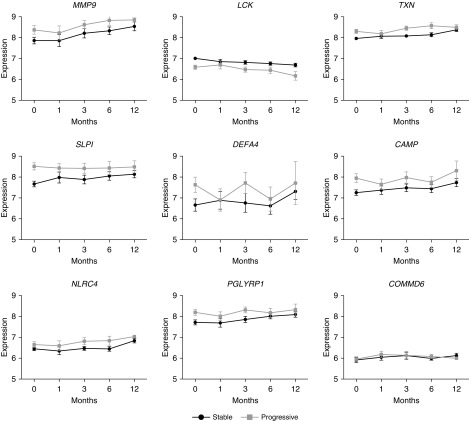
Measurements and main results: By network analysis of gene expression data, we identified two gene modules that strongly associated with a diagnosis of IPF, BAL bacterial burden (determined by 16S quantitative polymerase chain reaction), and specific microbial operational taxonomic units, as well as with lavage and peripheral blood neutrophilia. Genes within these modules that are involved in the host defense response include NLRC4, PGLYRP1, MMP9, and DEFA4. The modules also contain two genes encoding specific antimicrobial peptides (SLPI and CAMP). Many of these particular transcripts were associated with survival and showed longitudinal overexpression in subjects experiencing disease progression, further strengthening the relationship of the transcripts with disease.
Conclusions: Integrated analysis of the host transcriptome and microbial signatures demonstrated an apparent host response to the presence of an altered or more abundant microbiome. These responses remained elevated in longitudinal follow-up, suggesting that the bacterial communities of the lower airways may act as persistent stimuli for repetitive alveolar injury in IPF.
Keywords: acute lung injury; expression; idiopathic pulmonary fibrosis; microbiome; usual interstitial pneumonia.
Figures
Comment in
-
Host-Microbial Interactions: Idiopathic Pulmonary Fibrosis in Technicolor.Am J Respir Crit Care Med. 2017 Jun 15;195(12):1554-1556. doi: 10.1164/rccm.201701-0092ED. Am J Respir Crit Care Med. 2017. PMID: 28617080 Free PMC article. No abstract available.
-
IPF: Moving from Idiopathic to Infectious Pulmonary Fibrosis?Am J Respir Crit Care Med. 2017 Jul 15;196(2):125-127. doi: 10.1164/rccm.201702-0387ED. Am J Respir Crit Care Med. 2017. PMID: 28707969 No abstract available.
References
-
- Navaratnam V, Fleming KM, West J, Smith CJP, Jenkins RG, Fogarty A, Hubbard RB. The rising incidence of idiopathic pulmonary fibrosis in the U.K. Thorax. 2011;66:462–467. - PubMed
-
- Maher TM, Wells AU, Laurent GJ. Idiopathic pulmonary fibrosis: multiple causes and multiple mechanisms? Eur Respir J. 2007;30:835–839. - PubMed
-
- Song JW, Hong SB, Lim CM, Koh Y, Kim DS. Acute exacerbation of idiopathic pulmonary fibrosis: incidence, risk factors and outcome. Eur Respir J. 2011;37:356–363. - PubMed
Publication types
MeSH terms
Grants and funding
LinkOut - more resources
Full Text Sources
Other Literature Sources
Molecular Biology Databases
Miscellaneous
