. 2017 May;56(5):585-596.
doi: 10.1165/rcmb.2016-0118OC.

Age-Dependent Changes in AMPK Metabolic Pathways in the Lung in a Mouse Model of Hemorrhagic Shock

Affiliations

Age-Dependent Changes in AMPK Metabolic Pathways in the Lung in a Mouse Model of Hemorrhagic Shock

Lindsey R Klingbeil et al. Am J Respir Cell Mol Biol. 2017 May.

Abstract

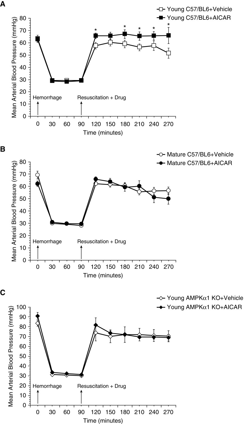
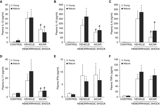
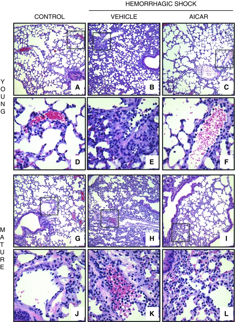
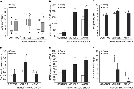
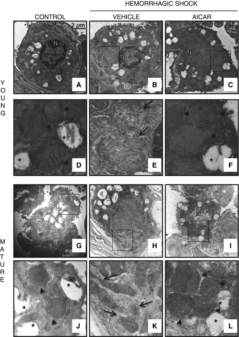
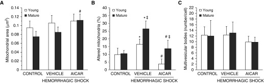
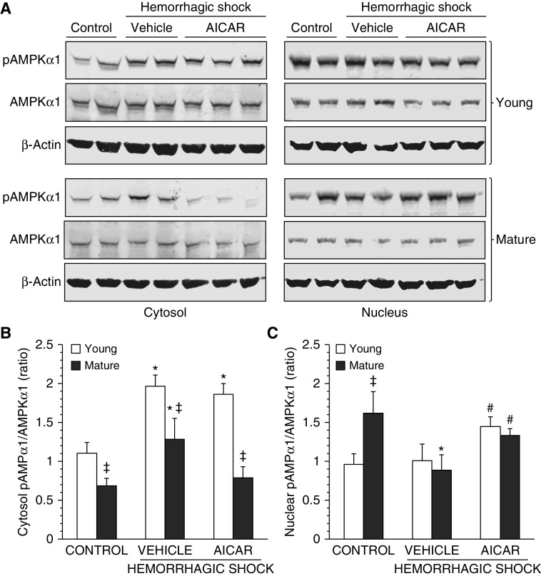
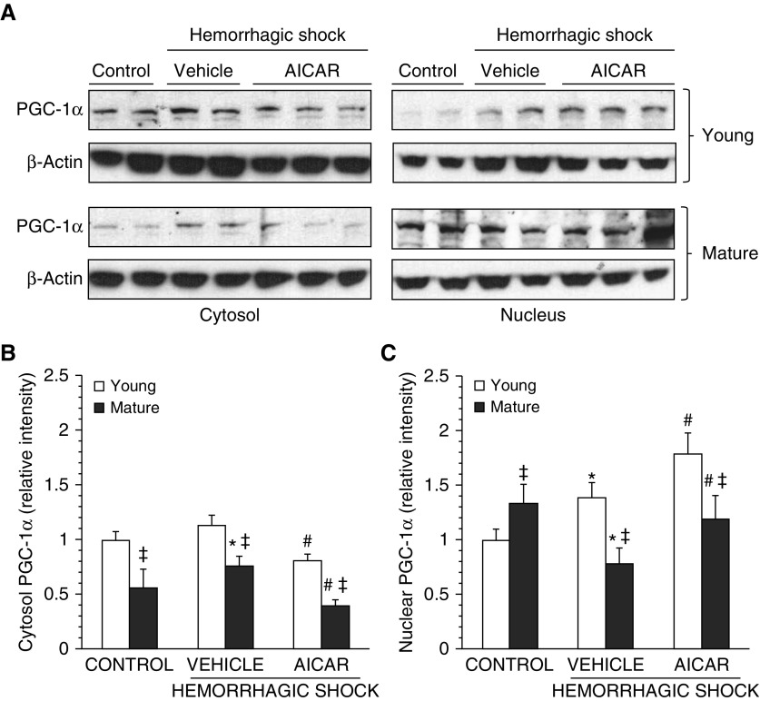
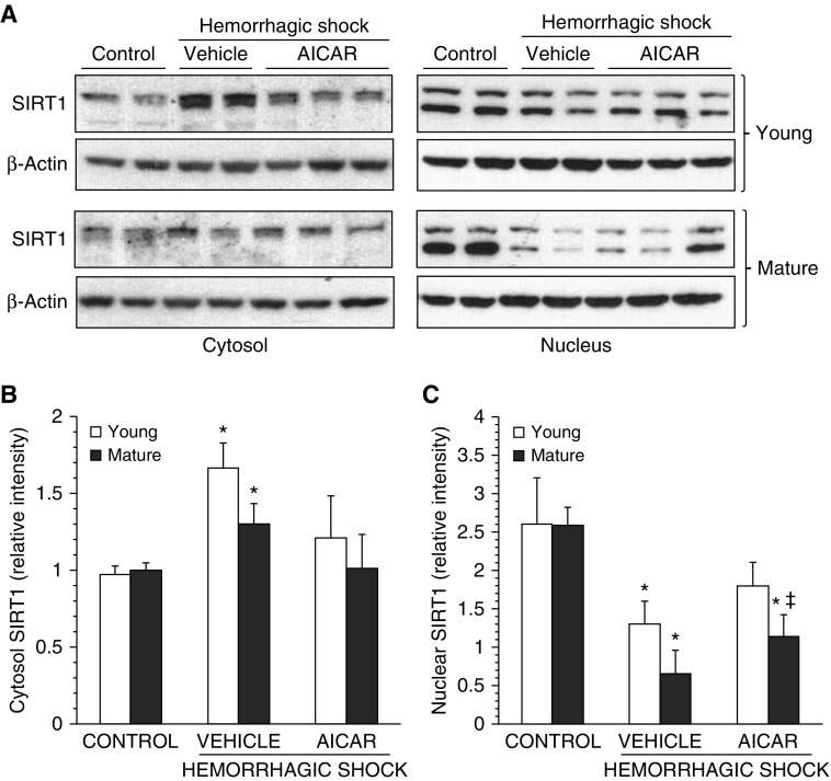
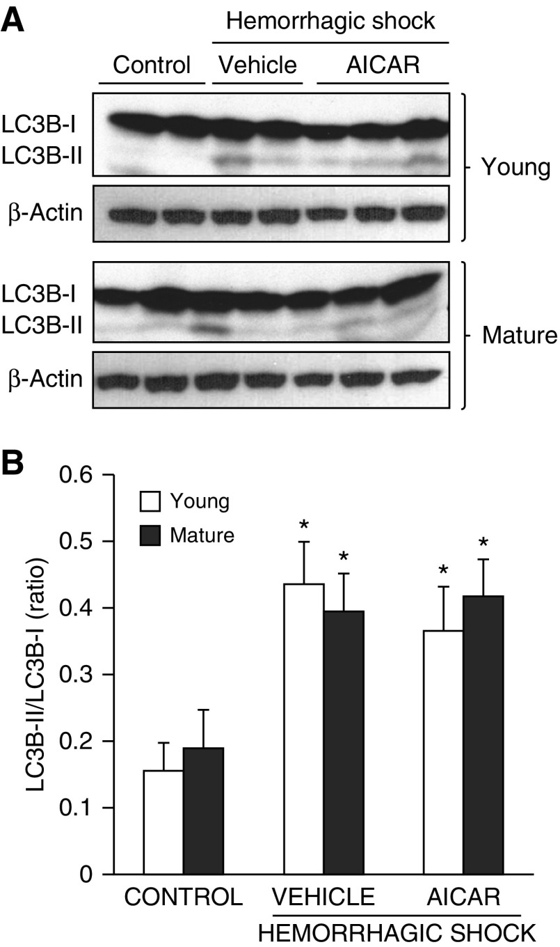
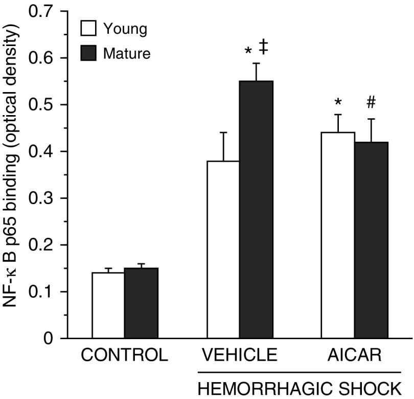
The development of multiple organ failure in patients with hemorrhagic shock is significantly influenced by patient age. Adenosine monophosphate-activated protein kinase (AMPK) is a crucial regulator of energy homeostasis, which coordinates metabolic repair during cellular stress. We investigated whether AMPK-regulated signaling pathways are age-dependent in hemorrhage-induced lung injury and whether AMPK activation by 5-amino-4-imidazole carboxamide riboside (AICAR) affords lung protective effects. Male C57/BL6 young mice (3-5 mo), mature adult mice (9-12 mo), and young AMPKα1 knockout mice (3-5 mo) were subjected to hemorrhagic shock by blood withdrawing, followed by resuscitation with shed blood and lactated Ringer's solution. Plasma proinflammatory cytokines were similarly elevated in C57/BL6 young and mature adult mice after hemorrhagic shock. However, mature adult mice exhibited more severe lung edema and neutrophil infiltration, and higher mitochondrial damage in alveolar epithelial type II cells, than did young mice. No change in autophagy was observed. At molecular analysis, the phosphorylation of the catalytic subunit AMPKα1 was associated with nuclear translocation of peroxisome proliferator-activated receptor γ co-activator-α in young, but not mature, adult mice. Treatment with AICAR ameliorated the disruption of lung architecture in mice of both ages; however, effects in mature adult mice were different than young mice and also involved inhibition of nuclear factor-κB. In young AMPKα1 knockout mice, AICAR failed to improve hypotension and lung neutrophil infiltration. Our data demonstrate that during hemorrhagic shock, AMPK-dependent metabolic repair mechanisms are important for mitigating lung injury. However, these mechanisms are less competent with age.

Keywords: AICAR; AMPK; autophagy; hemorrhagic shock; mitochondria.


Figures

Figure 1.
Figure 1.
Effect of in vivo treatment with 5-amino-4-imidazole carboxamide riboside (AICAR) on mean arterial blood pressure in (A) C57BL/6 wild-type young, (B) C57BL/6 wild-type mature, and (C) young AMP-activated protein kinase (AMPK) α1 knockout (KO) mice subjected to hemorrhage and resuscitation. Data represent the mean ± SD of 3 to 10 mice in each group. Vehicle (distilled water) or AICAR (100 mg/kg) was administered intraperitoneally at the time of resuscitation with shed blood and lactated Ringer’s solution (×2). Arrows indicate time of induction of hemorrhage and initiation of resuscitation and AICAR or vehicle administration. *P < 0.05 versus vehicle-treated group.
Figure 2.
Figure 2.
Effect of in vivo treatment with AICAR on plasma levels of (A) IL-1β, (B) IL-10, (C) IL-6, (D) IL-17, (E) IFNγ, and (F) TNF-α in C57BL/6 wild-type young and mature mice after hemorrhagic shock. Each data point represents the mean ± SEM of four to six animals for each group. *P < 0.05 versus age-matched control mice; #P < 0.05 versus vehicle-treated group of the same age.
Figure 3.
Figure 3.
Representative histology photomicrographs of lung sections. Normal lung architecture in control C57BL/6 wild-type (A) young and (G) mature mice presenting patent alveoli, and vessels with few or no adhering neutrophils (insets shown in D and J). Lung damage in vehicle-treated (B) young and (H) mature mice after hemorrhagic shock, with reduction of alveolar space and neutrophil adhesion along vascular wall and infiltration of inflammatory cells into parenchyma (insets shown in E and K). Improvement in lung architecture in AICAR-treated (C) young and (I) mature mice after hemorrhagic shock, with reduction of neutrophil infiltration in young mice (inset shown in F) but persistence of high neutrophil adhesion along vascular wall and parenchymal infiltration of inflammatory cell in mature mice (inset shown in L). Magnification × 100 for A, B, C, G, H, and I; magnification × 400 for D, E, F, J, K, and L. A similar pattern was seen in n = 4–6 different tissue sections in each experimental group.
Figure 4.
Figure 4.
Histopathologic scores of lung sections of C57BL/6 wild-type young and mature mice (n = 4–10 mice for each group). Lung injury was scored from 0 (no damage) to 16 (maximal damage). (A) Box plots represent 25th percentile, median, and 75th percentile; error bars define 10th and 90th percentiles; whiskers define minimal and maximal values. (B) Lung myeloperoxidase activity, (C) lung wet to dry weight ratio, and BAL fluid (BALF) analysis of (D) total protein content, (E) total count of viable inflammatory cells, and (F) IL-6 levels in C57BL/6 wild-type young and mature mice. Data represent the mean ± SEM of 4–10 animals per group. *P < 0.05 versus age-matched control mice; #P < 0.05 versus vehicle-treated group of the same age; P < 0.05 versus young group.
Figure 5.
Figure 5.
Transmission electronic microscopy of alveolar epithelial type II cells. Normal cellular structure of alveolar type II cells in (A) control young and (G) mature mice with normal electron dense mitochondria presenting organized cristae (arrowheads) and multivesicular bodies (*) (insets shown in D and J). Structural changes in vehicle-treated (B) young and (H) mature mice after hemorrhagic shock with mitochondria presenting translucent matrix, disrupted membrane and cristae and elongated shape (arrows; insets shown in E and K). Amelioration of cell structure in AICAR-treated (C) young and (I) mature mice after hemorrhagic shock with normal electron dense mitochondria (arrowheads; insets shown in F and L). Scale bar, 2 μm. N, nucleus.
Figure 6.
Figure 6.
Quantification of (A) average mitochondrial area, (B) altered mitochondria, and (C) multivesicular bodies in alveolar type II cells as determined by using the National Institutes of Health ImageJ software. Altered mitochondria were determined as percentage of total number of mitochondria; multivesicular bodies were determined as total number in a single cell. Data are expressed as mean ± SEM. *P < 0.05 versus age-matched control mice; #P < 0.05 versus vehicle-treated group of the same age; P < 0.05 versus young group.
Figure 7.
Figure 7.
(A) Western blot analysis of active phosphorylated AMPK (pAMPK) α1, AMPKα1, and β-actin (used as loading control protein) in lung cytosol and nuclear extracts. Image analyses of pAMPKα1/AMPKα1 ratio as determined by densitometry (B) in the cytosol and (C) in the nucleus. Data are expressed as mean ± SEM of four to six animals for each group and are expressed as ratio of relative intensity units. *P < 0.05 versus age-matched control mice; #P < 0.05 versus vehicle-treated group of the same age; P < 0.05 versus young group.
Figure 8.
Figure 8.
(A) Western blot analysis of proliferator-activated receptor-γ coactivator 1α (PGC1-α) and β-actin (used as loading control protein) in lung cytosol and nuclear extracts. Image analyses of PGC-1α expression as determined by densitometry (B) in the cytosol and (C) in the nucleus. Data are expressed as mean ± SEM of four to six animals for each group and are expressed as ratio of relative intensity units. *P < 0.05 versus age-matched control mice; #P < 0.05 versus vehicle-treated group of the same age; P < 0.05 versus young group.
Figure 9.
Figure 9.
(A) Western blot analysis of sirtuin 1 (SIRT1) and β-actin (used as loading control protein) in lung cytosol and nuclear extracts. Image analyses of SIRT1 expression as determined by densitometry (B) in the cytosol and (C) in the nucleus. Data are expressed as mean ± SEM of four to six animals for each group and are expressed as ratio of relative intensity units. *P < 0.05 versus age-matched control mice; P < 0.05 versus young group.
Figure 10.
Figure 10.
(A) Western blot analysis of light-chain 3B (LC3B)-I and LC3B-II and β-actin (used as loading control protein) in lung cytosol extracts. (B) Image analyses of LC3B-II/LC3B-I ratio as determined by densitometry. Data are expressed as mean ± SEM of four to six animals for each group and are expressed as ratio of relative intensity units. *P < 0.05 versus age-matched control mice.
Figure 11.
Figure 11.
Activity of the p65 subunit of nuclear factor (NF)-κB in lung nuclear extracts at 3 hours after resuscitation. Data are expressed as mean ± SEM of 5–13 animals for each group and are expressed as optical density units. *P < 0.05 versus age-matched control mice; #P < 0.05 versus vehicle-treated group of the same age; P < 0.05 versus young group.

References

    1. Kauvar DS, Lefering R, Wade CE. Impact of hemorrhage on trauma outcome: an overview of epidemiology, clinical presentations, and therapeutic considerations. J Trauma. 2006;60:S3–S11. - PubMed
    1. Lehmann R, Beekley A, Casey L, Salim A, Martin M. The impact of advanced age on trauma triage decisions and outcomes: a statewide analysis. Am J Surg. 2009;197:571–574, discussion 574–575. - PubMed
    1. Dewar DC, Tarrant SM, King KL, Balogh ZJ. Changes in the epidemiology and prediction of multiple-organ failure after injury. J Trauma Acute Care Surg. 2013;74:774–779. - PubMed
    1. Vanzant EL, Hilton RE, Lopez CM, Zhang J, Ungaro RF, Gentile LF, Szpila BE, Maier RV, Cuschieri J, Bihorac A, et al. Inflammation and Host Response to Injury Investigators. Advanced age is associated with worsened outcomes and a unique genomic response in severely injured patients with hemorrhagic shock. Crit Care. 2015;19:77. - PMC - PubMed
    1. Cairns CB, Moore FA, Haenel JB, Gallea BL, Ortner JP, Rose SJ, Moore EE. Evidence for early supply independent mitochondrial dysfunction in patients developing multiple organ failure after trauma. J Trauma. 1997;42:532–536. - PubMed

Publication types

MeSH terms