. 2017 Apr;33(3):149-160.
doi: 10.1089/jop.2016.0093. Epub 2017 Jan 13.

Photoelectric Dye Used for Okayama University-Type Retinal Prosthesis Reduces the Apoptosis of Photoreceptor Cells

Affiliations

Photoelectric Dye Used for Okayama University-Type Retinal Prosthesis Reduces the Apoptosis of Photoreceptor Cells

Shihui Liu et al. J Ocul Pharmacol Ther. 2017 Apr.

Abstract

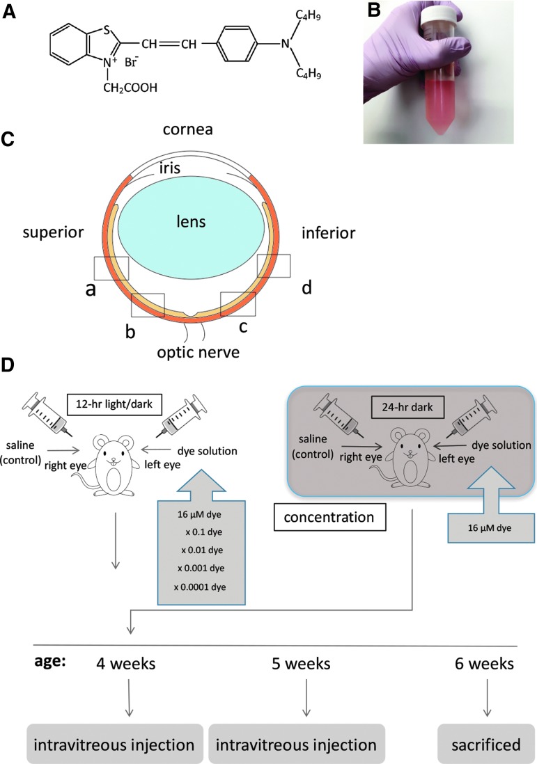
Purpose: Our previous study demonstrated that photoelectric dye-coupled polyethylene film (Okayama University-type retinal prosthesis), which was implanted in subretinal space of the eyes of Royal College of Surgeons (RCS) rats, prevented retinal neurons from apoptotic death. In this study, we aimed to examine whether photoelectric dye itself would protect retinal neurons from apoptosis in RCS rats.

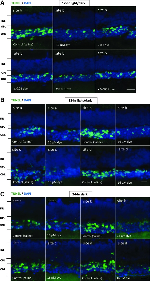
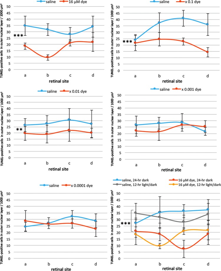
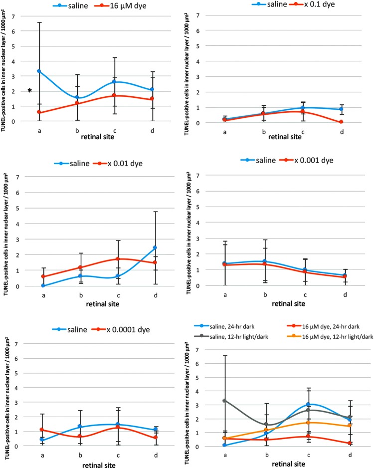
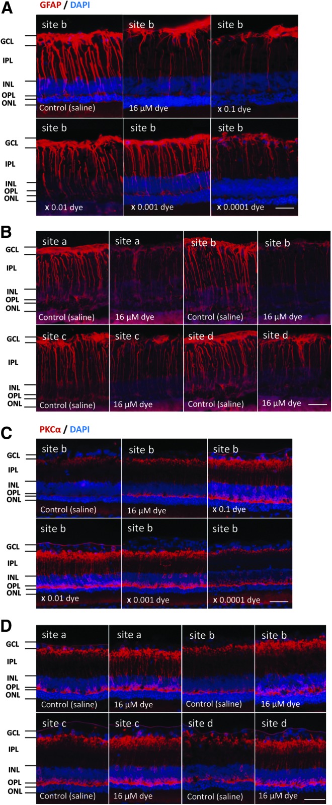
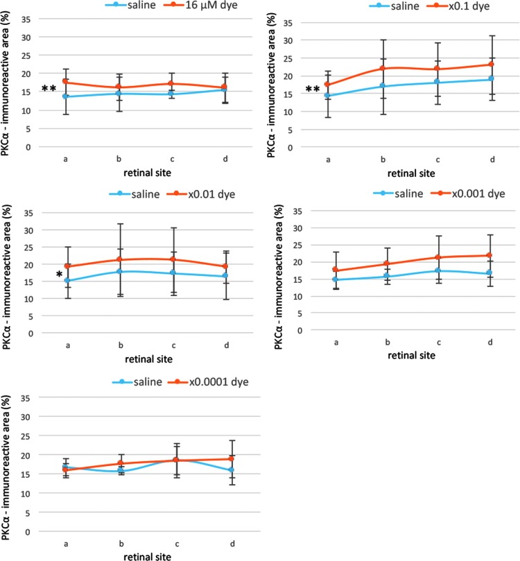
Methods: RCS rats received intravitreous injection of different concentrations of the dye in the left eye and housed under a 12-h light-dark cycle. Saline injection in the right eye served as control. In addition, RCS rats with dye injection were kept in 24-h daily dark condition. Sections were processed for terminal deoxynucleotidyl transferase-mediated fluorescein-conjugated-dUTP nick-end-labeling (TUNEL) assay and immunohistochemical staining of glial fibrillary acidic protein (GFAP) and protein kinase Cα (PKCα).

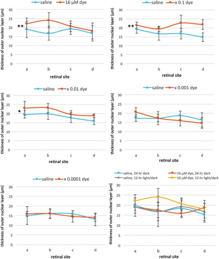
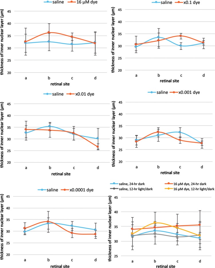
Results: The number of TUNEL-positive cells significantly decreased in the retina of dye-injected eyes compared with those in saline-injected eyes (P = 0.0001, 2-factor analysis of variance [ANOVA]), under 12-h light-dark cycle. Significant decrease of TUNEL-positive cells was noted in the retina of rats with dye injection compared with those with saline injection, kept under 24-h dark condition (P = 0.0001, 2-factor ANOVA). Immunoreactive area for GFAP decreased significantly in the retina of dye-injected eyes compared with that in controls (P = 0.0001, 2-factor ANOVA), whereas immunoreactive area for PKCα increased significantly in the retina of dye-injected eyes compared with that in controls (P = 0.01, 2-factor ANOVA).

Conclusions: Photoelectric dye inhibits apoptotic death of photoreceptor cells in RCS rats and downregulates GFAP expression in retinal Müller cells. Photoelectric dye may be a candidate agent for neuroprotection in retinitis pigmentosa and other retinal diseases.

Keywords: GFAP; PKCα; apoptosis; drug; photoreceptors; retina.


Conflict of interest statement

No competing financial interests exist.

Figures

<b>FIG. 1.</b>
FIG. 1.
Experimental design. (A) Molecular structure of photoelectric dye 2-[2-[4-(dibutylamino) phenyl] ethenyl]-3-carboxymethylbenzothiazolium. (B) Stock solution of the dye dissolved in water at a concentration of 8.2 μg/mL (16 μM). (C) Four retinal sites defined for immunohistochemical analysis. “a” and “b” begin at the straight distance of 373 and 160 μm, respectively, superior from the optic nerve head. “c” and “d” begin at the straight distance of 160 and 373 μm, respectively, inferior from the optic nerve head. (D) Time table for injections.
<b>FIG. 2.</b>
FIG. 2.
The detection of apoptosis in retinal sections of each group of rats. (A) TUNEL staining (green) of retinal sections (site b) of the eyes with intravitreous injection of 3 μL saline in the right eye or series of dilutions of the dye stock solution in the left eye under 12-h light–dark cycle. Eyes were enucleated 2 weeks after the first injection at the age of 4 weeks. The nuclei were counterstained with DAPI (blue). The number of TUNEL-positive cells in the outer nuclear layer (ONL) was less in the dye-injected eyes than in the saline-injected eyes. (B) TUNEL staining of retinal sections at 4 different retinal sites (a, b, c, and d) in the left eye with dye injection (16 μM) compared with that in the right eye with saline injection under 12-h light–dark cycle. (C) TUNEL staining of retinal sections at 4 different retinal sites (a, b, c, and d) in the left eye with dye injection (16 μM) compared with that in the right eye with saline injection under 24-h constant dark condition. INL, inner nuclear layer; OPL, outer plexiform layer. Scale bar: 10 μm. DAPI, 4′,6-diamidino-2-phenylindole.
<b>FIG. 3.</b>
FIG. 3.
Quantitative analysis of apoptotic cells in the ONL of each group of rats. TUNEL-positive cell counts per 1,000 μm2 in the ONL of 4 different retinal sites (a, b, c, and d) of the left eye with dye injection at 5 concentrations compared with those of the right eye with saline injection under 12-h light–dark cycle. TUNEL-positive cell counts were significantly different among different dye concentrations (P = 0.0001), but not significantly different among 4 different retinal sites (P = 0.144, 2-factor analysis of variance [ANOVA]). TUNEL-positive cell counts in dye-injected eyes were significantly less than in saline-injected eyes at the concentration of 16 μM (***P = 0.0001), 16 μM × 0.1 (***P = 0.0001), and 16 μM × 0.01 (**P = 0.002). The bottom right panel shows TUNEL-positive cell counts in the eyes injected with dye (16 μM) versus saline under 12-h light–dark cycle versus under 24-h constant dark condition. There was significant difference between dye-injected eyes and saline-injected eyes under 24-h dark condition (***P = 0.0001). T bars indicate standard deviation.
<b>FIG. 4.</b>
FIG. 4.
Quantitative analysis of apoptotic cells in the inner nuclear layer of each group of rats. TUNEL-positive cell counts per 1,000 μm2 in the inner nuclear layer of 4 different retinal sites (a, b, c, and d) of the left eye with dye injection at 5 concentrations compared with those of the right eye with saline injection under 12-h light–dark cycle. TUNEL-positive cell counts showed no significant differences among different dye concentrations (P = 0.084) and among 4 different retinal sites (P = 0.927, 2-factor ANOVA). However, TUNEL-positive cell counts in dye-injected eyes were significantly less than in saline-injected eyes at the concentration of 16 μM (*P = 0.043, post hoc test). The bottom right panel shows TUNEL-positive cell counts in the eyes injected with dye (16 μM) versus saline under 12-h light–dark cycle versus under 24-h constant dark condition. There was no significant difference between dye-injected eyes and saline-injected eyes under 24-h dark condition. T bars indicate standard deviation.
<b>FIG. 5.</b>
FIG. 5.
Quantitative analysis of the thickness of the ONL of each group of rats. The thickness of the ONL of 4 different retinal sites (a, b, c, and d) of the left eye with dye injection at 5 concentrations compared with that of the right eye with saline injection under 12-h light–dark cycle. The thickness showed significant differences among different concentrations of the dye (P = 0.0001) and among 4 different retinal sites (P = 0.014, 2-factor ANOVA). The ONL was significantly thicker with dye injection at 3 concentrations, 16 μM (**P = 0.001), 16 μM × 0.1 (**P = 0.001), and 16 μM × 0.01 (*P = 0.026), than with saline injection. The bottom right panel shows the ONL thickness of the eyes injected with dye (16 μM) versus saline under 12-h light–dark cycle versus under 24-h constant dark condition. There was no significant difference between dye-injected eyes and saline-injected eyes under 24-h dark condition. T bars indicate standard deviation.
<b>FIG. 6.</b>
FIG. 6.
Quantitative analysis of the thickness of the inner nuclear layer of each group of rats. The thickness of the inner nuclear layer of 4 different retinal sites (a, b, c, and d) of the left eye with dye injection at 5 concentrations compared with that of the right eye with saline injection under 12-h light–dark cycle. The thickness showed significant differences among different concentrations of the dye (P = 0.014) and among 4 different retinal sites (P = 0.0001, 2-factor ANOVA). However, post hoc test showed no significance at each concentration of the dye compared with that of saline. The bottom right panel shows the inner nuclear layer thickness of the eyes injected with dye (16 μM) versus saline under 12-h light–dark cycle versus under 24-h constant dark condition. There was no significant difference between dye-injected eyes and saline-injected eyes under 24-h dark condition. T bars indicate standard deviation.
<b>FIG. 7.</b>
FIG. 7.
Immunohistochemical staining of GFAP and PKCα in each group of rats. (A) GFAP staining (red) of the retina (site “b”) of RCS rats 2 weeks after intravitreous injection. The right eyes had saline injection and the left eyes had photoelectric dye injection at each concentration of 10-fold dilution series from 16 μM. (B) GFAP staining (red) of the retina at 4 different retinal sites (a, b, c, and d) in the left eye with dye injection (16 μM) compared with that in the right eye with saline injection under 12-h light–dark cycle. GFAP was downregulated with photoelectric dye injection. (C) PKCα staining (red) of the retina (site “b”) of RCS rats 2 weeks after intravitreous injection. The right eyes had saline injection and the left eyes had photoelectric dye injection at each concentration of 10-fold dilution series from 16 μM. (D) PKCα staining (red) of the retina at 4 different retinal sites (a, b, c, and d) in the left eye with dye injection (16 μM) compared with that in the right eye with saline injection under 12-h light–dark cycle. Dye injection led to enhanced staining in rod bipolar cells including their dendrites, soma, and axon terminals. The nuclei were counterstained with DAPI (blue). GCL, ganglion cell layer; IPL, inner plexiform layer; INL, inner nuclear layer; OPL, outer plexiform layer. Scale bar: 30 μm. GFAP, glial fibrillary acidic protein; PKCα, protein kinase Cα; RCS, Royal College of Surgeons.
<b>FIG. 8.</b>
FIG. 8.
Semiquantitative analysis of GFAP staining. GFAP immunoreactive areas were calculated for 4 different retinal sites (a, b, c, d) of the left eyes with dye injection at different concentrations and the right eyes with saline injection. GFAP staining showed significant differences among different concentrations of the dye (P = 0.0001) and among 4 different retinal sites (P = 0.0001, 2-factor ANOVA). GFAP staining was significantly higher with dye injection at 16 μM (*P = 0.023) and 16 μM × 0.01 (**P = 0.007) than with saline injection. T bars indicate standard deviation.
<b>FIG. 9.</b>
FIG. 9.
Semiquantitative analysis of PKCα staining. PKCα immunoreactive areas were calculated for 4 different retinal sites (a, b, c, and d) of the left eyes with dye injection at different concentrations and the right eyes with saline injection. PKCα staining showed significant differences among different concentrations of the dye (P = 0.01), but not among 4 different retinal sites (P = 0.413, 2-factor ANOVA). PKCα staining was significantly higher with dye injection at 16 μM (**P = 0.002), 16 μM × 0.1 (**P = 0.003), and 16 μM × 0.01 (*P = 0.014) than with saline injection. T bars indicate standard deviation.

Similar articles

Cited by

References

    1. Loewenstein J.I., Montezuma S.R., and Rizzo J.F. III. Outer retinal degeneration: an electronic retinal prosthesis as a treatment strategy. Arch. Ophthalmol. 122:587–596, 2004 - PubMed
    1. Tamaki M., and Matsuo T. Optical coherence tomographic parameters as objective signs for visual acuity in patients with retinitis pigmentosa, future candidates for retinal prostheses. J. Artif. Organs. 14:140–150, 2011 - PubMed
    1. Zrenner E. Will retinal implants restore vision? Science. 295:1022–1025, 2002 - PubMed
    1. Humayun M.S., Dorn J.D., da Cruz L., et al. . Interim results from the international trial of Second Sight's visual prosthesis. Ophthalmology. 119:779–788, 2012 - PMC - PubMed
    1. Chow A.Y., Chow V.Y., Packo K.H., et al. . The artificial silicon retina microchip for the treatment of vision loss from retinitis pigmentosa. Arch. Ophthalmol. 122:460–469, 2004 - PubMed

Publication types

LinkOut - more resources