. 2017 Feb;13(2):404-422.
doi: 10.1080/15548627.2016.1263776. Epub 2017 Jan 13.

Deletion of PRKAA triggers mitochondrial fission by inhibiting the autophagy-dependent degradation of DNM1L

Affiliations

Deletion of PRKAA triggers mitochondrial fission by inhibiting the autophagy-dependent degradation of DNM1L

Qilong Wang et al. Autophagy. 2017 Feb.

Abstract

PRKAA (protein kinase, AMP-activated, α catalytic subunit) regulates mitochondrial biogenesis, function, and turnover. However, the molecular mechanisms by which PRKAA regulates mitochondrial dynamics remain poorly characterized. Here, we report that PRKAA regulated mitochondrial fission via the autophagy-dependent degradation of DNM1L (dynamin 1-like). Deletion of Prkaa1/AMPKα1 or Prkaa2/AMPKα2 resulted in defective autophagy, DNM1L accumulation, and aberrant mitochondrial fragmentation in the mouse aortic endothelium. Furthermore, autophagy inhibition by chloroquine treatment or ATG7 small interfering RNA (siRNA) transfection, upregulated DNM1L expression and triggered DNM1L-mediated mitochondrial fragmentation. In contrast, autophagy activation by overexpression of ATG7 or chronic administration of rapamycin, the MTOR inhibitor, promoted DNM1L degradation and attenuated mitochondrial fragmentation in Prkaa2-deficient (prkaa2-/-) mice, suggesting that defective autophagy contributes to enhanced DNM1L expression and mitochondrial fragmentation. Additionally, the autophagic receptor protein SQSTM1/p62, which bound to DNM1L and led to its translocation into the autophagosome, was involved in DNM1L degradation by the autophagy-lysosome pathway. Gene silencing of SQSTM1 markedly reduced the association between SQSTM1 and DNM1L, impaired the degradation of DNM1L, and enhanced mitochondrial fragmentation in PRKAA-deficient endothelial cells. Finally, the genetic (DNM1L siRNA) or pharmacological (mdivi-1) inhibition of DNMA1L ablated mitochondrial fragmentation in the mouse aortic endothelium and prevented the acetylcholine-induced relaxation of isolated mouse aortas. This suggests that aberrant DNM1L is responsible for enhanced mitochondrial fragmentation and endothelial dysfunction in prkaa knockout mice. Overall, our results show that PRKAA deletion promoted mitochondrial fragmentation in vascular endothelial cells by inhibiting the autophagy-dependent degradation of DNM1L.

Keywords: AMPK; DNM1L; PRKAA/AMPK catalytic subunit α; autophagy; endothelial dysfunction; mitochondrial fission.


Figures
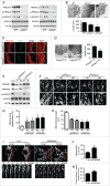
Figure 1.
Figure 1.
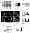
PRKAA deletion induces mitochondrial fragmentation in aortic endothelial cells. (A) Protein levels of PRKAA1/AMPKα1, PRKAA2/AMPKα2, ACACA, phosphorylation of PRKAA at Thr172 and ACACA at Ser79 in aortas isolated from wild-type (WT), prkaa1−/−, and prkaa2−/− mice were analyzed by western blotting. (B) Representative transmission electron micrographs of mitochondria and quantification of mitochondrial length in the aortic endothelium of WT, prkaa1−/−, and prkaa2−/− mice. Scale bars: 500 nm; L, lumen; M, mitochondria; E, endothelium. n = 5 mice, * P < 0.05 vs. WT. (C) Mouse aortas isolated from Prkaa2flox/flox (Prkaa2FloxCtrl) and endothelial-specific prkaa2 knockout (prkaa2Endoth−/−) mice were stained with antibodies against PRKAA2/AMPKα2 (Red) and PECAM1 (Green) and observed using fluorescence microscopy. (D) Representative electron micrographs of mitochondria and quantification of mitochondrial length in the aortic endothelium of Prkaa2FloxCtrl and prkaa2Endoth−/− mice. Scale bars: 500 nm. n = 5 mice, * P < 0.05 vs. Prkaa2FloxCtrl. (E and F) Human umbilical vein endothelial cells (HUVECs) were transfected with control small interfering RNA (siRNA; control, C-si), PRKAA1 siRNA (siPRKAA1), or PRKAA2 siRNA (siPRKAA2) for 48 h. (E) Protein levels of PRKAA1/AMPKα1, PRKAA2/AMPKα2, ACACA, and phosphorylation of ACACA at Ser79 were analyzed by western blotting. (F) Representative confocal images of the mitochondrial morphology in HUVECs stained with MitoTracker® Deep Red FM. Scale bars: 10 µm. Quantification of mitochondrial fragmentation of the cells (G) and average mitochondrial length (H). n ≥ 100, *P < 0.05 vs. C-si. (I to K) Mitochondrial morphology in live HUVECs stained with MitoTracker® Deep Red FM was captured using time-lapse confocal microscopy. Images were collected at 1 min intervals for 10 min. (I) Representative time-lapse confocal images. Scale bars: 10 µm. Quantification of mitochondrial fission (J) and the ratio of fission and fission-plus-fusion events (K) in live HUVECs. n = 15, *P < 0.05 vs. C-si.
Figure 2.
Figure 2.
Prkaa2 deletion increases DNM1L expression and DNM1L-dependent mitochondrial fission in aortic endothelial cells. (A) Western blot analysis of mitochondrial dynamics-related proteins, including DNM1L, FIS1 (fission, mitochondrial 1), MFN2 (mitofusin 2), and OPA1 (optic atrophy 1), in WT, prkaa1−/− and prkaa2−/− mouse aortas. n = 5 mice, *P < 0.05 vs. WT. (B) Representative images of immunohistochemical staining and quantification of positive staining for DNM1L in aortas from WT, prkaa1−/−, and prkaa2−/− mice. n = 5, *P < 0.05 vs. WT. (C) Immunohistochemical staining for DNM1L in aortas from Prkaa2FloxCtrl and prkaa2Endoth−/− mice. Positive staining for DNM1L was quantified, as described in Materials and Methods. n = 5 mice, *P < 0.05 vs. Prkaa2FloxCtrl. (D and E) WT and prkaa2−/− mice were retro-orbitally injected with control (C-si or Dnm1l siRNA (siDnm1l, 5mg/kg). (D) DNM1L protein levels in the aorta were analyzed by western blotting. (E) Representative transmission electron micrographs of mitochondria and quantification of mitochondrial length in mouse aortas. Scale bars: 500 nm. n = 6 mice, *P < 0.05 vs. WT, #P < 0.05 vs. C-si. (F) WT and prkaa2−/− mice were treated with mdivi-1 (1.2 mg/kg/d, osmotic pump) or vehicle (dimethyl sulfoxide [DMSO]) for 14 d. Representative transmission electron micrographs of mitochondria and quantification of mitochondrial length in mouse aortas. n = 6, *P < 0.05 vs. WT; #P < 0.05 vs. vehicle.
Figure 3.
Figure 3.
PRKAA deletion increases DNM1L-dependent mitochondrial fission via upregulation of DNM1L expression and translocation into mitochondria. (A and B) HUVECs were transfected with control (C-si), siPRKAA1, or siPRKAA2 for 48 h. Mitochondrial dynamics-related proteins were then analyzed by western blotting. n = 4 independent experiments, *P < 0.05 vs. C-si (C to F) HUVECs were transfected with C-si, siPRKAA2, or siPRKAA2 plus siDNM1L for 48 h. (C) Levels of PRKAA2/AMPKα2 and DNM1L were measured by western blotting. (D) Representative images of mitochondrial morphology. Scale bars: 10 µm. Quantification of mitochondrial fragmentation of the cells (E) and average mitochondrial length (F). n ≥ 100, *P < 0.05 vs. C-si, #P < 0.05 vs. siPrkaa2. (sG and H) Representative western blot (G) and quantification of DNM1L (H) in cytoplasmic and mitochondrial fractions from HUVECs transfection with Prkaa2 siRNA. COX4I1 and LDH were used as mitochondrial and cytosolic markers, respectively.
Figure 4.
Figure 4.
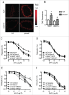
Inhibition of mitochondria fission attenuates oxidative stress and endothelial dysfunction in prkaa2−/− mice. (A) WT and prkaa2−/− mice were injected with control (siCtrl) or siDnm1l (5 mg/kg). Four d after treatment, frozen sections of carotid artery were incubated with dihydroethidium (DHE; 5 µM) for 30 min. Representative fluorescence microscopic images of DHE-stained artery (A) and quantification of intensity grade showing with color lookup table (B). n = 5; *P < 0.05 vs. WT; #P < 0.05 vs. siCtrl. (C) Aortas were contracted with U46619 (30 nM). Endothelium-dependent vasodilator responses were measured in the presence of acetylcholine (Ach, 10−9 to 10−5 M). (D) Endothelium-independent vasodilator responses were measured in the presence of sodium nitroprusside (SNP, 10−10 to 10−6 M). n = 6 to 8; *P < 0.05 vs. WT; #P < 0.05 vs. siCtrl. (E and F) WT and prkaa2−/− mice were treated with mdivi-1 (1.2 mg/kg/d) or vehicle (DMSO) for 14 d. (E) Aortas were contracted with U46619. Endothelium-dependent vasodilator responses were measured in the presence of Ach (10−9 to 10−4 M). (F) Endothelium-independent vasodilator responses were measured in the presence of SNP (10−10 to 10−6 M). n = 6 to 8; *P < 0.05 vs. WT; #P < 0.05 vs. vehicle.
Figure 5.
Figure 5.
Inhibited mitochondrial fusion impairs endothelial function. (A) HUVECs were transfected with control (siCtrl) or MFN2 siRNA (siMFN2) for 48 h, and mitochondria were labeled with MitoTracker® Deep Red FM. Mitochondrial morphology was analyzed using fluorescence microscopy. Scale bars: 5 µm. (B) Mitochondrial fragmentation was determined, as described in Materials and Methods. n ≥ 100; *P < 0.05 vs. siCtrl. (C) HUVECs were transfected with siCtrl or siMFN2 for 48 h and then treated with A23187 or DMSO for 30 min. Phosphorylation of NOS-3 was measured by western blotting. (D) Nitric oxide concentration in the cells was assessed using the DAF-2 fluorescence probe. *P < 0.05 vs. DMSO; #P < 0.05 vs. siCtrl. (E) WT mouse carotid arteries were transfected with siCtrl or siMfn2 using electroporation for 48 h, stained with antibodies against MFN2 (Red) and PECAM1 (Green), and observed using fluorescence microscopy. (F) Endothelium-dependent vasodilator responses were measured in the presence of Ach (10−9 to 10−5 M). (G) Endothelium-independent vasodilator responses were measured in the presence of SNP (10−10 to 10−6 M). n = 4, * P < 0.05 vs. siCtrl.
Figure 6.
Figure 6.
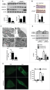
PRKAA deletion inhibits autophagic flux. (A) Levels of LC3B and SQSTM1 in WT and prkaa2−/− mouse aortas were determined by western blotting. n = 7 mice, *P < 0.05 vs. WT. (B) Immunohistochemical staining for SQSTM1 in WT and prkaa2−/− mouse aortas and quantification of positive staining for SQSTM1 in the aortas. n = 5, *P < 0.05 vs. WT. (C) Electron microscopic analysis of autophagic vacuoles (AV) and quantification of AV in WT and prkaa2−/− mouse aortas. n = 6 mice, *P < 0.05 vs. WT. (D) HUVECs were transfected with control (siCtrl) or siPRKAA2 for 24 h and then treated with bafilomycin A1 (BafA, 5 nM) for 24 h. Protein levels of LC3B and PRKAA2/AMPKα2 were measured by western blotting. n = 4. *P < 0.05 vs. siCtrl, #P < 0.05 vs. Veh. (E) HUVECs were cotransfected with siCtrl or siPRKAA2 and GFP-LC3B adenovirus for 24 h, and then treated with CQ for 24 h. Quantification of GFP-LC3B puncta formation in HUVECs was analyzed using fluorescence microscopy. n ≥ 50, *P < 0.05 vs. no CQ treatment; #P < 0.05 vs. siCtrl.
Figure 7.
Figure 7.
PRKAA deficiency-inhibited autophagy results in DNM1L accumulation. (A) HUVECs were treated with 3 µM CQ for 8 or 24 h. Protein levels of DNM1L and LC3B were analyzed by western blotting. n = 4. *P < 0.05 vs. no CQ. (B and D) HUVECs were transfected with control (siCtrl) or ATG7 siRNA (siATG7) for 48 h. (B) Protein levels of DNM1L, ATG7, and LC3B were analyzed by western blotting. n = 4. *P < 0.05 vs. siCtrl. (C) Mitochondrial morphology was analyzed by fluorescence microscopy. Scale bars: 5 µm. (D) Mitochondrial fission was determined, as described in Materials and Methods. n ≥ 100; *P < 0.05 vs. siCtrl. (E) HUVECs were incubated with cycloheximide (CHX, 1 µg/ml) for 4 to 24 h. DNM1L protein levels were analyzed by western blotting. n = 4. *P < 0.05 vs. no CHX. (F) HUVECs were transfected with C-si or siPRKAA2 for 24 h and then incubated with CHX for 8 or 24 h. After treatment, DNM1L protein levels were analyzed by western blotting. n = 4. *P < 0.05 vs. no CHX, #P < 0.05 vs. siCtrl. (G to I) HUVECs were transfected with siCtrl or siPRKAA2 for 24 h, after which they were transfected with adenoviruses encoding GFP (Ad-GFP) or ATG7 (Ad-ATG7) for 24 h. (G and H) Protein levels of DNM1L, p-ACACA, ATG7, LC3B, and SQSTM1 were measured by western blotting. n = 4. *P < 0.05 vs. siCtrl, #P < 0.05 vs. GFP (I) Mitochondrial morphology was analyzed using fluorescence microscopy. Scale bars: 10 µm. (J) Mitochondrial fission was determined, as described in Materials and Methods. n ≥ 100; *P < 0.05 vs. siCtrl, #P < 0.05 vs. Ad-GFP. (K) HUVECs were transfected with siCtrl, 2 independent siPRKAA1, or 2 independent siPRKAA2 for 48 h. Mitochondrial proteins, including VDAC, TOMM20 and CYCS/cytochrome C, were analyzed by western blotting.
Figure 8.
Figure 8.
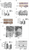
Activation of autophagy reduces DNM1L expression and mitochondrial fission in prkaa2−/− mice. WT and prkaa2−/− mice were orally administrated with rapamycin (Rapa, 14 mg/kg in diet) for 1 week. (A) Protein levels of phosphorylated MTOR at Ser2481 (p-MTOR) and LC3B in aortas were measured by western blotting. (B and C) Immunohistochemical staining for SQSTM1 in aortas from rapamycin-treated WT and prkaa2−/− mice, and quantification of positive staining for SQSTM1 in the aortas. *P < 0.05 vs. WT; #P < 0.05 vs. Vehicle. (D) Western blot analysis of DNM1L and p-ACACA protein expression in mouse aortas. (E and F) Immunohistochemical staining for DNM1L in aortas from rapamycin-treated WT and prkaa2−/− mice. Positive staining for DNM1L was quantified, as described in Materials and Methods. *P < 0.05 vs. WT; #P < 0.05 vs. Vehicle. (G) Representative electron micrographs of aortic endothelial mitochondria. Scale bars: 500 nm. (H) Mitochondrial length was quantified, as described in Materials and Methods. n = 6 mice, *P < 0.05 vs. WT, #P < 0.05 vs. Vehicle. (I) HUVECs were transfected with control (C-si) or siPRKAA for 24 h, after which they were treated with different concentrations of rapamycin for 24 h. Cell lysates were subjected to western blot analysis for DNM1L, PRKAA, p-MTOR, and phosphorylated RPS6KB at Thr389 (p-RPS6KB).
Figure 9.
Figure 9.
SQSTM1 is required for the autophagy-dependent degradation of DNM1L. (A and B) HUVECs were transfected with control (siCtrl) or siPRKAA2 for 48 h, and the interaction between DNM1L and SQSTM1 was determined by immunoprecipitation and western blotting. (C) The colocalization of DNM1L and SQSTM1 was assessed in HUVEC transfection with plasmid coding for cherry-DNM1L and GFP-SQSTM1. Scale bars: 5 µm. (D and E) HUVECs were transfected with or without BafA for 24 h, and the interaction between DNM1L and SQSTM1 was examined by immunoprecipitation and western blotting. (sF and G) The interaction between DNM1L and SQSTM1 in WT and atg7−/− MEFs was measured by immunoprecipitation and western blotting. (H and I) HUVECs were transfected with C-si and SQSTM1 siRNA (siSQSTM1) for 24 h, after which they were incubated with CHX for 8 to 24 h. DNM1L protein levels were analyzed by western blotting. (J) HUVECs were transfected with siCtrl and siSQSTM1 for 48 h. The interactions among SQSTM1, DNM1L, and LC3B were determined using immunoprecipitation and western blotting. (K to M) HUVECs were cotransfected with siPRKAA2 and siSQSTM1 for 48 h. (K) Protein levels of DNM1L, PRKAA, LC3B, and SQSTM1 were measured by western blotting. (L) Mitochondrial morphology was analyzed using fluorescence microscopy. Scale bars: 10 µm. (M) Mitochondrial fission was determined, as described in Materials and Methods. n = 100; *P < 0.05 vs. siCtrl.

Similar articles

Cited by

References

    1. Twig G, Hyde B, Shirihai OS. Mitochondrial fusion, fission and autophagy as a quality control axis: the bioenergetic view. Biochim Biophys Acta 2008; 1777:1092-7; PMID:18519024; http://dx.doi.org/10.1016/j.bbabio.2008.05.001 - DOI - PMC - PubMed
    1. Liesa M, Palacin M, Zorzano A. Mitochondrial dynamics in mammalian health and disease. Physiol Rev 2009; 89:799-845; PMID:19584314; http://dx.doi.org/10.1152/physrev.00030.2008 - DOI - PubMed
    1. Yu T, Robotham JL, Yoon Y. Increased production of reactive oxygen species in hyperglycemic conditions requires dynamic change of mitochondrial morphology. Proc Natl Acad Sci U S A 2006; 103:2653-8; PMID:16477035; http://dx.doi.org/10.1073/pnas.0511154103 - DOI - PMC - PubMed
    1. Burte F, Carelli V, Chinnery PF, Yu-Wai-Man P. Disturbed mitochondrial dynamics and neurodegenerative disorders. Nat Rev Neurol 2015; 11:11-24; PMID:25486875; http://dx.doi.org/10.1038/nrneurol.2014.228 - DOI - PubMed
    1. Shenouda SM, Widlansky ME, Chen K, Xu G, Holbrook M, Tabit CE, Hamburg NM, Frame AA, Caiano TL, Kluge MA, et al.. Altered mitochondrial dynamics contributes to endothelial dysfunction in diabetes mellitus. Circulation 2011; 124:444-53; PMID:21747057; http://dx.doi.org/10.1161/CIRCULATIONAHA.110.014506 - DOI - PMC - PubMed

MeSH terms