. 2017 Jan 13;13(1):e1006556.
doi: 10.1371/journal.pgen.1006556. eCollection 2017 Jan.

Acylation of the Type 3 Secretion System Translocon Using a Dedicated Acyl Carrier Protein

Affiliations

Acylation of the Type 3 Secretion System Translocon Using a Dedicated Acyl Carrier Protein

Julie P Viala et al. PLoS Genet. .

Abstract

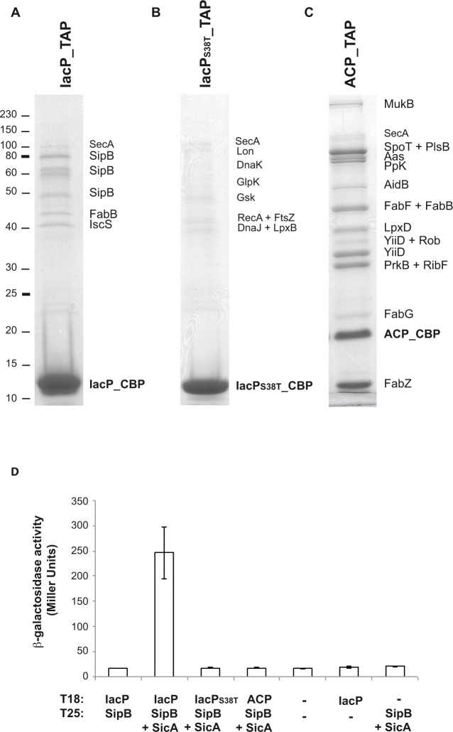
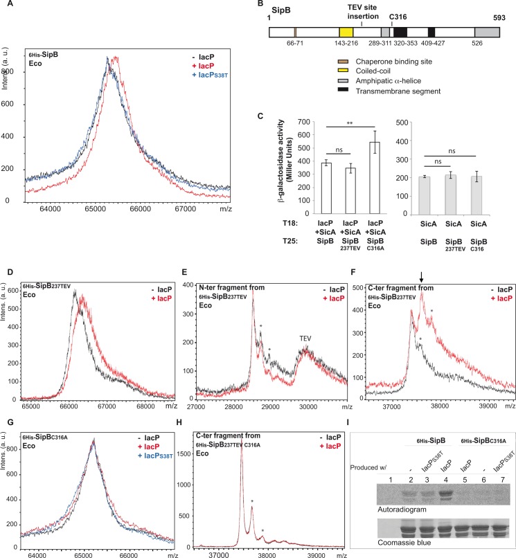
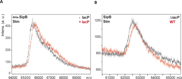
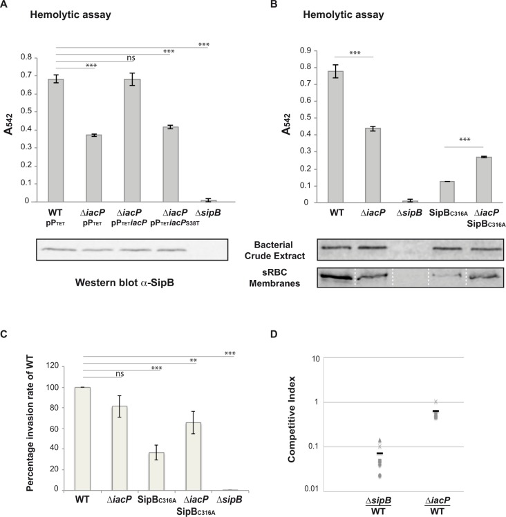
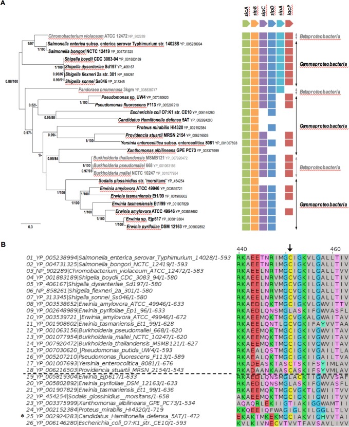
Bacterial pathogens often deliver effectors into host cells using type 3 secretion systems (T3SS), the extremity of which forms a translocon that perforates the host plasma membrane. The T3SS encoded by Salmonella pathogenicity island 1 (SPI-1) is genetically associated with an acyl carrier protein, IacP, whose role has remained enigmatic. In this study, using tandem affinity purification, we identify a direct protein-protein interaction between IacP and the translocon protein SipB. We show, by mass spectrometry and radiolabelling, that SipB is acylated, which provides evidence for a modification of the translocon that has not been described before. A unique and conserved cysteine residue of SipB is identified as crucial for this modification. Although acylation of SipB was not essential to virulence, we show that this posttranslational modification promoted SipB insertion into host-cell membranes and pore-forming activity linked to the SPI-1 T3SS. Cooccurrence of acyl carrier and translocon proteins in several γ- and β-proteobacteria suggests that acylation of the translocon is conserved in these other pathogenic bacteria. These results also indicate that acyl carrier proteins, known for their involvement in metabolic pathways, have also evolved as cofactors of new bacterial protein lipidation pathways.


Conflict of interest statement

The authors have declared that no competing interests exist.

Figures

Fig 1
Fig 1. Identification of protein partners of IacP and ACP in S. Typhimurium.
A, B, C: Tandem affinity purification experiments were performed with S. Typhimurium 12023 strains IacP_TAP, IacPS38T_TAP and ACP_TAP, respectively. The purified protein, still fused to the calmodulin binding peptide (CBP) part of the TAP tag (S1 Fig), and its protein partners were loaded on a 12% SDS-PAGE. After staining with Coomassie Blue, bands were cut from the gel and proteins were identified by mass spectrometry (S1 Fig). Proteins that were suspected to be non-specific partners are indicated in smaller characters. Markers of known molecular weight (kDa) are indicated on the left. D: Interactions between acyl carrier proteins and SipB were assayed by bacterial two-hybrid. Interactions between pairs of hybrid proteins, resulting from the fusion of the indicated protein with the T18 and T25 fragments of Bordetella pertussis adenylate cyclase, were assayed using the bacterial two-hybrid method in E. coli BTH101. A dash corresponds to an empty vector. When indicated, SicA was co-produced with T25_SipB from an artificial operon including T25_sipB and sicA on the two-hybrid vector. Interactions were assayed by β-galactosidase activity measurement. Values are the mean of three biological independent assays. Error bars stand for standard deviation.
Fig 2
Fig 2. Posttranslational modification of SipB analyzed by mass spectrometry and radiolabeling.
A and D to H: Mass spectrometry analysis of SipB produced with or without IacP. On the upper left of each graph is indicated the version of the purified SipB protein that has been analyzed by MALDI-TOF mass spectrometry. The SicA chaperone was always co-produced with SipB. Proteins were produced in E. coli (Eco). On the upper right of each graph is indicated whether or not the protein IacP had been co-produced with SipB (see S2B Fig for production plasmids). For example: (A) Mass analysis of full length 6His-SipB that had been produced without IacP (black spectrum), with IacP (red spectrum) or with IacPS38T (blue spectrum). (E, F) N-terminal and C-terminal fragments from the digestion of 6His-SipB237TEV by the TEV protease were first separated on cobalt beads. Both N-terminal fragments and TEV protease harbored a 6 His-tag and were retained on cobalt beads, which is why a peak close to 30 kDa corresponding to the TEV protease can be observed. (F) Arrow indicates the dominant peak observed for the analysis of the C-terminal fragment that appeared only when SipB had been produced with IacP (red spectrum). (H) C-terminal fragments from the digestion of 6His-SipB237TEV C316A by the TEV protease. (E, F, H) Minor peaks marked by an asterisk correspond to the analyzed protein fragments incremented by sinapinic matrix adducts. This sort of peaks are commonly observed by MALDI-TOF mass spectrometry when resolution allows their detection. B: Schematic of SipB regions according to the literature [,,–55]. The conserved translocator-chaperone binding motif is in brown, the coiled-coil region is in yellow, amphipatic α-helices are in grey, hydrophobic transmembrane segments are in black. Insertion of the TEV protease cleavage site at position 237 and the cysteine 316 residue are indicated. C: Interactions between pairs of hybrid proteins, resulting from the fusion of the indicated protein with the T18 and T25 fragments of Bordetella pertussis adenylate cyclase, were assayed using the bacterial two-hybrid method in E. coli BTH101. When indicated SicA was co-produced with T18_IacP from an artificial operon T18_iacP-sicA on the two-hybrid vector. Bacterial two-hybrid assays between SicA and the variant forms of SipB were performed to control the uniformity of the interaction signal with other partners of SipB. Hybrid proteins did not generate interaction when assayed against the corresponding empty two-hybrid vector (S3 Fig). Interactions were assayed by β-galactosidase activity measurement. The shown values are the mean of three biological independent assays. Error bars stand for standard deviation. Unpaired t-tests were used to determine whether the values were significantly different. p-values: ns, not significant; **, p ≤ 0.05. I: Radiolabelled acylation of SipB. The E. coli ΔgltA strain was transformed with the empty vector (pPTET) (lane 1) or with plasmids allowing expression of 6His-sipB and sicA without iacP, with iacPS38T, or with iacP (lanes 2, 3 and 4, respectively). Similarly 6His-sipBC316A and sicA were expressed with iacP, without iacP, or with iacPS38T (lanes 5, 6 and 7, respectively). Transformants were grown in M9 media; radiolabelled precursor of fatty acids, 14C-acetate, was provided when expression was triggered from the PTET promoter. Then, 6His-SipB and 6His-SipBC316A were purified on cobalt beads and loaded on 12% SDS-PAGE. Proteins were visualized by autoradiography after a 45 days exposure (top panel) and Coomassie Blue staining (bottom panel).
Fig 3
Fig 3. Mass spectrometry of 6His-SipB and endogenous SipB purified from S. Typhimurium.
MALDI-TOF mass spectrometry analysis of intact purified SipB. On the upper left of each graph is indicated the version of the purified SipB protein that has been analyzed by MALDI-TOF mass spectrometry and the organism from which it was purified: Stm for S. Typhimurium. A. The protein 6His-SipB was produced in the S. Typhimurium ΔiacP genetic background from production plasmids co-expressing iacP or not (S2B Fig for production plasmids); B. The intact native SipB was purified from S. Typhimurium WT and ΔiacP (strains JV1 and JV52) using an immunosorbent.
Fig 4
Fig 4. Hemolytic activity and invasiveness of S. Typhimurium SL1344 strains.
A and B: Sheep red blood cells were infected with S. Typhimurium SL1344 and derived mutant strains and hemolysis activity was followed measuring hemoglobin release at 542 nm. The hemolytic activity of the ΔsipB mutant strain is shown as a negative control. Hemolysis activity was assayed in triplicate and the error bar represents standard deviation. A representative experiment is shown. Unpaired t-tests were used to determine whether the values were significantly different. p-values: ns, not significant; ***, p ≤ 0.0005. A. Hemolytic activity of the wild-type and ΔiacP S. Typhimurium SL1344 strains that harbored the control empty plasmid pPTET or the corresponding plasmids containing iacP or iacPS38T. SipB was detected by western blot in bacterial crude extracts from the corresponding strains to show that similar amounts of SipB were produced (shown underneath the graph). B. Hemolytic activity of S. Typhimurium SL1344 strains, in which the indicated gene was deleted or modified by point substitution at the original locus. SipB was detected by western blot in bacterial crude extracts from the corresponding strains before starting the hemolysis assay (shown underneath the graph, top panel), and in sRBC membranes, isolated by sucrose density gradient, at the end of the hemolysis assay (shown underneath the graph, bottom panel). To load as much material as possible while ensuring that there was no leakage between the wells, the samples were separated by empty lanes. Those have been removed for the figure, which is symbolized by the white dotted lines. C: Invasion assays were performed on HeLa cells with S. Typhimurium SL1344 strains used for hemolysis assays. Invasion rates were normalized to the internalization level of WT, which was set to 100%. Each experiment was performed in triplicate and values are the mean of 6 independent experiments ± standard error of the mean. Unpaired t-tests were used to determine whether the values were significantly different. p-values: ns, not significant; **, p ≤ 0.05; ***, p ≤ 0.0001. D. Competitive index between the SL1344 wild-type strain (strain JV112) and ΔsipB (strain JV114) or between the SL1344 wild-type strain (strain JV112) and ΔiacPintra (strain JV129) in mice inoculated perorally. The black bar indicates the mean CI. Values for the mean CI ± SEM (t-test p-value) were 0.0716 ± 0.0225 (p-value < 0.0001) and 0.641 ± 0.1034 (p-value = 0.0255) for ΔsipB/WT and ΔiacP/WT, respectively.
Fig 5
Fig 5. Cooccurrence of SipB-like proteins and acyl carrier proteins in SPI-1-like T3SS.
A. Shown in the left is unrooted Bayesien phylogenetic tree of a selection of SipB homologs (26 sequences, 501 positions), which all belong to SPI-1 like T3SS (S5 Fig and S1 Table). Number at nodes indicates posterior probabilities (PP) and bootstrap support (BS) computed by Mrbayes and PhyMl, respectively. Only posterior probabilities and bootstrap values greater, respectively, than 0.5 and 50% are shown. The scale bars represent the number of substitutions per site. Shown in the right is the occurrence of each gene inferred from the genomic context analysis of SipB homologs. B. Zoom of the region of the multiple sequence alignment displayed in S5 Fig that includes the conserved cysteine residue (C316 in S. Typhimurium SipB). Sequences above the black dotted line are SipB and homologs that are genetically associated with an acyl carrier protein (both genes are localized less than five genes away), while below the black dotted line are SipB homologs that are not genetically associated with an acyl carrier protein (see Fig 5A and S1 Table). Black arrow indicates the cysteine residue corresponding to C316 of S. Typhimurium SipB, which is conserved in SipB and homologs that are genetically associated with an acyl carrier protein. * Although a gene encoding an acyl carrier protein was not found in proximity of the sipB-like gene in C. Halmitonella defensa, a gene encoding an acyl carrier protein was found associated with other genes of a SPI-1-type T3SS elsewhere in the genome.

Similar articles

Cited by

References

    1. de Souza Santos M, Orth K (2015) Subversion of the cytoskeleton by intracellular bacteria: lessons from Listeria, Salmonella and Vibrio. Cell Microbiol 17: 164–173. 10.1111/cmi.12399 - DOI - PMC - PubMed
    1. Ellermeier JR, Slauch JM (2007) Adaptation to the host environment: regulation of the SPI1 type III secretion system in Salmonella enterica serovar Typhimurium. Curr Opin Microbiol 10: 24–29. 10.1016/j.mib.2006.12.002 - DOI - PubMed
    1. Mattei PJ, Faudry E, Job V, Izore T, Attree I, Dessen A (2011) Membrane targeting and pore formation by the type III secretion system translocon. FEBS J 278: 414–426. 10.1111/j.1742-4658.2010.07974.x - DOI - PubMed
    1. Buttner D (2012) Protein export according to schedule: architecture, assembly, and regulation of type III secretion systems from plant- and animal-pathogenic bacteria. Microbiol Mol Biol Rev 76: 262–310. 10.1128/MMBR.05017-11 - DOI - PMC - PubMed
    1. Rathinavelan T, Lara-Tejero M, Lefebre M, Chatterjee S, McShan AC, Guo DC et al. (2014) NMR model of PrgI-SipD interaction and its implications in the needle-tip assembly of the Salmonella type III secretion system. J Mol Biol 426: 2958–2969. 10.1016/j.jmb.2014.06.009 - DOI - PMC - PubMed

Publication types

MeSH terms

Substances

LinkOut - more resources