. 2017 Jan 13;11(1):e0005294.
doi: 10.1371/journal.pntd.0005294. eCollection 2017 Jan.

European Aedes albopictus and Culex pipiens Are Competent Vectors for Japanese Encephalitis Virus

Affiliations

European Aedes albopictus and Culex pipiens Are Competent Vectors for Japanese Encephalitis Virus

Mélissanne de Wispelaere et al. PLoS Negl Trop Dis. .

Abstract

Background: Japanese encephalitis virus (JEV) is the causative agent of Japanese encephalitis, the leading cause of viral encephalitis in Asia. JEV transmission cycle involves mosquitoes and vertebrate hosts. The detection of JEV RNA in a pool of Culex pipiens caught in 2010 in Italy raised the concern of a putative emergence of the virus in Europe. We aimed to study the vector competence of European mosquito populations, such as Cx. pipiens and Aedes albopictus for JEV genotypes 3 and 5.

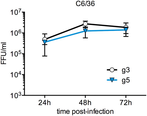
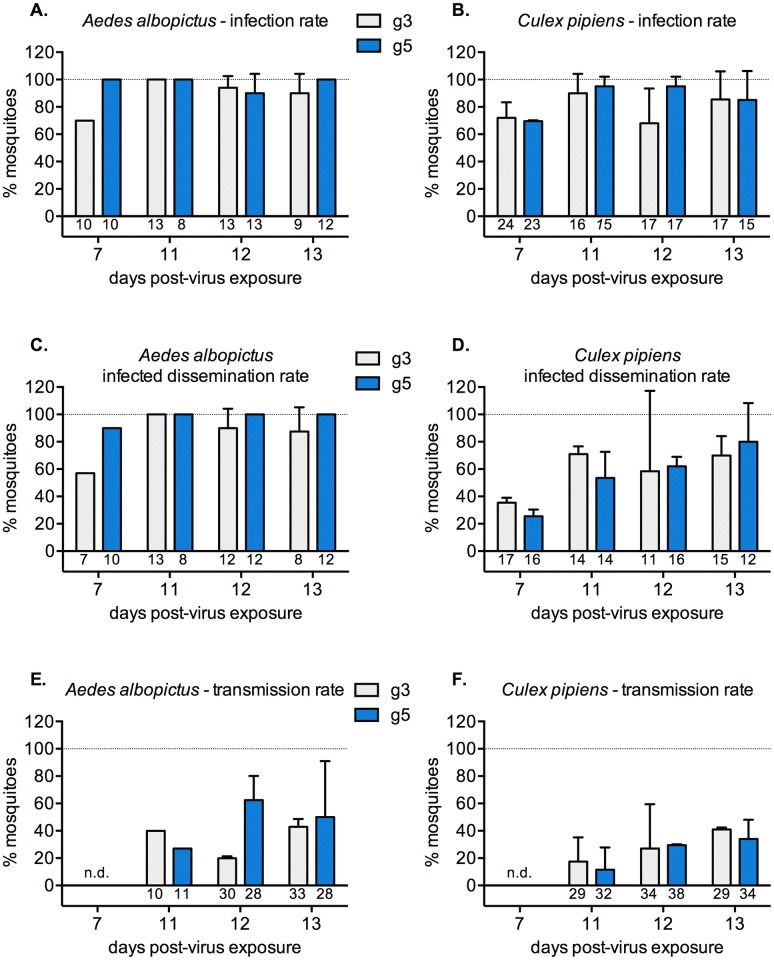
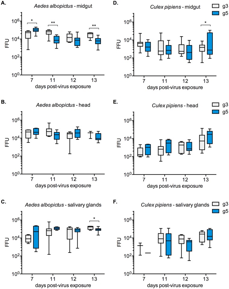
Findings: After oral feeding on an infectious blood meal, mosquitoes were dissected at various times post-virus exposure. We found that the peak for JEV infection and transmission was between 11 and 13 days post-virus exposure. We observed a faster dissemination of both JEV genotypes in Ae. albopictus mosquitoes, when compared with Cx. pipiens mosquitoes. We also dissected salivary glands and collected saliva from infected mosquitoes and showed that Ae. albopictus mosquitoes transmitted JEV earlier than Cx. pipiens. The virus collected from Ae. albopictus and Cx. pipiens saliva was competent at causing pathogenesis in a mouse model for JEV infection. Using this model, we found that mosquito saliva or salivary glands did not enhance the severity of the disease.

Conclusions: In this study, we demonstrated that European populations of Ae. albopictus and Cx. pipiens were efficient vectors for JEV transmission. Susceptible vertebrate species that develop high viremia are an obligatory part of the JEV transmission cycle. This study highlights the need to investigate the susceptibility of potential JEV reservoir hosts in Europe, notably amongst swine populations and local water birds.


Conflict of interest statement

The authors have declared that no competing interests exist.

Figures

Fig 1
Fig 1. Kinetics of JEV g3 and g5 infection in vitro.
Ae. albopictus derived C6/36 cells were infected with JEV g3 or g5 at a MOI of 1. The infectious virus released to the supernatants at 24, 48 and 72 h post-infection was quantified by FFA in BHK-21 cells. The error bars represent the standard deviation between two independent experiments (in each experiment, titrations were done on duplicate experimental samples). FFU, focus forming units.
Fig 2
Fig 2. Vector competence of European strains of mosquitoes for two different JEV genotypes following feeding on infectious bloodmeals.
Infection rates in Ae. albopictus (A) or Cx. pipiens (B) mosquitoes exposed to either JEV g3 or JEV g5 by feeding on blood meals containing 8 x 10⁶ FFU/ml of virus. The infection rates were determined after titration of midguts harvested from mosquitoes on days 7, 11, 12 and 13 post-virus exposure. The infected dissemination rates (C and D) were calculated by titration of the heads of infected mosquitoes. The transmission rates (E and F) were calculated by titration of the saliva collected from all blood-fed mosquitoes. The error bars represent the standard deviation between two independent experiments. The number of mosquitoes analyzed for each condition is indicated below each graph bar. An unpaired t test was employed to determine significant differences between JEV genotypes at each time point. No statistically significant differences were found (P≥0.08). n.d. not determined.
Fig 3
Fig 3. Kinetics of JEV infection in European strains of Ae. albopictus and Cx. pipiens mosquitoes.
Ae. albopictus (A, B and C) or Cx. pipiens (D, E and F) mosquitoes were exposed to either JEV g3 or JEV g5 by feeding on blood meals containing 8 x 10⁶ FFU/ml of virus. At 7, 11, 12 and 13 days post-virus exposure, the midguts (A and D), heads (B and E) and salivary glands (C and F) were harvested from individual mosquitoes and the levels of infectious virus in each organ was quantified by FFA in BHK-21 cells. The error bars represent the standard deviation amongst infected samples collected from two independent experiments. An unpaired t test was employed to determine significant differences between JEV genotypes at each time point (***, P < 0.001; **, 0.001 < P < 0.01; *, 0.01 < P < 0.05; only statistically significant differences are shown).
Fig 4
Fig 4. Analysis of JEV infection in mosquitoes.
A. Visualization of JEV envelope protein in the midgut (left panel) and salivary glands (right panel) of Ae. albopictus mosquitoes infected with JEV g3 at 14 days post-feeding on an infectious blood meal. Samples obtained from non-infected mosquitoes were used as a control (n.i.). Scale bars: 500 μm for the midgut samples, 50 μm for the salivary glands samples. B. Detection of JEV envelope protein in lysates obtained from midguts (top) and salivary glands (bottom) harvested from mosquitoes at 11 days post-feeding on an infectious blood meal. Samples from non-infected mosquitoes are used as a control (n.i.). The expected size for JEV envelope protein is indicated with an arrow.
Fig 5
Fig 5. Analysis of salivas collected from European mosquitoes infected by different genotypes of JEV.
At 11, 12 and 13 days post-virus exposure, salivas were collected from mosquitoes via forced salivation in pipette tips. A. Individual salivation samples were analyzed in a dot-blot assay using an anti-saliva antibody, to determine the salivation efficiency for each mosquito species. The rates of salivation obtained from two independent salivation assays are given, and a representative dot blot assay is shown. B. and C. The levels of infectious virus in JEV-positive saliva were measured by FFA in BHK-21 cells.
Fig 6
Fig 6. Effect of mosquito saliva and salivary glands on JEV infection in a mouse model.
Groups of 3-week-old BALB/c mice were monitored for survival after intradermal injection with JEV g5 (n = 6 per group). A. Mice were injected with JEV-positive saliva collected from infected mosquitoes. The levels of virus in saliva were between 7 and 98 FFU (Ae. albopictus: 9, 15, 22, 28, 60 and 70 FFU; Cx. pipiens: 7, 16, 24, 30, 51 and 98 FFU). Control mice were injected with 50 FFU of virus grown in C6/36 cells (C6/36 supernatants). B. Mice were injected with 50 FFU of virus, in presence or absence of one pair of salivary glands (SGP) harvested from either Ae. albopictus or Cx. pipiens mosquitoes. A Log-rank (Mantel-Cox) test was employed to determine significant differences between inocula. No statistically significant differences were found (P≥0.6).

Similar articles

Cited by

References

    1. Campbell GL, Hills SL, Fischer M, Jacobson JA, Hoke CH, Hombach JM, et al. Estimated global incidence of Japanese encephalitis: a systematic review. Bull World Health Organ. 2011;89(10):766–74, 74A–74E. 10.2471/BLT.10.085233 - DOI - PMC - PubMed
    1. Fischer M, Hills S, Staples E, Johnson B, Yaich M, Solomon T, et al. Japanese encephalitis prevention and control: advances, challenges, and new initiatives,. In: W M H S, H JM, editors. Emerging infections. 8. Washington, DC: American Society for Microbiology; 2008. p. 93–124.
    1. Schuh AJ, Ward MJ, Brown AJ, Barrett AD. Phylogeography of Japanese encephalitis virus: genotype is associated with climate. PLoS Negl Trop Dis. 2013;7(8):e2411 10.1371/journal.pntd.0002411 - DOI - PMC - PubMed
    1. Schuh AJ, Guzman H, Tesh RB, Barrett AD. Genetic diversity of Japanese encephalitis virus isolates obtained from the Indonesian archipelago between 1974 and 1987. Vector Borne Zoonotic Dis. 2013;13(7):479–88. 10.1089/vbz.2011.0870 - DOI - PMC - PubMed
    1. Gao X, Liu H, Wang H, Fu S, Guo Z, Liang G. Southernmost Asia is the source of Japanese encephalitis virus (genotype 1) diversity from which the viruses disperse and evolve throughout Asia. PLoS Negl Trop Dis. 2013;7(9):e2459 10.1371/journal.pntd.0002459 - DOI - PMC - PubMed

Publication types

Substances