Review
. 2017 Jun:46:9-16.
doi: 10.1016/j.ceb.2016.12.007. Epub 2017 Jan 10.

Getting to and through the inner nuclear membrane during herpesvirus nuclear egress

Affiliations
Review

Getting to and through the inner nuclear membrane during herpesvirus nuclear egress

Ming F Lye et al. Curr Opin Cell Biol. 2017 Jun.

Abstract

Herpesviruses, like most DNA viruses, replicate and package their genomes into capsids in the host cell nucleus. Capsids then transit to the cytoplasm in a fascinating process called nuclear egress, which includes several unusual steps: Movement of capsids from the nuclear interior to the periphery, disruption of the nuclear lamina, capsid budding through the inner nuclear membrane, and fusion of enveloped particles with the outer nuclear membrane. Here, we review recent advances and emerging questions relating to herpesvirus nuclear egress, emphasizing controversies regarding mechanisms for capsid trafficking to the nuclear periphery, and implications of recent structures of the two-subunit, viral nuclear egress complex for the process, particularly at the step of budding through the inner nuclear membrane.


Figures

Fig 1
Fig 1. Overview of nuclear egress
1) Newly assembled herpesvirus capsids migrate from the nuclear interior to the nuclear rim. 2) The NEC recruits viral and/or cellular kinases for phosphorylation and disruption of the nuclear lamina. 3) Capsids engage the NEC and bud through the inner nuclear membrane (primary envelopment). 4) The perinuclear capsid-containing vesicle (primary enveloped particle) fuses with the outer nuclear membrane and releases capsid into the cytoplasm (de-envelopment). The inset shows the structure of an NEC (that of HCMV) and summarizes four distinctive structural features: 1) The N-terminal heterodimerization domain on the nucleoplasmic NEC subunit (in blue) composed of two helices angled to form a V shape, 2) A vise-like heterodimerization mechanism on the INM-anchored NEC subunit whereby a C-terminal helix (light purple) acts as a moveable jaw and swings out (red arrow) to accommodate the V-shaped heterodimerization domain of the nucleoplasmic subunit and clamp it to the rest of the INM-anchored globular body. The form of the INM-anchored subunit not bound to the nucleoplasmic subunit is shown in gray (from the structure of MCMV M50) and superimposed onto the bound form shown in light purple. The C-terminal helix which swings out in the unbound versus NEC-bound form of the transmembrane subunit is indicated with a red star. 3) The nucleoplasmic subunit includes a zinc finger, and 4) Both subunits have a Bergerat fold shown in wheat. The INM-anchored subunit attaches to the inner nuclear membrane through a C-terminal extension that is predicted to be unstructured (light purple dashed lines), and to make a single pass through the INM with a very short segment in the perinuclear space.
Fig 2
Fig 2. Structures of the nuclear egress complex (NEC) and its subunits
A. NEC structures from (left to right) Pseudorabies virus (PrV; two crystal forms shown in shades of red [29] and yellow [30] respectively), herpes simplex virus-1 (HSV-1; shades of green [30]), and human cytomegalovirus (HCMV; two crystal forms shown in shades of blue [28] and purple [7] respectively). The PDB accession codes are listed under each structure. In the right-most structure, the Bergerat fold found in both subunits is shown in wheat. All structures display a zinc ion represented as a gray sphere. The nucleoplasmic (UL31/UL53) and INM-anchored (UL34/UL50) subunits are labeled in the same color code as in the structures. B. (Left) Structural alignment of the nucleoplasmic (UL31/UL53) subunit from PrV (red and yellow), HSV-1 (green), and HCMV (blue and purple). The UL53 monomeric subunit [7] is also superimposed, and shown in orange. View from a 90 degree rotation from the left is also shown, highlighting the cis-proline and β-strand (insert) preceding the diverging V-shaped heterodimerization domains of the nucleoplasmic (UL31/UL53) subunit structures. The stable core and heterodimerization domain that flank the linker region of the nucleoplasmic (UL31/UL53) subunit are labeled. (Right) Structural alignment of INM-anchored (UL34/UL50) subunit structures in the bound state, but shown without the nucleoplasmic subunit. C. HCMV NEC (PDB ID: 5D5N) [28] shown with the NMR structure of the unbound state of the MCMV INM-anchored subunit M50 (PDB ID: 5A3G) [8]. M50 is superimposed onto UL50 in the bound state. The red arrow indicates the movement of the C-terminal helix of UL50 that is required for binding to UL53.
Fig 3
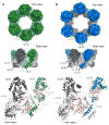
Fig 3. The NEC hexameric interface
(A). Hexameric arrangement of the HSV-1 NEC shown from the top (top panel) and side (middle panel) [30]. A single heterodimer is shown in dark and light green for the HSV nucleoplasmic (UL31) and INM-anchored (UL34) subunits, respectively. For clarity when comparing interactions between neighboring heterodimers (middle and bottom panels), the alternating subunits are shown in dark and light gray for the UL31 and UL34 subunits respectively. The linker region in UL31 is labeled with a pink arrow, and selected interacting residues at the hexameric interface shown, specifically focusing on interactions with the linker region. The Bergerat fold in both subunits is shown in wheat (B). Hexameric arrangement of the HCMV with the nucleoplasmic (UL53) and INM-anchored (UL50) subunits in dark and light blue respectively [28]. Representations of the structure follow the same format as in A.

Similar articles

Cited by

References

    1. Pellett PE, Roizman B. Herpesviridae. In: Knipe DM, Howley PM, editors. Fields Virology. 2013. pp. 1802–1822.
    1. Speese SD, Ashley J, Jokhi V, Nunnari J, Barria R, Li Y, Ataman B, Koon A, Chang Y-T, Li Q, et al. Nuclear envelope budding enables large ribonucleoprotein particle export during synaptic Wnt signaling. Cell. 2012;149:832–846. - PMC - PubMed
    1. Jokhi V, Ashley J, Nunnari J, Noma A, Ito N, Wakabayashi-Ito N, Moore MJ, Budnik V. Torsin mediates primary envelopment of large ribonucleoprotein granules at the nuclear envelope. Cell Rep. 2013;3:988–995. - PMC - PubMed
    1. Sharma M, Bender BJ, Kamil JP, Lye MF, Pesola JM, Reim NI, Hogle JM, Coen DM. Human cytomegalovirus UL97 phosphorylates the viral nuclear egress complex. J Virol. 2015;89:523–534. - PMC - PubMed
    1. Mou F, Wills EG, Park R, Baines JD. Effects of lamin A/C, lamin B1, and viral US3 kinase activity on viral infectivity, virion egress, and the targeting of herpes simplex virus U(L)34-encoded protein to the inner nuclear membrane. J Virol. 2008;82:8094–8104. - PMC - PubMed

LinkOut - more resources