Review
. 2017 Apr:11:482-501.
doi: 10.1016/j.redox.2016.12.001. Epub 2016 Dec 7.

Happily (n)ever after: Aging in the context of oxidative stress, proteostasis loss and cellular senescence

Affiliations
Review

Happily (n)ever after: Aging in the context of oxidative stress, proteostasis loss and cellular senescence

Annika Höhn et al. Redox Biol. 2017 Apr.

Abstract

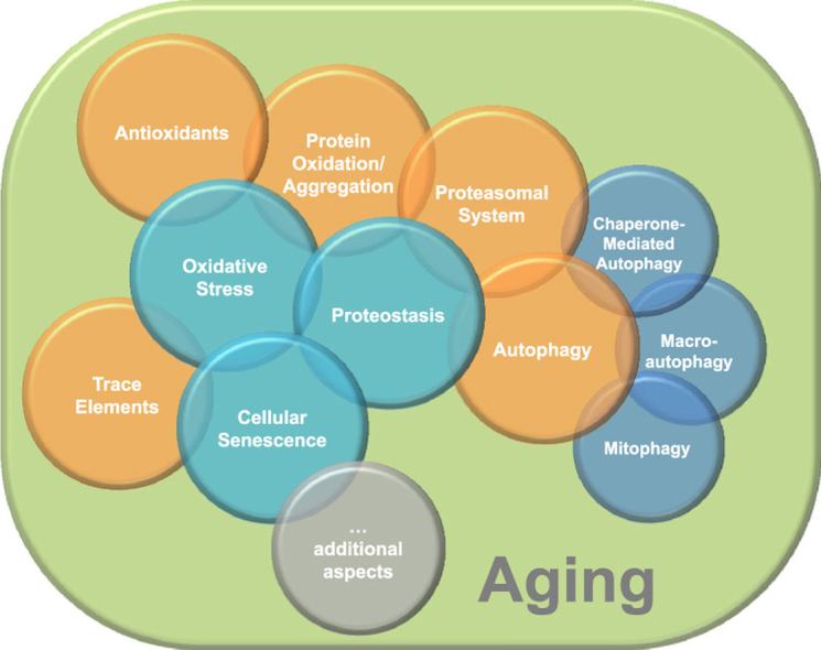
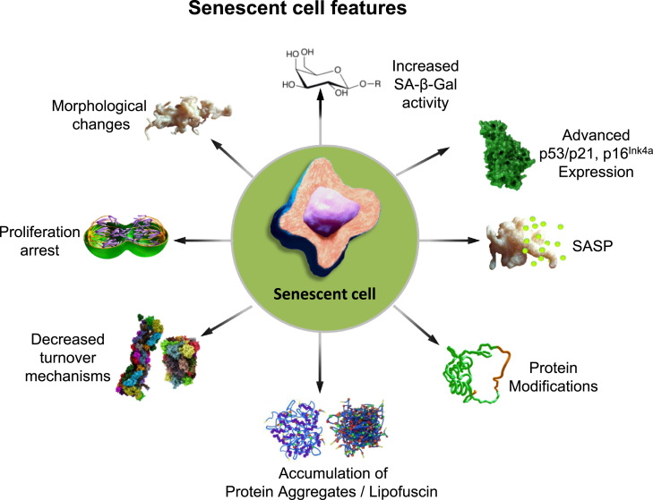
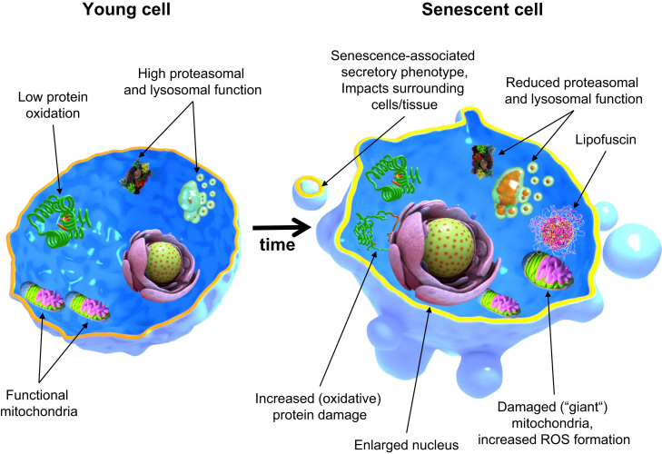
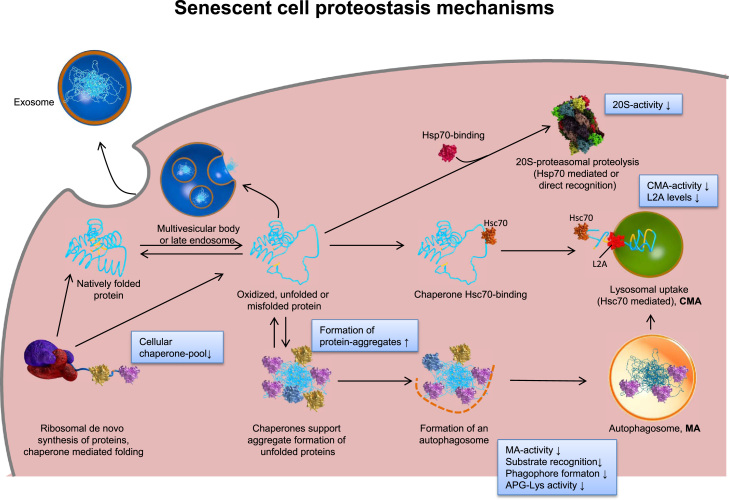
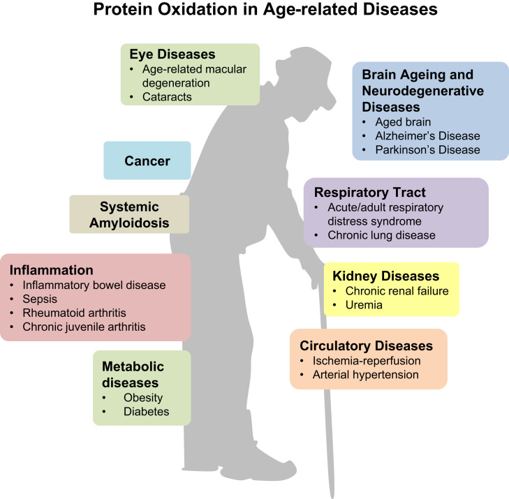
Aging is a complex phenomenon and its impact is becoming more relevant due to the rising life expectancy and because aging itself is the basis for the development of age-related diseases such as cancer, neurodegenerative diseases and type 2 diabetes. Recent years of scientific research have brought up different theories that attempt to explain the aging process. So far, there is no single theory that fully explains all facets of aging. The damage accumulation theory is one of the most accepted theories due to the large body of evidence found over the years. Damage accumulation is thought to be driven, among others, by oxidative stress. This condition results in an excess attack of oxidants on biomolecules, which lead to damage accumulation over time and contribute to the functional involution of cells, tissues and organisms. If oxidative stress persists, cellular senescence is a likely outcome and an important hallmark of aging. Therefore, it becomes crucial to understand how senescent cells function and how they contribute to the aging process. This review will cover cellular senescence features related to the protein pool such as morphological and molecular hallmarks, how oxidative stress promotes protein modifications, how senescent cells cope with them by proteostasis mechanisms, including antioxidant enzymes and proteolytic systems. We will also highlight the nutritional status of senescent cells and aged organisms (including human clinical studies) by exploring trace elements and micronutrients and on their importance to develop strategies that might increase both, life and health span and postpone aging onset.

Keywords: Aging; Antioxidants; Micronutrients; Protein oxidation; Proteostasis; Senescence; Trace elements.


Figures

fx1
Graphical abstract
Fig. 1
Fig. 1
Publications per year related to “aging”. The search term “aging” was entered on PubMed on October 7th, 2016. Results were plotted as publications per year.
Fig. 2
Fig. 2
Features of senescent cells: Several markers were identified to characterize the senescent state in relation to morphology and proteostasis. During the development of senescence, cells show morphological changes by extension of their size and protein content or nuclei enlargement. Also, their lysosomes size and number increase resulting in an elevated activity of SA-β-Gal, the most widely used marker for senescence. The cells enter a proliferative arrest state, detected by cell cycle inhibitor levels such as p53/p21 and tumor suppressor p16INK4a, the latter is correlated with the formation of the SAHF. Other factors secreted during senescence are cytokines and chemokines, growth factors, proteases, fibronectin as well as ROS and RNS, altogether these are summarized as the SASP. Additionally, proteostasis changes during senescence shown by an increase in modified proteins, accumulation of protein aggregates and reduced functionality of the proteasomal and autophagy systems.
Fig. 3
Fig. 3
Cellular and molecular features of young and aged cells. Comparing to young, aged cells often exhibit marked features. For example, it is known that their nucleus is often enlarged. Their proteolysis mechanisms suffer a major reduction in functionality, found by evaluating the activity of both proteasomal and lysosomal mechanisms. The consequence is the accumulation of oxidized proteins and the formation of insoluble material such as lipofuscin, a hallmark of aged cells as well and altogether contributing to a loss in cellular proteostasis. Furthermore, recent years have brought us evidence of a secretory phenotype acquired by senescent cells (SASP), which is characterized by the release of several inflammatory cytokines into the surrounding cells and tissues, resulting in low-grade chronic inflammation over time causing tissue and organism dysfunction.
Fig. 4
Fig. 4
Proteostasis changes in a senescent cell. The scheme shows the overall changes of the cellular systems maintaining protein homeostasis (proteostasis) and thus, cellular functionality during aging. The main proteolytic systems, responsible for recognition and degradation of un/misfolded or oxidatively damaged proteins are the proteasomal system, involving the ATP-dependent 26S proteasome as well as the ATP-independent 20S proteasome, and autophagy (including both autophagy (MA) as well as the chaperone mediated one (CMA)). Damaged proteins can be directly recognized as substrates by the 20S proteasome, resulting in proteolytic removal from the cell. Furthermore, they can be recognized by chaperones or heat shock proteins that keep their substrates in a soluble state, preventing the formation of aggregates. Another fate might be the formation of aggregates, driven by hydrophobic residues exposure. Such aggregates can be incorporated into an autophagosome and fusing with lysosomes, resulting in proteolytic degradation by lysosomal proteases, or they can be removed from the cell via excretion as exosome. Modified from and according to .
Fig. 5
Fig. 5
Cellular aging models. Primary cells can be isolated from young and old donors. (1.) Cells isolated from an old donor are called in vivo aged cells and can be investigated directly to study aging. In case of using cells from a young donor there a several options to study aging: (2.) After a multitude of subcultures, the young cells reach their replicative limit and stop dividing. These cells are called senescent or in vitro aged cells and are the most frequently used cellular aging model. (3.) Treatment of young cells with different oxidants or stressors for several days leads to the so-called stress-induced premature senescence (SIPS). After a short recovery phase these SIPS cells can be used for aging research. (4.) Finally young cells can be incubated with aggregates such as AGEs or lipofuscin. These cells mirror several important features of aged cells. Therefore, these aggregate-fed cells are an additional useful aging model.
Fig. 6
Fig. 6
Overview of endogenous and exogenous antioxidants. This figure gives a broad overview of antioxidants. Endogenous antioxidants comprise proteins, low-molecular weight molecules and enzymes, among others. Exogenous (dietary) sources of antioxidants include animal products, fruits, vegetables and grains (see chapter on micronutrients).
Fig. 7
Fig. 7
Lipofuscin accumulation in young, aged and SIPS fibroblasts. One hallmark of aged cells is the formation of lipofuscin, which consists of highly oxidized proteins and lipids. A special characteristic of lipofuscin is its stable autofluorescence which can be used for the detection as well as quantification. Aged human dermal fibroblasts, obtained from an 81-year old donor (panel B) are marked by a strong accumulation of lipofuscin compared to fibroblasts from a 1-year old donor (panel A). To investigate the aging process in cell culture systems the so-called model of stress-induced premature senescence (SIPS) can be used. During SIPS, cells are treated chronically with a low dose of an oxidant to generate cells with a senescent phenotype. Panel C shows fibroblasts which were incubated with 40 µM paraquat as stressor for 10 days leading to the accumulation of lipofuscin. Lipofuscin autofluorescence was measured at 408 nm excitation and 420 nm emission wavelengths using a laser scanning microscope. Fibroblasts are cultivated as described in König et al. .
Fig. 8
Fig. 8
Age-related changes in the autophagy-lysosome pathway. The autophagy-lysosome pathway (ALP) is one of the main intracellular degradation systems, responsible for the removal of dysfunctional cell constituents and their recycling. One part of the ALP, macroautophagy can be subdivided into different phases, each of them can be negatively affected in aging. Phase I includes different autophagy-related genes (ATGs), which are mainly responsible for the initiation and development of the autophagophore. During the aging process, ATGs, such as Beclin-1, ULK1, ATG5 and ATG12 decrease; resulting in a decline in the initial steps of autophagy. In addition up-regulation of Bcl-2 and mammalian target of rapamycin (mTOR), enhanced by decreased levels of Sestrin1, involved in AMPK activation, aggravate the impaired initiation of autophagy. The potential decrease of p62 and Parkin, both involved in the delivery of either dysfunctional, ubiquitinated proteins or Pink1-tagged mitochondria, can support the accumulation of dysfunctional proteins and organelles in aging. Finally, reduced conversion of unbound LC3-I into the membrane-bound LC3-II demonstrates the impairment of the initiation phase. In the second phase, the number of autophagosomes and lysosomes increase in aging, reported by several studies, analyzing lysosomal-associated membrane protein 1 (LAMP1) and the autophagosomal marker monodansylcadaverine (MDC), related to total cellular protein. But an increased autophagosome and lysosome number is not able to explain decreased protein degradation and increased protein aggregation as well as accumulation in aging, assuming that fusion of both is likely to be impaired, a process which is not known yet and needs further investigation. In addition to impaired mito- and macroautophagy also chaperone-mediated autophagy is reduced in aging, demonstrated by reduced levels of Lamp2a and HSC70.
Fig. 9
Fig. 9
Protein oxidation in age-related diseases. This figure shows the numerous diseases in which protein oxidation has been demonstrated so far. Protein oxidation may be the cause or consequence of these diseases which affect nearly all organ systems.

References

    1. C. López-Otín, M. Blasco, L. Partridge, M. Serrano, G. Kroemer, The Hallmarks of Aging. Cell153(6):1194–1217 - PMC - PubMed
    1. Flatt T. A new definition of aging? Front. Genet. 2012;3:148. - PMC - PubMed
    1. Hayflick L., Moorhead P.S. The serial cultivation of human diploid cell strains. Exp. Cell Res. 1961;25:585–621. - PubMed
    1. Harley C.B., Futcher A.B., Greider C.W. Telomeres shorten during ageing of human fibroblasts. Nature. 1990;345(6274):458–460. - PubMed
    1. As I.J., Greider C.W. Short telomeres induce a DNA damage response in Saccharomyces cerevisiae. Mol. Biol. cell. 2003;14(3):987–1001. - PMC - PubMed