. 2017 Jan 13;17(1):46.
doi: 10.1186/s12906-016-1501-6.

Cheongsangbangpung-tang ameliorated the acute inflammatory response via the inhibition of NF-κB activation and MAPK phosphorylation

Affiliations

Cheongsangbangpung-tang ameliorated the acute inflammatory response via the inhibition of NF-κB activation and MAPK phosphorylation

Seon Young Kim et al. BMC Complement Altern Med. .

Abstract

Background: Cheongsangbangpung-tang (CBT) is a traditional herbal formula used in Eastern Asia to treat heat-related diseases and swellings in the skin. The present study was conducted to evaluate the anti-inflammatory effects of cheongsangbangpung-tang extract (CBTE) both in vitro and in vivo.

Methods: The in vitro effects of CBTE on the lipopolysaccharide (LPS)-induced production of inflammation-related proteins were examined in RAW 264.7 cells. The levels of nitric oxide (NO) were measured with the Griess reagent. Inflammatory cytokines and prostaglandin E2 (PGE2) were detected using the enzyme-linked immunosorbent assay (ELISA) method. Inflammation-related proteins were detected by Western blot. The effect of CBTE on acute inflammation in vivo was evaluated using carrageenan (CA)-induced paw oedema. To evaluate the anti-inflammatory effect, paw oedema volume, thickness of the dorsum and ventrum pedis skin, number of infiltrated inflammatory cells, and number of COX-2-, iNOS-immunoreactive cells were measured.

Results: In an in vitro study, CBTE inhibited the production of NO and PGE2 and also decreased the expression of inducible nitric oxide synthase (iNOS), cyclooxygenase-2 (COX-2) activity, interleukin (IL)-1β, IL-6 and tumuor necrosis factor-α. In LPS-activated macrophages, nuclear factor-kappaB (NF-κB) and mitogen-activated protein kinase (MAPK) signalling is a pivotal pathway in the inflammatory process. These plausible molecular mechanisms increased the phosphorylation of I-κBα, while the activation of NF-κB and the phosphorylation of MAPK by LPS were blocked by CBTE treatment. In our in vivo study, a CA-induced acute oedematous paw inflammation rat model was used to evaluate the anti-inflammatory effect of CBTE. CBTE significantly reduced the increases in paw swelling, skin thicknesses, infiltrated inflammatory cells and iNOS-, COX-2 positive cells induced by CA injection.

Conclusions: Based on these results, CBTE should favourably inhibit the acute inflammatory response through modulation of NF-κB activation and MAPK phosphorylation. Furthermore, the inhibition of CBTE in rat paw oedema induced by CA is considered to be clear evidence that CBTE may be a useful source to treat inflammation.

Keywords: Cheongsangbangpung-tang; Inflammation; Mitogen-activated protein kinase; Nuclear factor-kappaB; Paw oedema.


Figures

Fig. 1
Fig. 1
Analysis of seven standard compounds in CBTE by UPLC-PDA. Ultra performance liquid chromatography (UPLC) chromatograms of seven standard compounds: glycyrrhizic acid, liquiritigenin, berberine, baicalin, baicalein, forsythiaside-A and poncirin a UPLC chromatograms of glycyrrhizic acid, liquiritigenin, berberine, baicalin, baicalein, forsythiaside-A and poncirin in CBTE (glycyrrhizic acid and liquiritigenin; 254 nm, berberine; 345 nm, baicalin and baicalein; 277 nm, forsythiaside-A; 280 nm, poncirin; 230 nm) b CBTE, Cheongsangbangpung-tang extract
Fig. 2
Fig. 2
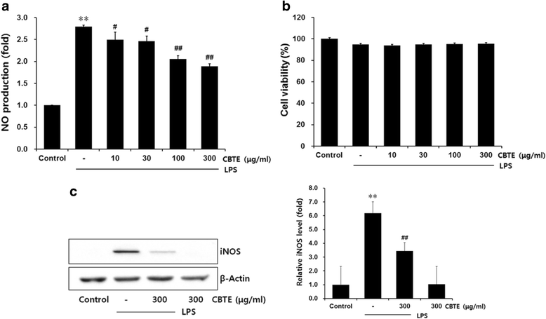
The inhibitory effects of CBTE on NO production and iNOS expression. Raw 264.7 cells were treated with 10–300 μg/ml of CBTE for 1 h prior to the addition of lipopolysaccharide (LPS) (1 μg/ml), and further incubated for 12 h. Cell viability was measured by 3-(4,5-dimethylthiazol-2-yl)-2,5-diphenyltetrazolium bromide assay. The concentrations of nitrite in the culture medium were assessed using the Griess reagent as described in the Methods section (a). The effect of CBTE plus LPS on cell viability was determined after 12 h of incubation (b). Expression of the iNOS protein was assessed by immunoblotting using a specific anti-iNOS antibody (c). Equal amounts of the total protein (50 μg/lane) were separated by SDS-PAGE. β-actin was used as a loading control, and the relative levels of protein bands were measured by scanning densitometry. Values represent the mean ± S.D. of three independent experiments (significant compared to the control, **P < 0.01, significant as compared to LPS alone, ## P < 0.01). CBTE, Cheongsangbangpung-tang extract
Fig. 3
Fig. 3
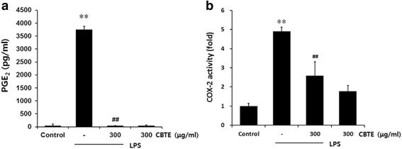
The inhibitory effect of CBTE on the LPS-induced PGE2 secretion and COX-2 activity. RAW264.7 cells were treated with CBTE for 1 h prior to the addition of LPS (1 μg/ml), and the cells were further incubated for 12 h. The levels of PGE2 in the culture medium were measured with an assay kit as described in the Methods section (a). COX-2 enzyme activity was analysed using an assay kit also as described in the Methods section (b). Values represent the mean ± S.D. of three independent experiments (significant compared to the control, **P < 0.01, significant as compared to LPS alone, ## P < 0.01). CBTE, Cheongsangbangpung-tang extract; PGE2, prostaglandin E2; COX-2, cyclooxygenase-2
Fig. 4
Fig. 4
The inhibitory effect of CBTE on the LPS-induced secretion of pro-inflammatory cytokines. RAW264.7 cells were treated with CBTE for 1 h prior to the addition of LPS (1 μg/ml), and the cells were further incubated for 12 h. The concentrations of TNF-α (a) IL-6 (b) and IL-1β (c) in the culture medium were measured using an assay kit as detailed in the Methods section. Values represent the mean ± S.D. of three independent experiments (significant compared to the control, **P < 0.01, significant as compared to LPS alone, ## P < 0.01). CBTE, Cheongsangbangpung-tang extract; TNF, tumor necrosis factor; IL, interleukin
Fig. 5
Fig. 5
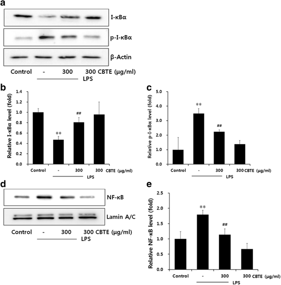
The inhibitory effect of CBTE on the LPS-induced nuclear factor-kappa B protein expressions in RAW264.7 cells. The levels of I-κBα, p-I-κBα (30 min in the cytosol fraction) and NF-κB (1 h in the nuclear fraction) protein were monitored with or without CBTE pre-treatment (i.e. 1 h before LPS). β-actin and lamin A/C were used as a loading control (a and d). The relative levels of I-κBα (b) p-I-κBα (c) and NF-κB (e) were measured by scanning densitometry. Values represent the mean ± S.D. of three independent experiments (significant as compared to the control, **P < 0.01, significant as compared to LPS alone, ## P < 0.01). CBTE, Cheongsangbangpung-tang extract; NF-κB, Nuclear factor-kappa B; I-κB, Inhibitor-kappa B
Fig. 6
Fig. 6
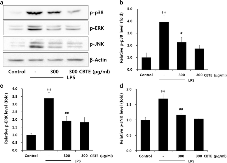
The inhibitory effect of CBTE on the LPS-induced phosphorylation of MAPKs in RAW264.7 cells. The levels of phosphorylation of MAPK proteins were monitored 1 h after treatment of cells with LPS (1 μg/ml) either with or without CBTE pre-treatment (i.e. 1 h before LPS). Expressions of the MAPKs protein were determined by immunoblotting using specific anti-p-p38, anti-p-ERK and anti-p-JNK antibodies. An antibody against β-actin was used to verify equal protein loading of the cell lysate (a). The relative levels of the MAPKs were measured by scanning densitometry (b c d). Values represent the mean ± S.D. of three independent experiments (significant as compared to the control, **P < 0.01, significant as compared to LPS alone, ## P < 0.01). CBTE, Cheongsangbangpung-tang extract; MAPK, Mitogen-activated protein kinases; ERK, extracellular signal-regulated kinase; JNK, Jun N-terminal kinase
Fig. 7
Fig. 7
The inhibitory effect of CBTE on carrageenan-induced paw oedema. CBTE was orally administered to rats at 0.3 or 1.0 g/kg/day prior to the induction of paw oedema for 3 days. Paw oedema was induced by the subcutaneous injection of 1% carrageenan solution as described in the Materials and Methods section. The swelling volume of the paw was measured up to 4 h after the carrageenan injection at intervals of 1 h using a plethysmometer. DEXA (1 mg/kg, p.o.) was used as a positive control drug. Values represent the mean ± S.D. of three independent experiments (significant as compared to the control, **P < 0.01, significant as compared to CA alone, ## P < 0.01). CBTE, Cheongsangbangpung-tang extract; CA, carrageenan; DEXA, dexamethasone
Fig. 8
Fig. 8
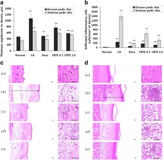
The inhibitory effect of CBTE on paw skin thickness and infiltrated inflammatory cells. Changes in histological profiles, paw skin thickness (a) and infiltrated inflammatory cells (b) of the dorsum and ventrum pedis skin in normal a, carrageenan b, dexamethasone c, 0.3 g/kg CBTE d and 1.0 g/kg CBTE e treated groups (c d). After 4 h of carrageenan treatment, the dorsum and ventrum pedis skins were separated and fixed in 10% neutral buffered formalin then embedded in paraffin, sectioned and stained with haematoxylin and eosin. Note that marked increases of dorsum (c) and ventrum (d) pedis skin thicknesses (arrow) due to oedematous changes were detected following carrageenan treatment with increases in inflammatory cell infiltrations compared with the normal skin. However, these increases in skin thicknesses and inflammatory cell infiltrations were effectively inhibited by treatment with dexamethasone and were also dose-dependently affected by treatment with two different dosages of CBTE 0.3 and 1.0 g/kg. Scale bars = 60 μm. CBTE, Cheongsangbangpung-tang extract; CA, carrageenan; DEXA, dexamethasone
Fig. 9
Fig. 9
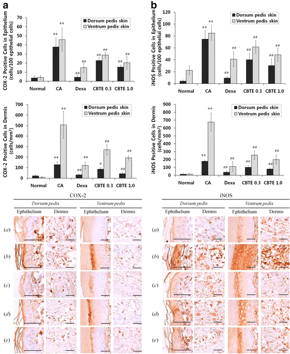
Representative immunohistochemical profiles of COX-2 and iNOS on the paw skins. Note that marked increases of COX-2 (a)- and iNOS (b)-positive cells were detected on the epithelium and dermis of the dorsum and ventrum pedis skin tissues in CA rats compared with normal rats, respectively. However, these increases of epidermal and dermal COX-2- and iNOS-immunolabelled cells were effectively inhibited by treatment with dexamethasone and were also dose-dependently affected by treatment with CBTE at two different dosages: 0.3 and 1.0 g/kg. Immunoreactive cells were stained using ABC methods where a = Normal; b = Carrageenan (CA); c = CA and dexamethasone; d = CA and CBTE 0.3 g/kg; e = CA and CBTE 1.0 g/kg. Scale bars = 60 μm. CBTE, Cheongsangbangpung-tang extract; DEXA, dexamethasone

References

    1. Heo J. Donguibogam (in Korean) Seoul: Namsandang; 1983. p. 211.
    1. Kim K, Kim KI, Lee J. Inhibitory effects of cheongsangbangpoong-tang on both inflammatory acne lesions and facial heat in patients with acne vulgaris: a randomized controlled trial protocol. BMC complement altern med. 2016;16:21. doi: 10.1186/s12906-016-1000-9. - DOI - PMC - PubMed
    1. Neutra MR, Pringault E, Kraehenbuhl JP. Antigen sampling across epithelial barriers and induction of mucosal immune responses. Annu rev immunol. 1996;14:275–300. doi: 10.1146/annurev.immunol.14.1.275. - DOI - PubMed
    1. Akira S, Uematsu S, Takeuchi O. Pathogen recognition and innate immunity. Cell. 2006;124:783–801. doi: 10.1016/j.cell.2006.02.015. - DOI - PubMed
    1. Stephensen CB. Vitamin A, infection, and immune function. Annu rev nutr. 2001;21:167–192. doi: 10.1146/annurev.nutr.21.1.167. - DOI - PubMed

MeSH terms