. 2017 Mar;187(3):570-580.
doi: 10.1016/j.ajpath.2016.10.021. Epub 2017 Jan 11.

miR-24 Inhibition Increases Menin Expression and Decreases Cholangiocarcinoma Proliferation

Affiliations

miR-24 Inhibition Increases Menin Expression and Decreases Cholangiocarcinoma Proliferation

Laurent Ehrlich et al. Am J Pathol. 2017 Mar.

Abstract

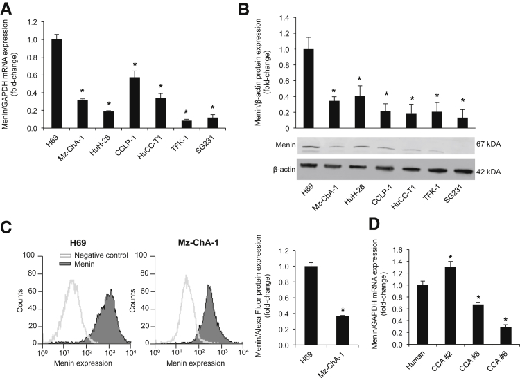
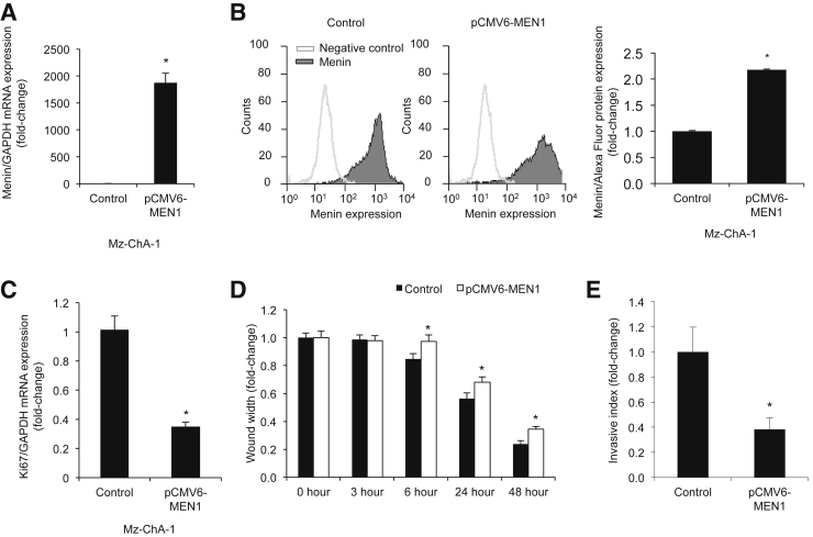
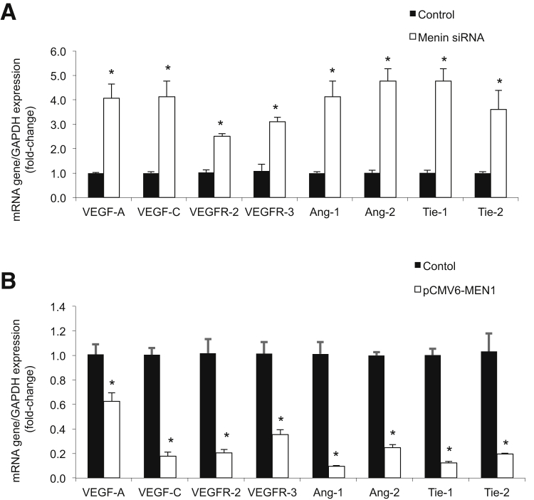
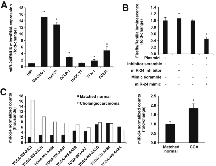
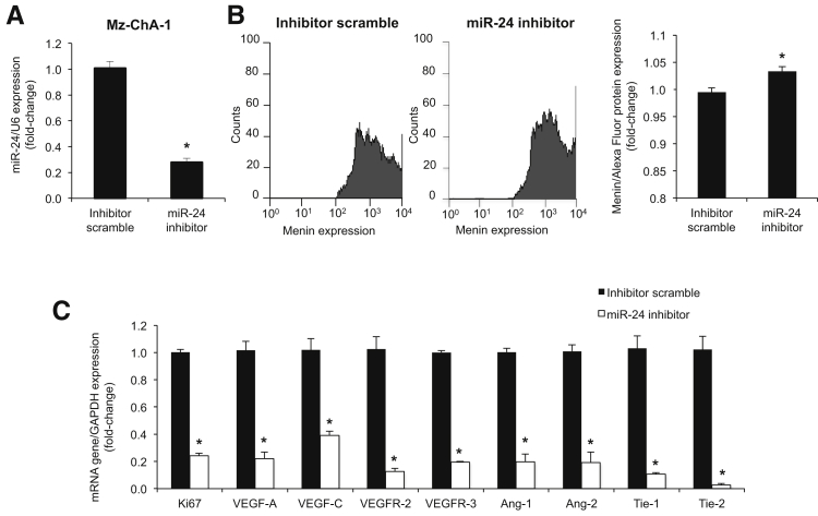
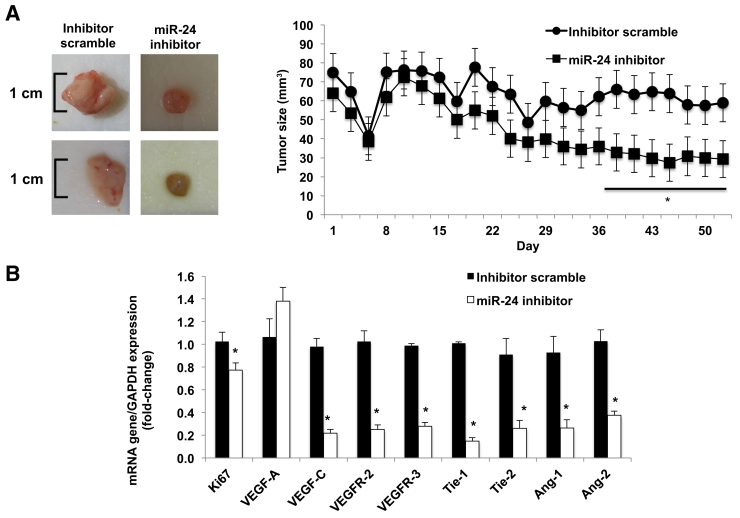
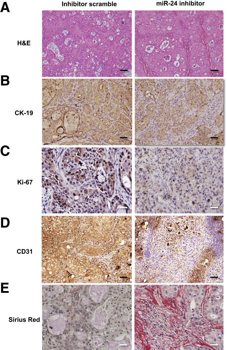
Menin (MEN1) is a tumor-suppressor protein in neuroendocrine tissue. Therefore, we tested the novel hypothesis that menin regulates cholangiocarcinoma proliferation. Menin and miR-24 expression levels were measured in the following intrahepatic and extrahepatic cholangiocarcinoma (CCA) cell lines, Mz-ChA-1, TFK-1, SG231, CCLP, HuCCT-1, and HuH-28, as well as the nonmalignant human intrahepatic biliary line, H69. miR-24 miRNA and menin protein levels were manipulated in vitro in Mz-ChA-1 cell lines. Markers of proliferation and angiogenesis (Ki-67, vascular endothelial growth factors A/C, vascular endothelial growth factor receptors 2/3, angiopoietin 1/2, and angiopoietin receptors 1/2) were evaluated. Mz-ChA-1 cells were injected into the flanks of nude mice and treated with miR-24 inhibitor or inhibitor scramble. Menin expression was decreased in advanced CCA specimens, whereas miR-24 expression was increased in CCA. Menin overexpression decreased proliferation, angiogenesis, migration, and invasion. Inhibition of miR-24 increased menin protein expression while decreasing proliferation, angiogenesis, migration, and invasion. miR-24 was shown to negatively regulate menin expression by luciferase assay. Tumor burden and expression of proliferative and angiogenic markers was decreased in the miR-24 inhibited tumor group compared to controls. Interestingly, treated tumors were more fibrotic than the control group. miR-24-dependent expression of menin may be important in the regulation of nonmalignant and CCA proliferation and may be an additional therapeutic tool for managing CCA progression.


Figures

Figure 1
Figure 1
Menin is down-regulated in CCA. A and B: By real-time PCR and immunoblots, menin expression is decreased in CCA cell lines compared to H69. Significance is shown versus H69 cells. C: Flow cytometry analysis demonstrated a decrease in menin protein expression in Mz-ChA-1 cells compared to H69 cells. D: By real-time PCR, menin expression decreased in advanced-stage human CCA tissue biopsy specimens compared with normal control. Data are expressed as means ± SEM performed in triplicate (AD). n = 3 independent samples (A and B). P < 0.05 versus H69 or human control. GAPDH, glyceraldehyde-3-phosphate dehydrogenase.
Figure 2
Figure 2
Increased menin expression decreases proliferation. Mz-ChA-1 cells overexpressing menin with pCMV6-MEN1 vector exhibit a decrease in Ki-67 proliferative marker expression. AC: Increased menin expression in pCMV6-MEN1 Mz-ChA-1 cells by real-time PCR (A) and flow cytometry (B) decreased Ki-67 proliferative marker expression by real-time PCR (C). D: Decreased cell migration as measured by wound healing assay. E: Decreased cell invasion as measured by Boyden chamber assay in pCMV6-MEN1 Mz-ChA-1 cells. Data are expressed as means ± SEM performed in triplicate (A–E). P < 0.05 versus Mz-ChA-1 control cells. GAPDH, glyceraldehyde-3-phosphate dehydrogenase.
Figure 3
Figure 3
Menin expression negatively regulates angiogenesis. A: By real-time PCR, Mz-ChA-1 MEN1 knockout cells increased expression of angiogenic factors compared to Mz-ChA-1 control cells. B: By real-time PCR, pCMV6-MEN1 Mz-ChA-1 cells decreased expression of angiogenic factors compared to Mz-ChA-1 control cells. Data are expressed as means ± SEM performed in triplicate (A and B). P < 0.05 versus Mz-ChA-1 control cells. GAPDH, glyceraldehyde-3-phosphate dehydrogenase.
Figure 4
Figure 4
miR-24 negatively regulates menin. A: Real-time PCR evaluation of miR-24 expression in CCA and H69 cell lines demonstrates increased levels in CCA lines compared to H69 cells. B: Luciferase luminescence shows decreased menin expression with miR-24 mimic treatment. C: Left panel: miRNA-sequence data demonstrate increased expression of miR-24 in human CCA tumors compared with matched normal tissue. Right panel: Statistical significance of increased miR-24 expression is validated with an unpaired t-test. Data are expressed as means ± SEM (A–C). n = 3 (A and B); n = 9 (C). P < 0.05 versus normal matched human control.
Figure 5
Figure 5
miR-24 drives proliferation. A: Real-time PCR confirmed knockdown of miR-24 in Mz-ChA-1 cells by hairpin inhibitor. miR-24 knockdown increased menin expression via fluorescence-activated cell sorting (B) and decreased expression of angiogenic factors via real-time PCR (C). Data are expressed as means ± SEM performed in triplicate unless otherwise stated (AC). P < 0.05 versus Mz-ChA-1 control cells. GAPDH, glyceraldehyde-3-phosphate dehydrogenase.
Figure 6
Figure 6
In vivo miR-24 inhibition decreases growth. A: Ten weeks after Mz-ChA-1 implantation into the flanks of nude mice, there was a significant reduction in tumor size in athymic mice treated with the miR-24 hairpin inhibitor compared to scramble mismatch-treated mice. Significance is shown versus mice treated with scramble mismatch. B: Real-time PCR performed on total RNA isolated from both tumor groups revealed decreased expression of proliferative and angiogenic factors. Significance is shown versus tumor samples from mismatch-treated mice. Data are expressed as means ± SEM of tumor size evaluations. n = 4 mice per each group (A); n = 3 experiments (B). P < 0.05 versus scramble mismatch control. GAPDH, glyceraldehyde-3-phosphate dehydrogenase.
Figure 7
Figure 7
In vivo miR-24 inhibition decreases growth. A and B: Immunohistochemical images of formalin-fixed, paraffin-embedded sections confirm expected histological depictions of Mz-ChA-1 CCA tumor via hematoxylin and eosin (H&E) and CK-19 staining. CE: miR-24 inhibition decreased nuclear Ki-67 staining from 1 ± 0.15 inhibitor scramble to 0.4 ± 0.05 miR-24 inhibition (P = 0.001; C), decreased CD31 staining from 1 ± 0.29 inhibitor scramble to 0.1 ± 0.05 miR-24 inhibition (P = 0.02; D), and increased Sirius Red staining from 1 ± 0.47 inhibitor scramble to 10.7 ± 1.68 miR-24 inhibition (P = 0.003; E). Data are expressed as means ± SEM. n = 3 independent samples (A–E). P < 0.05 versus control inhibitor scramble. Original magnification: ×20 (A, B, and D); ×40 (C and E).

Similar articles

Cited by

References

    1. Razumilava N., Gores G.J. Cholangiocarcinoma. Lancet. 2014;383:2168–2179. - PMC - PubMed
    1. Komuta M., Govaere O., Vandecaveye V., Akiba J., Van Steenbergen W., Verslype C., Laleman W., Pirenne J., Aerts R., Yano H., Nevens F., Topal B., Roskams T. Histological diversity in cholangiocellular carcinoma reflects the different cholangiocyte phenotypes. Hepatology. 2012;55:1876–1888. - PubMed
    1. Francis H., Alpini G., DeMorrow S. Recent advances in the regulation of cholangiocarcinoma growth. Am J Physiol Gastrointest Liver Physiol. 2010;299:G1–G9. - PMC - PubMed
    1. Alvaro D., Mancino M.G., Glaser S., Gaudio E., Marzioni M., Francis H., Alpini G. Proliferating cholangiocytes: a neuroendocrine compartment in the diseased liver. Gastroenterology. 2007;132:415–431. - PubMed
    1. Chandrasekharappa S.C., Guru S.C., Manickam P., Olufemi S.-E., Collins F.S., Emmert-Buck M.R., Debelenko L.V., Zhuang Z., Lubensky I.A., Liotta L.A., Crabtree J.S., Wang Y., Roe B.A., Weisemann J., Boguski M.S., Agarwal S.K., Kester M.B., Kim Y.S., Heppner C., Dong Q., Spiegel A.M., Burns A.L., Marx S.J. Positional cloning of the gene for multiple endocrine neoplasia-type 1. Science. 1997;276:404–407. - PubMed

MeSH terms