. 2017 Feb:103:65-73.
doi: 10.1016/j.yjmcc.2017.01.004. Epub 2017 Jan 11.

Myocardial relaxation is accelerated by fast stretch, not reduced afterload

Affiliations

Myocardial relaxation is accelerated by fast stretch, not reduced afterload

Charles S Chung et al. J Mol Cell Cardiol. 2017 Feb.

Abstract

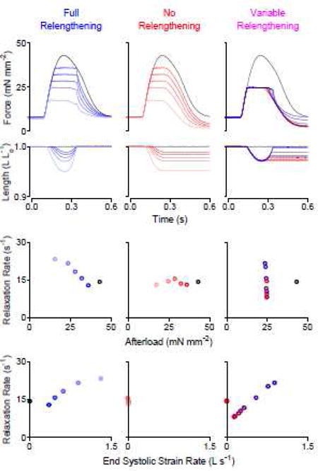
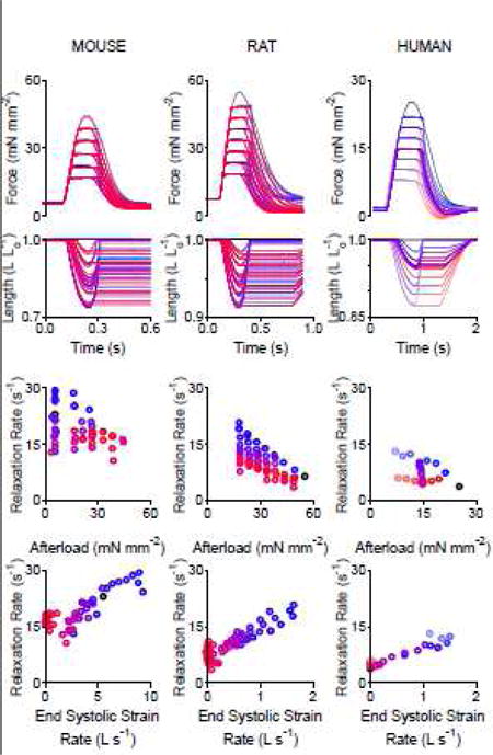
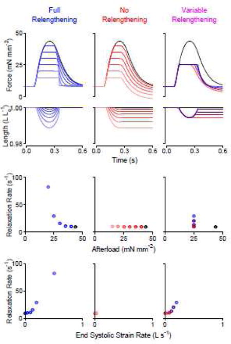
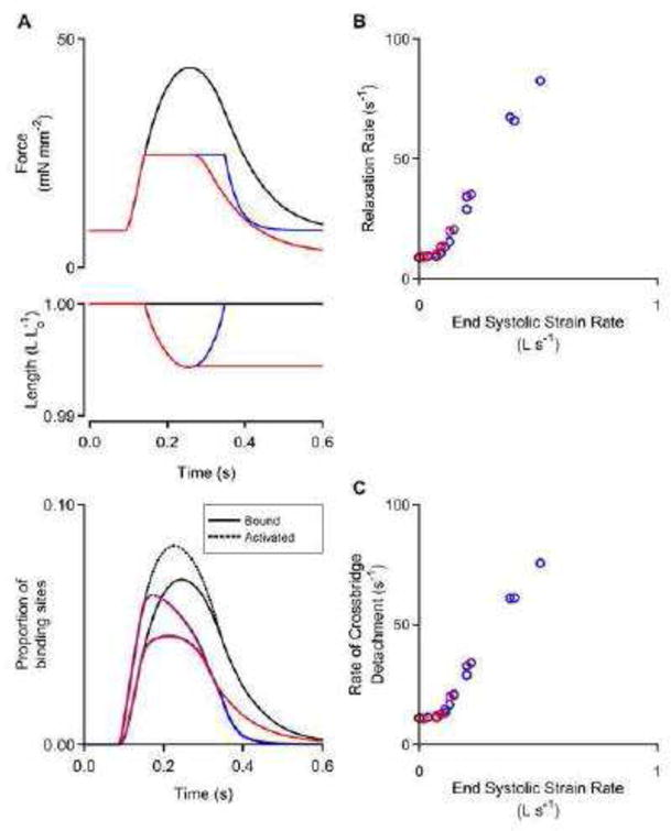
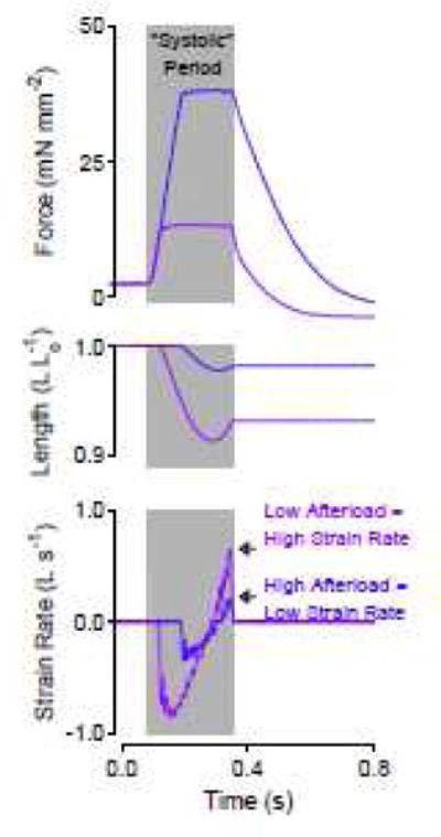
Fast relaxation of cross-bridge generated force in the myocardium facilitates efficient diastolic function. Recently published research studying mechanisms that modulate the relaxation rate has focused on molecular factors. Mechanical factors have received less attention since the 1980s when seminal work established the theory that reducing afterload accelerates the relaxation rate. Clinical trials using afterload reducing drugs, partially based on this theory, have thus far failed to improve outcomes for patients with diastolic dysfunction. Therefore, we reevaluated the protocols that suggest reducing afterload accelerates the relaxation rate and identified that myocardial relengthening was a potential confounding factor. We hypothesized that the speed of myocardial relengthening at end systole (end systolic strain rate), and not afterload, modulates relaxation rate and tested this hypothesis using electrically-stimulated trabeculae from mice, rats, and humans. We used load-clamp techniques to vary afterload and end systolic strain rate independently. Our data show that the rate of relaxation increases monotonically with end systolic strain rate but is not altered by afterload. Computer simulations mimic this behavior and suggest that fast relengthening quickens relaxation by accelerating the detachment of cross-bridges. The relationship between relaxation rate and strain rate is novel and upends the prevailing theory that afterload modifies relaxation. In conclusion, myocardial relaxation is mechanically modified by the rate of stretch at end systole. The rate of myocardial relengthening at end systole may be a new diagnostic indicator or target for treatment of diastolic dysfunction.

Keywords: Afterload; Cross-bridge; Diastole; Myocardium; Relaxation; Strain rate.


Conflict of interest statement

DISCLOSURES/CONFLICTS OF INTEREST

None

Figures

Figure 1
Figure 1
Load-clamp protocols indicate that relaxation rate depends on the end systolic strain rate, not afterload. Top: Force and length versus time traces; Middle: Relationship between relaxation rate and afterload; Bottom: Relationship between relaxation rate and end systolic strain rate. Left (blue): overlay of 6 twitches that were load-clamped at various afterloads; the muscle was relengthened back to its original length before being allowed to relax. Center (red): overlay of 6 twitches; the muscle relaxed while being held at the minimum attained length. Right: overlay of 9 twitches that were load-clamped at ~50% of the peak isometric force; the load-clamp was stopped with variable amounts of relengthening before being allowed to relax. Muscle relengthening was necessary to obtain an inverse relationship between relaxation rate and afterload. Relengthening alone (without modifying afterload) was sufficient to modify the relaxation rate. All data in this figure measured from the same rat trabecula.
Figure 2
Figure 2
Relaxation rate is dependent on the end systolic strain rate in mouse, rat, and human myocardial trabeculae. Top: Force and length versus time traces. Bottom: Relationship between relaxation rate and end systolic strain rate. Each column shows multiple afterload-clamped twitches overlaid from a single mouse trabecula (53 twitches), rat trabecula (62 twitches), and human (25 twitches) trabecula. Six additional rat trabeculae shown in Supplementary Figure S2.
Figure 3
Figure 3
Model simulation mimics load-clamp experiments from a rat cardiac trabecula. Top: Mathematical modeling simulated force and length versus time traces; Middle: Relationship between relaxation rate and afterload, and bottom: Relationship between relaxation rate and end systolic strain rate observed in simulated data. These data show that relaxation rate is 1) only dependent on afterload only when the muscle relengthens and 2) dependent on the end systolic strain rate.
Figure 4
Figure 4
Mathematical modeling reveals molecular mechanisms. A) Model predicted force, length, and acto-myosin binding site status as a function of time. Black: isometric twitch. Red: load-clamped twitch held at minimum length. Blue: load-clamp twitch allowed to fully relengthen before relaxation. B) Relaxation rate versus end systolic strain rate and C) cross-bridge detachment rate versus end systolic strain rate for multiple simulated twitches. This modeling data set shows that cross-bridge detachment rates are slow unless a fast stretch is applied before relaxation.
Figure 5
Figure 5
A short, quick relengthening explains why relaxation rate can be faster at reduced afterloads. Force, length, and strain rate versus time for two twitches load-clamped at different afterloads but allowed to relengthen for equal durations. Afterload may have been mistaken for the mechanical factor that modified relaxation rate in intact hearts because strain and strain rate were not measured.

References

    1. Borlaug BA, Jaber WA, Ommen SR, Lam CS, Redfield MM, Nishimura RA. Diastolic relaxation and compliance reserve during dynamic exercise in heart failure with preserved ejection fraction. Heart. 2011;97:964–9. - PMC - PubMed
    1. Paulus WJ, Tschope C, Sanderson JE, Rusconi C, Flachskampf FA, Rademakers FE, et al. How to diagnose diastolic heart failure: a consensus statement on the diagnosis of heart failure with normal left ventricular ejection fraction by the Heart Failure and Echocardiography Associations of the European Society of Cardiology. Eur Heart J. 2007;28:2539–50. - PubMed
    1. Biesiadecki BJ, Davis JP, Ziolo MT, Janssen PM. Tri-modal regulation of cardiac muscle relaxation; intracellular calcium decline, thin filament deactivation, and cross-bridge cycling kinetics. Biophys Rev. 2014;6:273–89. - PMC - PubMed
    1. He H, Giordano FJ, Hilal-Dandan R, Choi DJ, Rockman HA, McDonough PM, et al. Overexpression of the rat sarcoplasmic reticulum Ca2+ ATPase gene in the heart of transgenic mice accelerates calcium transients and cardiac relaxation. J Clin Invest. 1997;100:380–9. - PMC - PubMed
    1. Babu GJ, Bhupathy P, Petrashevskaya NN, Wang H, Raman S, Wheeler D, et al. Targeted overexpression of sarcolipin in the mouse heart decreases sarcoplasmic reticulum calcium transport and cardiac contractility. J Biol Chem. 2006;281:3972–9. - PubMed

Publication types

LinkOut - more resources