Review
. 2017 Jun:349:138-147.
doi: 10.1016/j.heares.2017.01.003. Epub 2017 Jan 10.

Cochlear synaptopathy in acquired sensorineural hearing loss: Manifestations and mechanisms

Affiliations
Review

Cochlear synaptopathy in acquired sensorineural hearing loss: Manifestations and mechanisms

M Charles Liberman et al. Hear Res. 2017 Jun.

Abstract

Common causes of hearing loss in humans - exposure to loud noise or ototoxic drugs and aging - often damage sensory hair cells, reflected as elevated thresholds on the clinical audiogram. Recent studies in animal models suggest, however, that well before this overt hearing loss can be seen, a more insidious, but likely more common, process is taking place that permanently interrupts synaptic communication between sensory inner hair cells and subsets of cochlear nerve fibers. The silencing of affected neurons alters auditory information processing, whether accompanied by threshold elevations or not, and is a likely contributor to a variety of perceptual abnormalities, including speech-in-noise difficulties, tinnitus and hyperacusis. Work described here will review structural and functional manifestations of this cochlear synaptopathy and will consider possible mechanisms underlying its appearance and progression in ears with and without traditional 'hearing loss' arising from several common causes in humans.

Keywords: Auditory nerve; Cochlear neuropathy; Cochlear synaptopathy; Hidden hearing loss; Noise-induced hearing loss.


Figures

Fig. 1
Fig. 1. Noise-induced and age-related loss of synapses and SGNs
Evaluating synaptopathy by triple-staining cochlear whole mounts for a pre-synaptic marker (CtBP2-red), a postsynaptic marker (GluA2-green) and a hair cell marker (Myosin VIIa-blue). Confocal z-stacks in the IHC area from a control (A) and a noise-exposed mouse (B), 2 wks post exposure. Light micrographs of osmium-stained plastic sections from noise-exposed ears, 2 wks (C) or 2 yrs (D) post exposure. Exposure in B and D was 8–16 kHz, 2 h, 100 dB SPL, delivered at 16 wk to CBA/CaJ mice. (E) In aging ears from the same inbred strain, synaptic counts at IHCs decrease steadily from 4 to 144 wks and parallel ganglion cell loss follows whereas, (F) threshold loss begins comparatively later and accelerates beyond 80 wks, mirrored by accelerating loss of OHCs. IHC loss is trivial at any age. Red symbols flag 80 wk data points for all measurements. After Kujawa and Liberman 2006, ; Sergeyenko et al. (2013).
Fig. 2
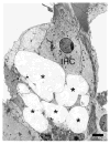
Fig. 2. Excitotoxic swelling in the cochlea
Infusion of AMPA (200 μM) in the cochlea triggers massive swelling of afferent endings (*) underneath the inner hair cell (IHC). Scale bar = 1 μM. From Ruel et al. (2007), with permission.
Fig. 3
Fig. 3. Response amplitudes and synapse counts
Permanent reductions in ABR, but not DPOAE amplitudes in ears with recovered thresholds after noise. Shown are DPOAE (A) and ABR wave 1 (B) response growth functions in the region of maximum acute TTS 1 d and 2 wk after exposure (as in Fig. 1) to 16 wk CBA/CaJ mice; unexposed controls shown for comparison. Neural response amplitude declines are proportional to synaptic and neural losses in aging CBA/CaJ, where synapses are plotted vs mean wave 1 amplitudes (at 80 dB SPL in 4–128 wk animals (C). Panels A,B from Fernandez et al., 2015; Panel C from Sergeyenko et al., 2013.
Fig. 4
Fig. 4. Low-SR neuron loss after noise
Single unit recordings were made in guinea pigs 10 days after a TTS-producing noise exposure that resulted in permanent ABR amplitude declines and synapse loss but no hair cell loss. Spontaneous rate distributions suggest selective loss of low-SR fibers in the high-frequency region of maximum noise-induced injury (A). In the same animals, thresholds and tuning of surviving nerve fibers, matched for CF, were not altered in noise exposed ears compared to controls (B). The single-fiber database included 367 fibers from 14 control animals, and 382 fibers from 9 exposed animals. After Furman et al., 2013.
Fig. 5
Fig. 5. Gradients in synaptic and afferent fiber morphology
IHC synapses in confocal z-stacks, acquired in the x-y plane (A) and re-projected into the y-z (B) plane. A Pre- and post-synaptic elements in the IHC area are counted in cochlear whole mounts quadruple-immunostained for CtBP2 (red), GluA2 (green), NaK ATPase (blue), and myosin VIIa (white). B Size gradients in pre- and post-synaptic elements are quantified according to location along habenular-cuticular and modiolar-pillar axes (Liberman et al., 2015). (C) Tracing of peripheral axons from a cross section through the osseous spiral lamina (OSL; D) in a normal cat shows the SR-based gradient from thin (low-SR) to thick (high-SR) fibers (Kawase and Liberman, 1992).
Fig. 6
Fig. 6. Cochlear de-afferentation in human temporal bones
Cochlear de-afferentation is seen in human temporal bones as a function of age (A) and cochlear location (B) in cases with no hair cell loss and no explicit otologic disease. The small pink symbols (A) are estimated total SGC counts from archival sections (Makary et al., 2011); the five large symbols (A) are the estimated total peripheral axon counts from the same five cases shown in B. Counts of cochlear nerve terminals per IHC (B) in 5 normal aging temporal bones (55–89 yrs) with no history of otologic disease (Viana et al., 2015). Blue symbols are counts of synapses per IHC from an electron microscopic study of a normal middle-aged human (Nadol, 1988).

References

    1. ACOEM Task Force on Occupational Hearing Loss. Kirchner DB, Evenson E, Dobie RA, Rabinowitz P, Crawford J, Kopke R, Hudson TW. Occupational noise-induced hearing loss: ACOEM Task Force on Occupational Hearing Loss. J Occup Environ Med. 2012;54:106–108. - PubMed
    1. American Academy of Audiology Position Statement and Clinical Practice Guidelines. Ototoxicity Monitoring. 2009 Oct; Available at: http://www.audiology.org/publications-resources/document-library/ototoxi....
    1. American Speech-Language-Hearing Association. Audiologic Management of Individuals Receiving Cochleotoxic Drug Therapy [Guidelines] 1994 Available at: www.asha.org/policy.
    1. Antoli-Candela F, Jr, Kiang NYS. Unit activity underlying the N1 potential. In: Naunton RF, Fernandez C, editors. Evoked Electrical Activity in the Auditory Nervous System. Academic Press; New York: 1978. pp. 165–191.
    1. Bae WY, Kim LS, Hur DY, Jeong SW, Kim JR. Secondary apoptosis of spiral ganglion cells induced by aminoglycoside: Fas-Fas ligand signaling pathway. Laryngoscope. 2008;118(9):1659–1668. - PubMed

Publication types

MeSH terms