. 2017 Feb:143:32-41.
doi: 10.1016/j.mod.2017.01.003. Epub 2017 Jan 10.

Wnt signaling balances specification of the cardiac and pharyngeal muscle fields

Affiliations

Wnt signaling balances specification of the cardiac and pharyngeal muscle fields

Amrita Mandal et al. Mech Dev. 2017 Feb.

Abstract

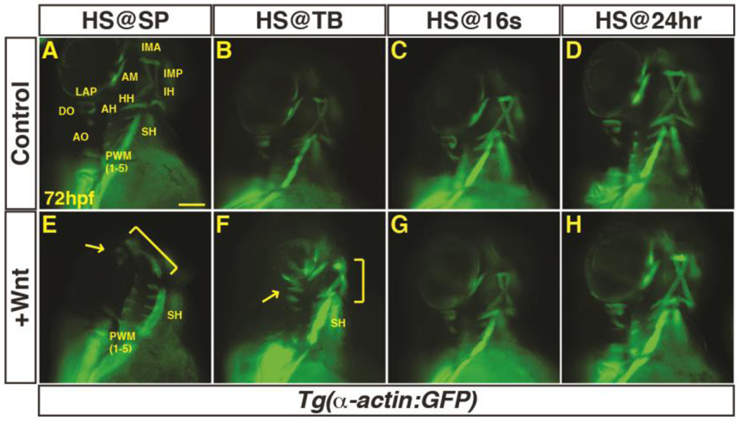
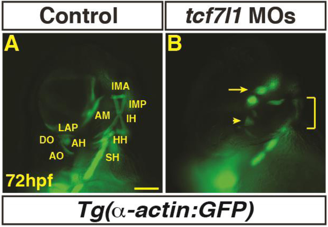
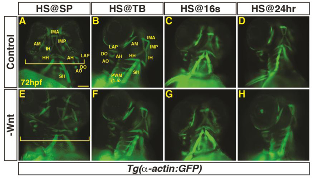
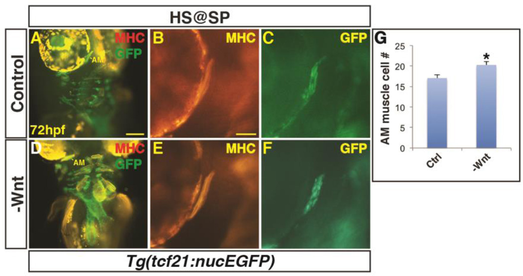
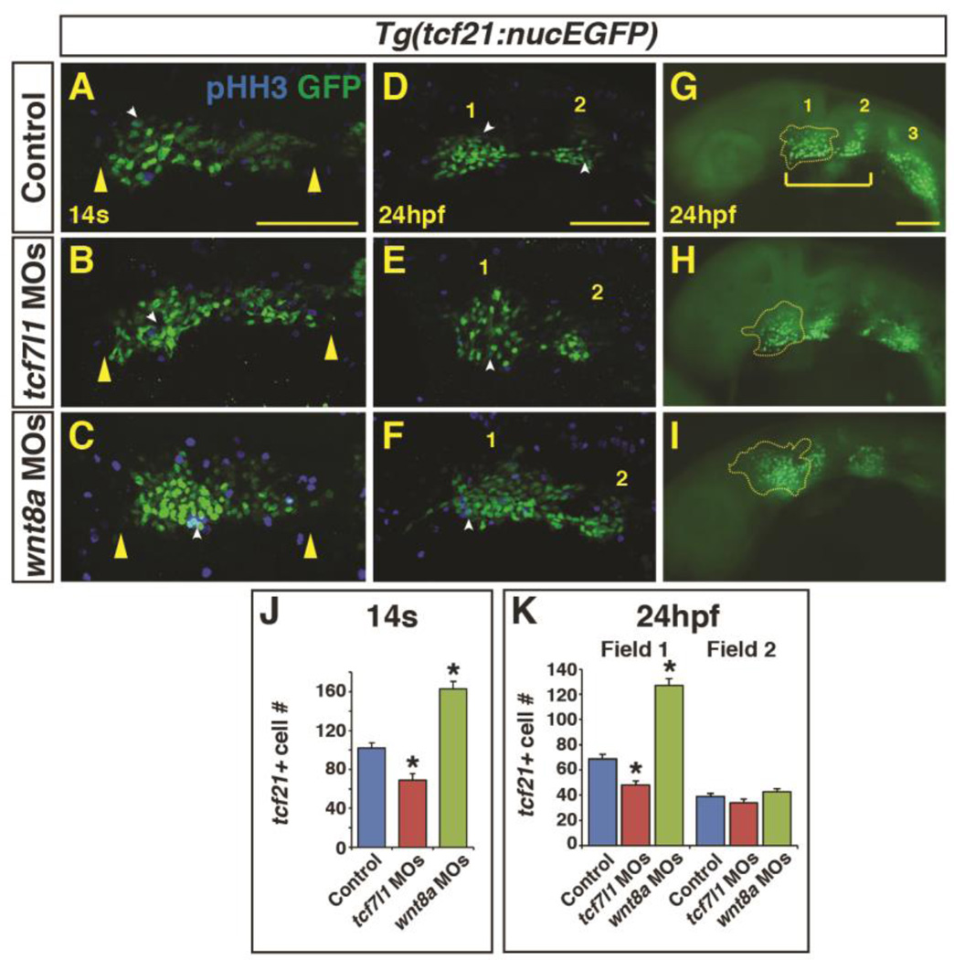
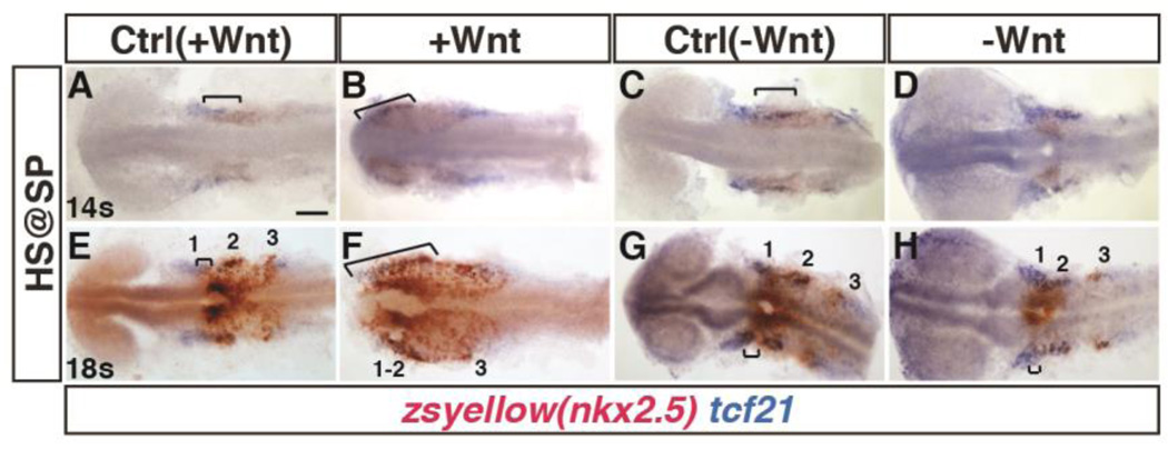
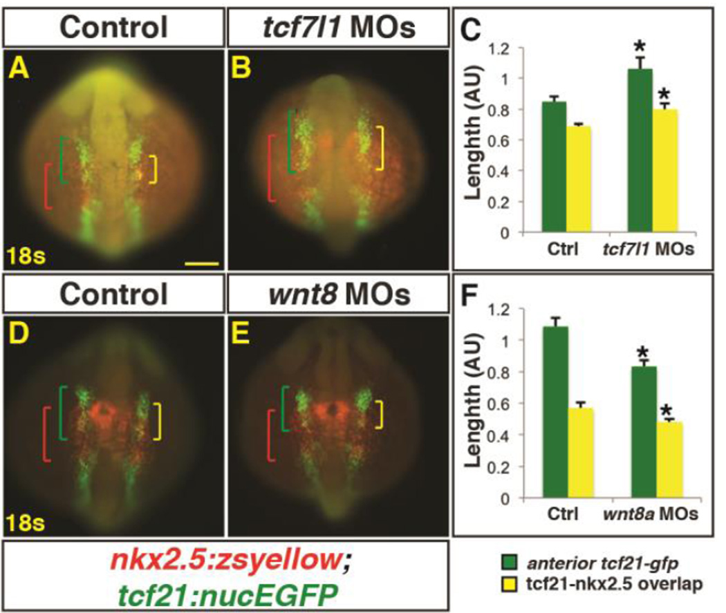
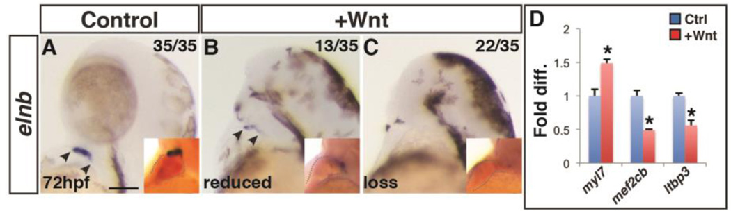
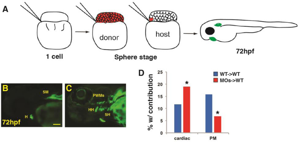
Canonical Wnt/β-catenin (Wnt) signaling plays multiple conserved roles during fate specification of cardiac progenitors in developing vertebrate embryos. Although lineage analysis in ascidians and mice has indicated there is a close relationship between the cardiac second heart field (SHF) and pharyngeal muscle (PM) progenitors, the signals underlying directional fate decisions of the cells within the cardio-pharyngeal muscle field in vertebrates are not yet understood. Here, we examined the temporal requirements of Wnt signaling in cardiac and PM development. In contrast to a previous report in chicken embryos that suggested Wnt inhibits PM development during somitogenesis, we find that in zebrafish embryos Wnt signaling is sufficient to repress PM development during anterior-posterior patterning. Importantly, the temporal sensitivity of dorso-anterior PMs to increased Wnt signaling largely overlaps with when Wnt signaling promotes specification of the adjacent cardiac progenitors. Furthermore, we find that excess early Wnt signaling can cell autonomously promote expansion of the first heart field (FHF) progenitors at the expense of PM and SHF within the anterior lateral plate mesoderm (ALPM). Our study provides insight into an antagonistic developmental mechanism that balances the sizes of the adjacent cardiac and PM progenitor fields in early vertebrate embryos.

Keywords: Cardiac; Mesoderm patterning; Organ fields; Pharyngeal muscle; Wnt signaling; Zebrafish.


Figures

Figure 1
Figure 1. Excess Wnt signaling inhibits PM development during gastrulation
(A–P) Tg(α-actin:GFP) control sibling embryos and embryos with increased Wnt signaling (hsp70l:wnt8a-GFP+). (A,E) After increasing Wnt signaling from heat-shock (HS) at the SP stage, the EOMs, dorsal 1st and 2nd arch PMs are lost (arrow), while the ventral 1st and 2nd arch PMs are reduced (bracket). PW muscles are also reduced, while the SH was overtly unaffected. (B,F) After increasing Wnt signaling at the TB stage, there is a similar but less severe loss of EOMs, 1st and 2nd arch PMs (dorsal PMs - arrow; ventral PMs bracket). (C, G, D, H) Wnt signaling does not overtly affect PM development at later stages. Muscle nomenclature used is from (Schilling and Kimmel, 1997). 1st arch muscles - intermandibularis anterior (IMA), intermandibularis posterior (IMP), adductor mandibulae (AM), levator arcus palatine (LAP), dilatator opercula (DO). 2nd (hyoid) arch muscles - hyohyal (HH), interhyal (IH), adductor hyomandibulae (AH), adductor opercula (AO), and levator opercula (LO). Embryos are at 72 hpf. Images are anterior ventrolateral views. Anterior is up in all images. Heat-shock (HS). >15 embryos were examined for each condition. Scale bar indicates 100 µm.
Figure 2
Figure 2. Tcf7l1 depletion causes loss of dorso-anterior PMs
(A–B) Control and Tcf7l1 depleted Tg(α-actin:GFP) embryos at 72 hpf. Tcf7l1 depletion resulted in similar loss of dorso-anterior PMs as increasing Wnt signaling prior to gastrulation. EOMs (arrow), 1st and 2nd PMs are severely reduced and misorganized or lost (arrowhead). Ventral 1st and 2nd arch muscles are reduced (bracket). Views are anterior ventrolateral. >15 embryos were examined for each condition. Scale bar indicates 100 µm.
Figure 3
Figure 3. Decreased Wnt signaling during gastrulation increases head size with minimal effect on PM morphology
(A–H) Tg(α-actin:GFP) control sibling embryos and embryos with decreased Wnt signaling (hsp70l:dkk1-GFP+). (A,E) After decreasing Wnt signaling at the SP stage, the embryos have enlarged heads (brackets). (B–D, F–H) After decreasing Wnt signaling at later stages, there was no discernible effect on pharyngeal muscles. Embryos are at 72 hpf. Images in A,E are anterior ventral views. Images in B–D, F–H are anterior ventro-lateral views. Anterior is up in all images. Heat-shock (HS). >15 embryos were examined for each condition. Scale bar indicates 100 µm.
Figure 4
Figure 4. Decreased Wnt signaling causes an increase in 1st arch PM cells
(A, B) Tg(tcf21:nucEGFP) control sibling embryos and embryos with decreased Wnt signaling at the SP stage. Decreased Wnt signaling causes enlargement of the 1st arch AM muscle (boxes). Sarcomeric myosin (MHC; red). NucGFP (green). (B, E) MHC alone of AM muscles. (C, F) NucEGFP alone of AM. (G) Graph depicting quantification of nuclei in the AM muscles. Decreasing Wnt signaling at the SP stage produces a modest, but significant increase in AM muscle nuclei. Control embryos n=15, Tg(hsp70l:dkk1-GFP) embryos n=7. Asterisks in all graphs indicate p<0.05. Error bars in all graphs indicate S.E.M. Scale bar in A indicates 100 µm. Scale bar in B indicates 50 µm.
Figure 5
Figure 5. Endogenous Wnt signaling components restrict tcf21+ PM progenitor development
(A–C) Tcf21+ (GFP+) cells within the ALPM of Tg(tcf21:nucEGFP) control, Tcf7l1 depleted, and Wnt8a depleted embryos at the 14s stage. Images are dorsal views of the left side of the embryos with anterior left. Yellow arrowheads indicate size of field. (D-I) Tcf21+ (GFP+) cells within the ALPM of Tg(tcf21:nucEGFP) control, Tcf7l1 depleted, and Wnt8a depleted embryos at 24 hpf. Images are lateral views with anterior left. (E,H) Tcf7l1 depletion produces a loss of the anterior most tcf21+ progenitors (yellow outline). (F,I) Wnt8a depletion results in expansion of the anterior most pharyngeal tcf21+ progenitors. White arrowheads in A-F indicate overlap between GFP+ and phospho-histone H3 (pHH3)+ cells (see Supplementary Material – Figure S3 for analysis of pHH3). Bracket in G indicates the anterior tcf21+ field of cells that give rise to the 1st and 2nd arch muscles. Numbers 1 and 2 in D-G indicate the two anterior tcf21+ fields of cells. Number 3 in G indicates the more posterior tcf21+ population that gives rise to PM in arches 3–7 (Nagelberg et al., 2015). Images A-F are confocal images. (J) Graph depicting quantification of tcf21+ progenitors at the 14s stage. Tg(tcf21:nucEGFP) control n=15, Tcf7l1 depleted n=10, and Wnt8a depleted embryos n=10. (K) Graph depicting quantification of tcf21+ progenitors at the 24hpf. Tg(tcf21:nucEGFP) control n=10, Tcf7l1 depleted n=13, and Wnt8a depleted embryos n=10. Cell counts are from confocal images of the tcf21+ field on one side of the embryo. There is a significant decrease in tcf21+ progenitors with Tcf7l1 depletion and conversely a significant increase in tcf21+ progenitors following Wnt8a depletion in the total field at the 14s stage and anterior field at 24 hpf. Scale bars indicates 100 µm.
Figure 6
Figure 6. Manipulation of Wnt signaling affects the overlap of nkx2.5+ and tcf21+ domains within the ALPM
(A–H) Tg(nkx2.5:ZsYellow) control sibling embryos and embryos with increased and decreased Wnt signaling at the SP stage. (A,B,E,F) Two-color ISH of zsyellow (nkx2.5; brownish red) and tcf21 (blue) at the 14s and 18s stages shows expansion of the lateral nkx2.5+ domains into the anterior tcf21+ domain after increased Wnt signaling. Numbers in E indicate the three tcf21+ fields depicted in Figure 5. (C,D,G,H) Decreasing Wnt signaling prior to gastrulation causes a decrease in the overlap of nkx2.5 and tcf21 at the 14s and 18s stages. Significant overlap between nkx2.5 and tcf21 could not be detected at the 14s stage in embryos when with Wnt signaling was inhibited prior to gastrulaiton. Images are dorsal views with anterior to left. Brackets indicate overlap of nkx2.5+ and tcf21+ domains. For each group >15 embryos were examined. Scale bar indicates 100 µm.
Figure 7
Figure 7. Manipulation of endogenous Wnt signaling affects the size of the nkx2.5+ and tcf21+ domains
(A,B,D,E) Tg(nkx2.5:ZsYellow);Tg(tcf21:nucEGFP) control, Tcf7l1 depleted, and Wnt8a depleted embryos at the 18s stage. Tcf7l1 depletion expands the total anterior tcf21+ progenitor domain length (green brackets) as well as the overlapping expression (yellow brackets) within the ALPM. (C) Graph depicting measurements of the total tcf21+ progenitors and overlap with nkx2.5+ progenitors from Tcf7l1 depleted embryos. Control embryos n=10, Tcf7l1 depleted embryos n=9. (D, E) Wnt8a depletion reduces the overall length of the tcf21+ domain and the overlap with the nkx2.5+ progenitor domain. (F) Graph depicting measurements of the total tcf21+ progenitors and overlap with nkx2.5+ progenitors from Wnt8a depleted embryos. Control embryos n=8, Wnt8a depleted embryos n=10. Green brackets - anterior tcf21+ progenitors. Red brackets - nkx2.5+ progenitors. Yellow brackets - regions of overlap between tcf21+ and nkx2.5+ domains. Scale bar indicates 100 µm.
Figure 8
Figure 8. Increased Wnt signaling promotes loss of the SHF
(A–C) Control sibling embryos and embryos after increased Wnt signaling at the SP stage. Increasing Wnt signaling prior to gastrulation leads to loss of the SHF-derived OFT smooth muscle (elnb). Inserts indicate embryos with two-color ISH for myl7 and elnb. Outlines indicate the hearts. (D) RT-qPCR for cardiac differentiation and SHF progenitor marker expression at 24 hpf. Increasing Wnt signaling prior to gastrulation promotes increased expression of the pan-cardiac differentiation marker myl7 and decreased expression of the SHF markers mef2cb and ltbp3 compared to WT sibling controls. Images are lateral views with anterior upward. Scale bar indicates 100 µm.
Figure 9
Figure 9. Wnt signaling cell-autonomously inhibits PM specification
(A) Schematic of blastula cell transplantation method. (B, C) Representative donor cells that incorporated into the heart and PMs at 72 hpf. (D) Graph indicating the frequency of donor cell contribution in embryos to the heart and the PMs. WT donor cells ->WT host transplants n=240. Tcf7l depleted donor cells ->WT host em bryos n=221. Tcf7l1 depleted donor cells contributed significantly more to the cardiomyocytes and less frequently to the PMs compared to WT donors. Images are lateral views with anterior leftwards. Scale bar indicates 100 µm.

References

    1. Baldini A. DiGeorge syndrome: the use of model organisms to dissect complex genetics. Hum Mol Genet. 2002;11:2363–2369. - PubMed
    1. D’Aniello E, Rydeen AB, Anderson JL, Mandal A, Waxman JS. Depletion of Retinoic Acid Receptors Initiates a Novel Positive Feedback Mechanism that Promotes Teratogenic Increases in Retinoic Acid. PLoS genetics. 2013;9:e1003689. - PMC - PubMed
    1. de Pater E, Clijsters L, Marques SR, Lin YF, Garavito-Aguilar ZV, Yelon D, Bakkers J. Distinct phases of cardiomyocyte differentiation regulate growth of the zebrafish heart. Development. 2009;136:1633–1641. - PMC - PubMed
    1. Devine WP, Wythe JD, George M, Koshiba-Takeuchi K, Bruneau BG. Early patterning and specification of cardiac progenitors in gastrulating mesoderm. Elife. 2014:3. - PMC - PubMed
    1. Diogo R, Kelly RG, Christiaen L, Levine M, Ziermann JM, Molnar JL, Noden DM, Tzahor E. A new heart for a new head in vertebrate cardiopharyngeal evolution. Nature. 2015;520:466–473. - PMC - PubMed

MeSH terms

LinkOut - more resources