. 2017 Oct 1:276:31-39.
doi: 10.1016/j.cbi.2017.01.002. Epub 2017 Jan 10.

Impact of disease-Linked mutations targeting the oligomerization interfaces of aldehyde dehydrogenase 7A1

Affiliations

Impact of disease-Linked mutations targeting the oligomerization interfaces of aldehyde dehydrogenase 7A1

David A Korasick et al. Chem Biol Interact. .

Abstract

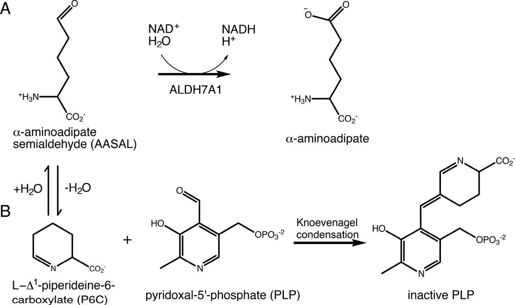
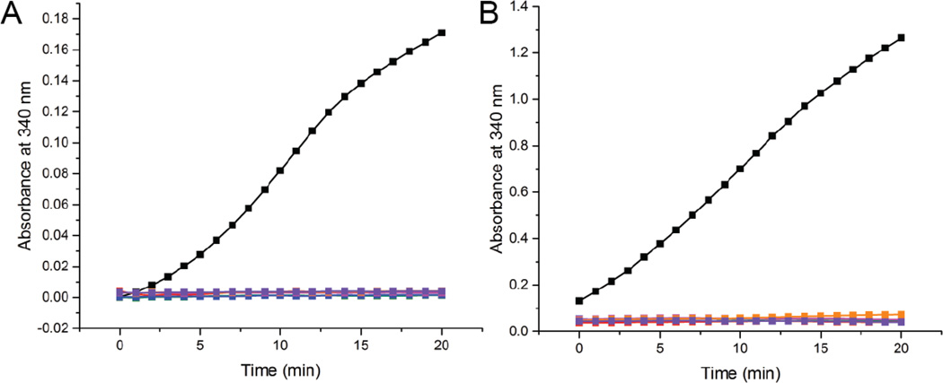
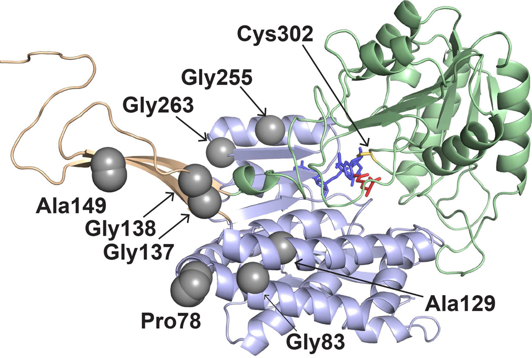
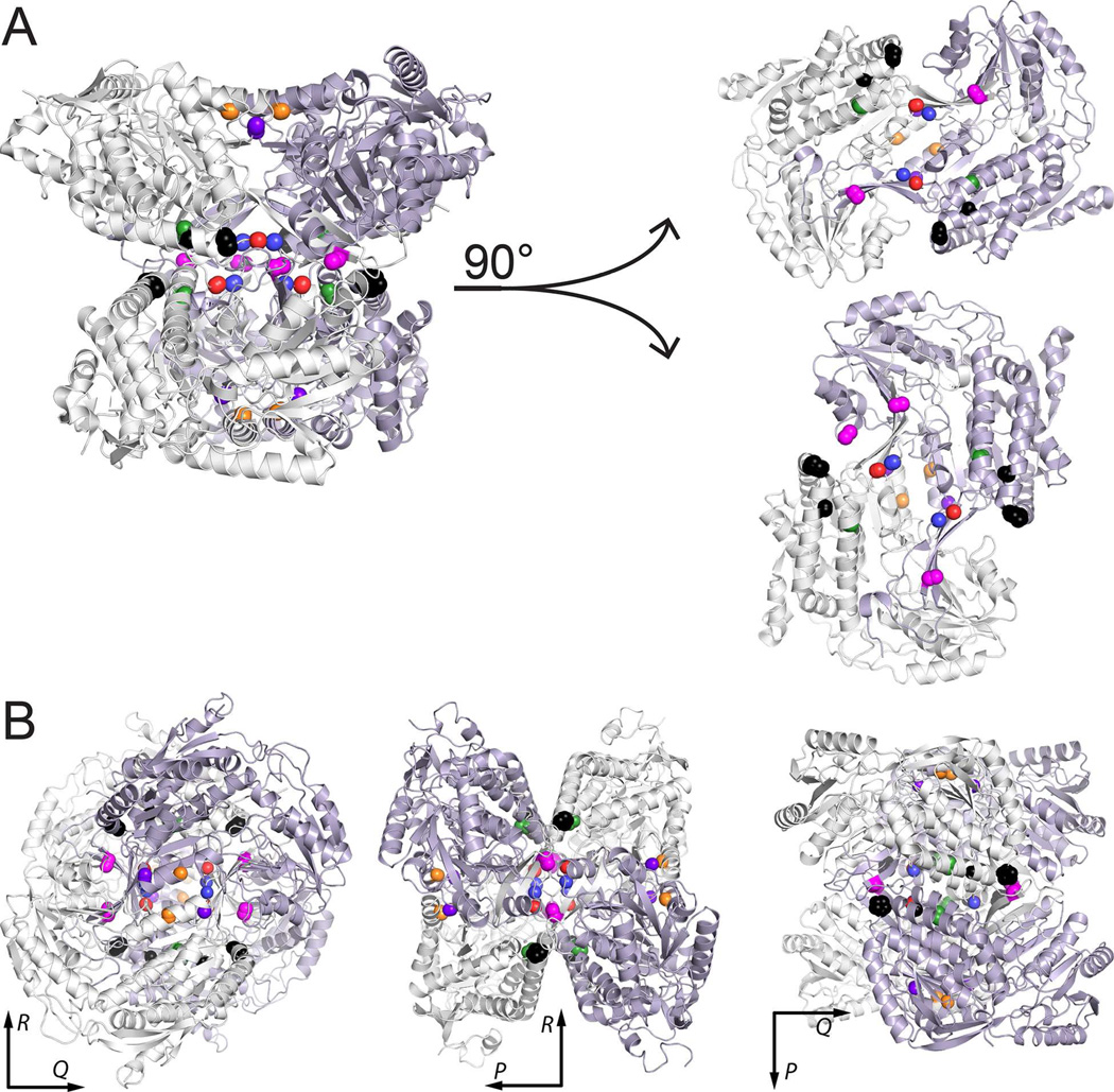
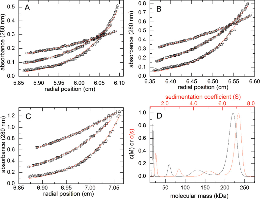
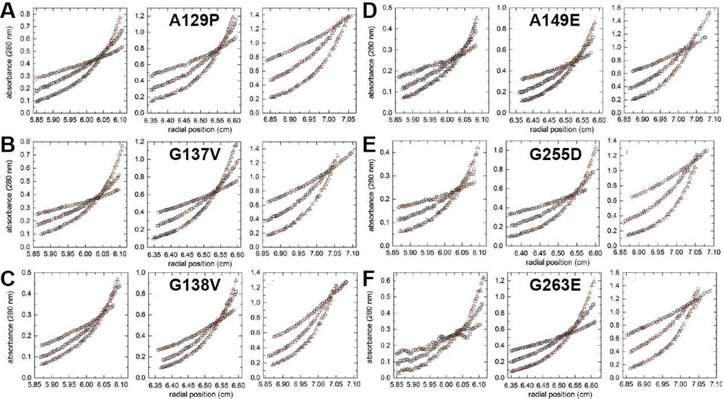
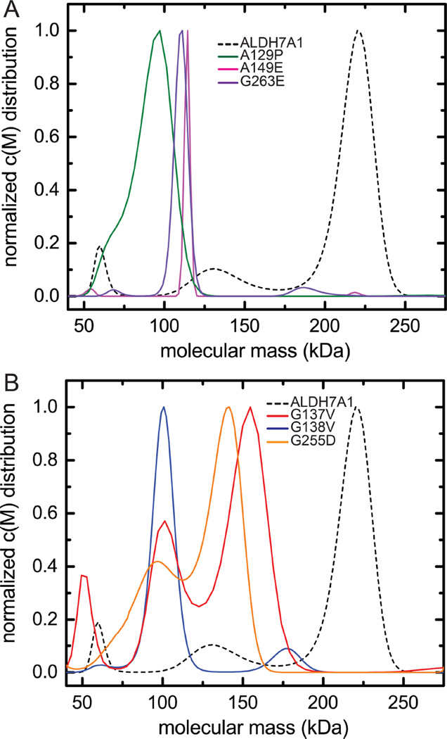
Aldehyde dehydrogenase 7A1 (ALDH7A1) is involved in lysine catabolism, catalyzing the oxidation of α-aminoadipate semialdehyde to α-aminoadipate. Certain mutations in the ALDH7A1 gene, which are presumed to reduce catalytic activity, cause an autosomal recessive seizure disorder known as pyridoxine-dependent epilepsy (PDE). Although the genetic association between ALDH7A1 and PDE is well established, little is known about the impact of PDE-mutations on the structure and catalytic function of the enzyme. Herein we report the first study of the molecular consequences of PDE mutations using purified ALDH7A1 variants. Eight variants, with mutations in the oligomer interfaces, were expressed in Escherichia coli: P78L, G83E, A129P, G137V, G138V, A149E, G255D, and G263E. All but P78L and G83E were soluble and could be purified. All six soluble mutants were catalytically inactive. The impact of the mutations on oligomerization was assessed by analytical ultracentrifugation. Wild-type ALDH7A1 is shown to exist in a dimer-tetramer equilibrium with a dissociation constant of 16 μM. In contrast to the wild-type enzyme, the variants reside in monomer-dimer equilibria and are apparently incapable of forming a tetrameric species, even at high enzyme concentration. The available evidence suggests that they are misfolded assemblies lacking the three-dimensional structure required for catalysis.

Keywords: ALDH7A1; Aldehyde dehydrogenase; Analytical ultracentrifugation; Lysine catabolism; Protein oligomerization; Pyridoxine-dependent epilepsy.


Conflict of interest statement

Conflict of interest

The authors declare no conflict of interest.

Figures

Figure 1
Figure 1
Chemical reactions relevant to ALDH7A1 and PDE. (A) The reaction catalyzed by ALDH7A1. (B) The reaction between P6C and PLP, which results in covalent inactivation of PLP. P6C forms a nonenzymatic equilibrium with AASAL.
Figure 2
Figure 2. Enzymatic activity of interface mutants compare to wild-type ALDH7A1
NADH formation was monitored at 340 nm using either 0.5 µM protein and 2 mM AASAL (A) or 4 µM protein and 30 mM AASAL (B). The color scheme in both panels is identical: wild type ALDH7A1 (black), A129P (green), G137V (red), G138V (blue), A149E (magenta), G255D (orange), and G263E (purple). The NAD+ concentration was 2.5 mM. Note that the vertical scales are different in the two panels. The enzyme concentrations were calculated using the molecular weight of an ALDH7A1 monomer.
Figure 3
Figure 3
The locations of PDE-associated mutations in the ALDH7A1 protomer. The three structural domains are represented as light green (catalytic domain), light blue (NAD+-binding domain), and light orange (oligomerization domain). The active site is indicated by catalytic Cys302, NAD+ (blue), and the product α-aminoadipate (red). The gray spheres indicate the residues mutated in this study.
Figure 4
Figure 4
Locations of PDE-associated mutations in the protein-protein interfaces of ALDH7A1. (A) Depiction of the tetramer as a dimer of dimers, with the two dimers separated to show the interfacial locations of the mutated residues. (B) Three views of the tetramer aligned along the P, Q, and R 2-fold axes. In both panels, the locations of the residues mutated in this report are indicated by color-coded spheres: P78L (black), G83E (black), A129P (green), G137V (red), G138V (blue), A149E (magenta), G255D (orange), and G263E (purple).
Figure 5
Figure 5. Analytical ultracentrifugation analysis of wild-type ALDH7A1
(A-C) Sedimentation equilibrium data collected at three different protein concentrations: 0.2 mg/ml (A), 0.4 mg/ml (B), and 0.8 mg/ml (C). The symbols in panels A-C correspond to different centrifugation speeds: 6000 rpm (circles), 9000 rpm (squares), and 12,000 rpm (triangles). The curves represent a global fit of the data to a dimer-tetramer equilibrium model. (D) Sedimentation velocity experiment performed at 4.5 mg/ml (~80 µM, based on monomer molecular weight). The red curve corresponds to the continuous c(s) distribution, whereas the black curve corresponds to the continuous c(M) distribution. For reference, the molecular weights of the dimer and tetramer are 111 and 222 kDa.
Figure 6
Figure 6. Sedimentation equilibrium analysis for the mutant enzymes
For each protein, the left, middle, and right graphs show three different concentrations: 0.2 mg/ml (left), 0.4 mg/ml (middle), and 0.8 mg/ml (right). Within each graph, the three data sets correspond centrifugation speeds of 6000 rpm (circles), 9000 rpm (squares), and 12,000 rpm (triangles). The red curves represent global fits of the data to an oligomerization equilibrium model.
Figure 7
Figure 7
Sedimentation velocity analysis of mutant ALDH7A1 conducted at high concentration (~80 µM, monomer molecular weight). (A) Sedimentation velocity data for A129P (green), A149E (magenta), and G263E (purple). The result for wild-type ALDH7A1 (also ~80 µM) is shown for comparison (dashed black). (B) Sedimentation velocity data for G137V (red), G138V (blue), and G255D (orange). The result for wild-type ALDH7A1 is shown for comparison (dashed black).

Similar articles

Cited by

References

    1. Ma I, Allan AL. The role of human aldehyde dehydrogenase in normal and cancer stem cells. Stem. Cell. Rev. 2011;7:292–306. - PubMed
    1. Muzio G, Maggiora M, Paiuzzi E, Oraldi M, Canuto RA. Aldehyde dehydrogenases and cell proliferation. Free Radic. Biol. Med. 2012;52:735–746. - PubMed
    1. Mills PB, Struys E, Jakobs C, Plecko B, Baxter P, Baumgartner M, Willemsen MA, Omran H, Tacke U, Uhlenberg B, Weschke B, Clayton PT. Mutations in antiquitin in individuals with pyridoxine-dependent seizures. Nat. Med. 2006;12:307–309. - PubMed
    1. Stockler S, Plecko B, Gospe SM, Jr, Coulter-Mackie M, Connolly M, van Karnebeek C, Mercimek-Mahmutoglu S, Hartmann H, Scharer G, Struijs E, Tein I, Jakobs C, Clayton P, Van Hove JL. Pyridoxine dependent epilepsy and antiquitin deficiency: clinical and molecular characteristics and recommendations for diagnosis, treatment and follow-up. Mol. Genet. Metab. 2011;104:48–60. - PubMed
    1. Stenson PD, Ball E, Howells K, Phillips A, Mort M, Cooper DN. Human Gene Mutation Database: towards a comprehensive central mutation database. J Med Genet. 2008;45:124–126. - PubMed

Supplementary concepts