. 2017 Apr 1:52:60-73.
doi: 10.1016/j.actbio.2017.01.032. Epub 2017 Jan 10.

Cathepsin K-targeted sub-micron particles for regenerative repair of vascular elastic matrix

Affiliations

Cathepsin K-targeted sub-micron particles for regenerative repair of vascular elastic matrix

Brenton Jennewine et al. Acta Biomater. .

Abstract

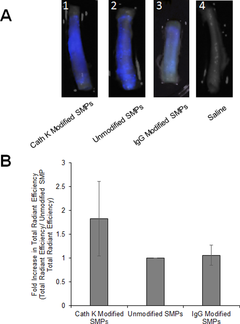
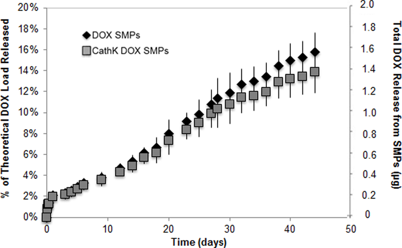
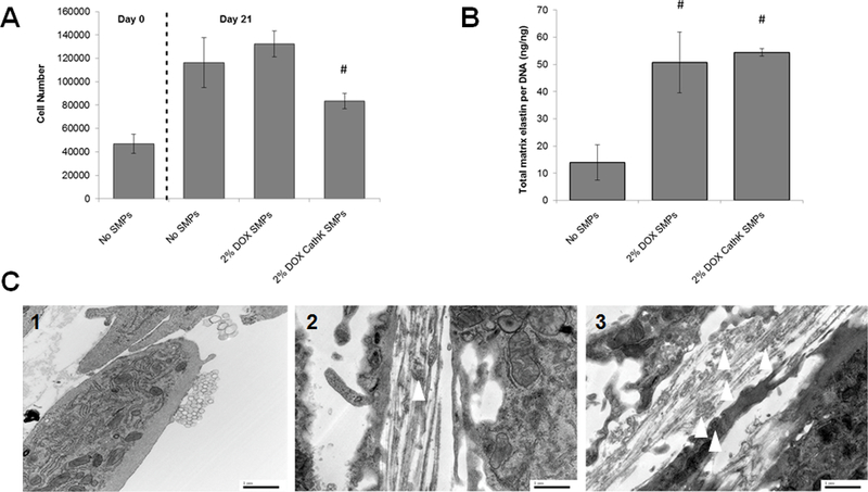
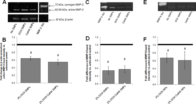
Abdominal Aortic Aneurysms (AAA) involve slow dilation and weakening of the aortic wall due to breakdown of structural matrix components, such as elastic fibers by chronically overexpressed matrix metalloproteinases (MMPs), primarily, MMPs-2 and -9. Auto-regenerative repair of disrupted elastic fibers by smooth muscle cells (SMCs) at the AAA site is intrinsically poor and together with chronic proteolysis prevents restoration of elastin homeostasis, necessary to enable AAA growth arrest or regression to a healthy state. Oral doxycycline (DOX) therapy can inhibit MMPs to slow AAA growth, but has systemwide side-effects and inhibits new elastin deposition within AAA tissue, diminishing prospects for restoring elastin homeostasis preventing the arrest/regression of AAA growth. We have thus developed cationic amphiphile (DMAB)-modified submicron particles (SMPs) that uniquely exhibit pro-elastogenic and anti-proteolytic properties, separate from similar effects of the encapsulated drug. These SMPs can enable sustained, low dose DOX delivery within AAA tissue to augment elastin regenerative repair. To provide greater specificity of SMP targeting, we have conjugated the DOX-SMP surface with an antibody against cathepsin K, a lysosomal protease that is highly overexpressed within AAA tissue. We have determined conditions for efficient cathepsin K Ab conjugation onto the SMPs, improved SMP binding to aneurysmal SMCs in culture and to injured vessel walls ex vivo, conjugation did not affect DOX release from the SMPs, and improved pro-elastogenic and anti-proteolytic effects due to the SMPs likely due to their increased proximity to cells via binding. Our study results suggest that cathepsin K Ab conjugation is a useful targeting modality for our pro-regenerative SMPs. Future studies will investigate SMP retention and biodistribution following targeting to induced AAAs in rat models through intravenous or catheter-based aortal infusion and thereafter their efficacy for regenerative elastic matrix repair in the AAA wall.

Statement of significance: Proactive screening of high risk elderly patients now enables early detection of Abdominal Aortic Aneurysms (AAAs). Current management of small, growing AAAs is limited to passive, imaging based growth monitoring. There are also no established drug-based therapeutic alternatives to surgery for AAAs, which is unsuitable for many elderly patients, and none which can achieve restore disrupted and lost elastic matrix in the AAA wall, which is essential to achieve growth arrest or regression. We seek to test the feasibility of a regenerative therapy based on localized, one time delivery of drug-releasing Sub-Micron-sized drug delivery polymer Particles (SMPs) that are also uniquely chemically functionalized on their surface to also provide them pro-elastin-regenerative & anti-matrix degradative properties, and also conjugated with antibodies targeting cathepsin K, an elastolytic enzyme that is highly overexpressed in AAA tissues; the latter serves as a modality to enable targeted binding of the SMPs to the AAA wall following intravenous infusion, or intraoartal, catheter-based delivery. Such SMPs can potentially stimulate structural repair in the AAA wall following one time infusion to delay or prevent AAA growth to rupture. The therapy can provide a non-surgical treatment option for high risk AAA patients.

Keywords: Cathepsin K; Drug delivery; Elastic matrix; Matrix metalloproteinases (MMPs); Regenerative matrix repair; Submicron particles.


Figures

Figure 1.
Figure 1.
UV spectroscopy analysis of PLGA SMPs conjugated with cathepsin K antibody. Panel 1A shows effect of incubation time on conjugation of the antibody to SMPs. The cathepsin K antibody was detected with a fluorescein-tagged secondary Ab. A higher fluorescence intensity (RFU) indicates more effective antibody conjugation. Panel 1B compares relative abundance of conjugated antibodies on the SMPs. Cathepsin K antibodies were conjugated onto fluorescein-loaded SMPs over 5 hours, and were detected with secondary antibodies tagged with AF546. Values shown indicate mean ± SD of RFUs (Panel A) or of ratios of RFUs due to the fluorescein and AF546; n = 3 per case; # denotes significance of differences versus 2 h of incubation, deemed for p < 0.05; * denotes significance of differences versus control FITC SMPs treated with the AF-546-tagged secondary antibody, deemed for p< 0.05. Panels 1C, 1D show results of fluorescence microscopy analysis of cathepsin K surface modification to AF633-loaded SMPs (red). A fluorescein antibody (green) was added to visualize the cathepsin K modification. Panel C shows representative images for the adsorption and conjugation methods at day 1 and day 14. The green fluorescence demonstrates successful cathepsin K conjugation to the SMP surface. At day 14, green fluorescence associated with SMPs modified using Ab-adsorption was much lower compared to SMPs chemically conjugated with the Abs. Panel 1D shows the ratio of FITC intensity to AF633 intensity (mean ± SE; adsorption n=132, n=130 and conjugation n=154, n=207). The conjugation method bound more cathepsin K to the SMP surface for a longer period of time. # denotes significance of differences between adsorption and conjugation on day 14 deemed for p<0.05. * denotes significance of differences between day 1 and day 14 for the adsorption method deemed for p<0.05. In panel 1E, confocal micrographs compare cathepsin K antibody bound to SMPs via adsorption and covalent conjugation methods (see quantitative data in panel 1B). Conjugation was performed over 5 hours. Fluorescein (green) was encapsulated within the SMPs and the cathepsin K antibody was detected with an AF546-tagged secondary antibody (red). Panel 1E1 shows lack of red auto-fluorescence from cathepsin K antibody-conjugated SMPs not treated with the AF546-tagged secondary antibody. Panels E2 and E3 show that cathepsin K antibody was successfully conjugated to the SMPs using the adsorption- and covalent binding methods respectively. Scale bar: 100 μm (panel C), 100 μm (panel E).
Figure 2.
Figure 2.
(A). IF images showing relative expression of cathepsin K by healthy, and aneurysmal SMCs, without and with TNF-α stimulation. Cathepsin K, visualized with AF-546-tagged secondary antibody, appears red while the cytoskeletal actin filaments stained with AF488 phalloidin appear green, and DAPI-stained nuclei appear blue. Scale bar: 100 μm. (B) High magnification view of EaRASMCs stimulated with TNF-α and cathepsin K visualized with AF-546-tagged secondary antibody and cytoskeletal actin stained with AF488 phalliodin. Grid: 23 μm x 23 μm. (C). Images of the EaRASMCs at different z-axis heights. (1) The bottom of the cell layer which shows minimal cathepsin K. (2) The middle of the cell layer in which cathepsin K begins to appear. (3) The top of the cells where the most cathepsin K is found. Scale bar for panels 2C1–3: 50 μm. (4) Schematic of the z-axis heights for images 2C1–3. (D). Western blot analysis for relative expression of cathepsin K by healthy, and aneurysmal SMCs, without and with TNF-α stimulation. The figure shows representative blot, indicating bands for the cathepsin K zymogen and β-actin (loading control). The plot shows β-actin normalized cathepsin K band intensity (mean ± SD; n = 3 per case; # denotes p < 0.05 compared to control RASMCs); * denotes p < 0.05 compared to TNF-α-unstimulated EaRASMCs. # indicates significance of differences versus RASMCs, deemed for p < 0.05.
Figure 3.
Figure 3.
Western blot analysis for cathepsin K expression in saline and elastase treated porcine carotid arteries. Panel A shows a representative blot, showing the active cathepsin K form (38 kDa) and β-actin (loading control). Panel B shows β-actin normalized cathepsin K band intensity (mean ± SEM; n = 9 per case; # denotes significance of difference, deemed for p < 0.05.
Figure 4.
Figure 4.
(A) Confocal micrographs showing binding of cathepsin K antibody-conjugated SMPs to healthy, and aneurysmal SMCs, without and with TNF-α stimulation. All SMPs were encapsulated with AF546, causing them to fluoresce red. A significantly higher number of SMPs bound to EaRASMCs than to the RASMCs and more still in EaRASMC cultures stimulated with TNF-α (white arrows). Cytoskeletal actin filaments, stained with AF488 phalloidin fluoresce green and DAPI-stained nuclei appear blue. (B). High magnification image of SMP localization to TNF-α stimulated EaRASMCs. (C). Images of EaRASMCs with SMPs at various z-axis heights. (1) Bottom of cell layer with minimal SMP bound. (2) and (3) Middle layers of the cell where SMPs begin to appear around the cell. (4) Top of the cell layer where SMPs are bound to the cell surface. (5) Schematic of the various z-axis heights for images 1–4. Scale bars represent 50 μm (A), 23 μm x 23 μm (B), and 50 μm (C).
Figure 5.
Figure 5.
Targeting of cathepsin K Ab-conjugated SMPs to wall of elastase injured porcine carotid arteries. (A) Pseudocolor of SMP localization in elastase treated arteries with cathepsin K-Ab conjugated AF633 SMPs (1), unconjugated AF633 SMPs (2), IgG-Ab conjugated AF633 SMPs (3), and saline (4). (B) Fold difference in binding of cathepsin K Ab-conjugated SMPs, and non-specific IgG conjugated SMPs to elastase treated arteries compared to binding of unmodified SMPs (mean ± SEM; n = 6 and n = 3 for IgG-Ab conjugation). * indicates significance of differences between the cases deemed for p < 0.05.
Figure 6.
Figure 6.
In vitro DOX release profiles from cathepsin K antibody-conjugated and unconjugated PLGA SMPs (0.5 mg/ml) loaded with 2% w/w DOX (mean ± SD; n = 3 per group).
Figure 7.
Figure 7.
(7A) Proliferation of EaRASMCs is not impacted by coculture with DOX-SMPs (cathepsin K Ab-conjugated or unmodified), although cell proliferation in the cultures treated with the Ab-conjugated SMPs was significantly lower than that cultured with the unmodified SMPs. DOX loading within the SMPs was 2% w/w. SMP concentration in the cultures was 0.2 mg/ml. Cells were harvested at 1 and 21 days after initial seeding (mean ± SD; n = 3 cultures per group; # denotes significance of differences versus unconjugated DOX SMPs, deemed for p< 0.05). (7B) Cathepsin K antibody conjugation of DOX-SMPs does not alter their pro-elastogenic effects on cultured EaRASMCs. SMP-untreated EaRASMC cultures were investigated as treatment controls. All cell layers were cultured for 21 days. Amounts of deposited elastic matrix, comprised of both the alkali-soluble and alkali-insoluble elastin fractions were normalized to DNA content of the respective cell layers (mean ± SD; n =3 cultures per group; # denotes significance of differences relative to treatment controls deemed for p < 0.05). (7C) Transmission electron micrographs showing effects of cathepsin K-Ab-conjugated and unconjugated DOX-SMPs on elastic matrix deposition in TNF-α activated SMC cultures. Elastic matrix deposition was sparse in the SMP-untreated cultures and few amorphous elastin deposits and no mature fibers were seen (1). Numerous forming elastic fibers were seen in the SMP-treated cultures (2), with a greater number of amorphous elastin deposits (white arrows) associated with the microfibrillar components in the cultures that received the cathepsin-K Ab-modified DOX-SMPs (3). Scale bars: 1 μm.
Figure 8.
Figure 8.
Panels 8A and 8B show effects of co-culture with unconjugated and cathepsin K antibody-conjugated DOX-SMPs on MMP-2 protein synthesis in TNF-α-activated EaRASMCs cultures, as analyzed by western blots. Panel A shows a representative blot. Panel B shows fold difference in β-actin normalized band intensity for active MMP-2 protein in DOX-SMP-treated EaRASMC layers, relative to control cultures cultured with no SMPs (mean ± SD; n = 3 cultures/condition). # indicates significant differences versus controls (assigned a value of 1.0) deemed for a p value < 0.05. Panels 8C-F show effects of co-culture with unconjugated and cathepsin K antibody-conjugated DOX-SMPs on MMP-2 and MMP-9 activity in TNF-α-activated EaRASMCs cultures, as analyzed by gel zymography. Panel C shows representative image of gel zymogram for MMP-2. Panel D shows fold difference in β-actin-normalized MMP2 band intensities compared to SMP-free control cultures (assigned value of 1.0; dotted line). Panel E shows representative gel zymogram for MMP-9. Panel F shows the fold difference in β-actin-normalized MMP9 band intensities versus SMP-free control cultures (assigned value of 1.0) Values shown indicate mean ± SD based on analysis of n = 3 cultures per condition. # denotes significance of differences versus control cultures, deemed for p < 0.05.

References

    1. Lederle FA, Johnson GR, Wilson SE, Chute EP, Littooy FN, Bandyk D, Krupski WC, Barone GW, Acher CW, Ballard DJ. Prevalence and associations of abdominal aortic aneurysm detection through screening. Ann Intern Med 1997;126;441–9. - PubMed
    1. Tanaka A, Hasegawa T, Chen Z, Okita Y, Okada K. A novel rat model of abdominal aortic aneurysm using a combination of intraluminal elastase infusion and extraluminal calcium chloride exposure. Journal for Vascular Surgery 2009;50;6;1423–32. - PubMed
    1. Thompson SG, Ashton HA, Gao L, Scott RAP. Screening men for abdominal aortic aneurysm: 10 year mortality and cost effectiveness results from the randomized multicenter aneurysm screening study. BMJ 2009;338;2307. - PMC - PubMed
    1. Upchurch GR Jr, Schaub TA. Abdominal Aortic Aneurysm. Am Fam Physician 2006;73;1198–204. - PubMed
    1. Bartoli MA, Parodi FE, Chu J, Pagano MB, Mao DL, Baxter BT, Buckley C, Ennis TL, Thompson RW. Localized administration of doxycycline suppresses aortic dilation in an experimental mouse model of abdominal aortic aneurysm. Ann Vasc Surg 2006;20;228–36. - PubMed

Publication types

LinkOut - more resources