. 2017 Feb 15;144(4):677-686.
doi: 10.1242/dev.146076. Epub 2017 Jan 13.

Reconstitution of Torso signaling in cultured cells suggests a role for both Trunk and Torso-like in receptor activation

Affiliations

Reconstitution of Torso signaling in cultured cells suggests a role for both Trunk and Torso-like in receptor activation

Smita Amarnath et al. Development. .

Abstract

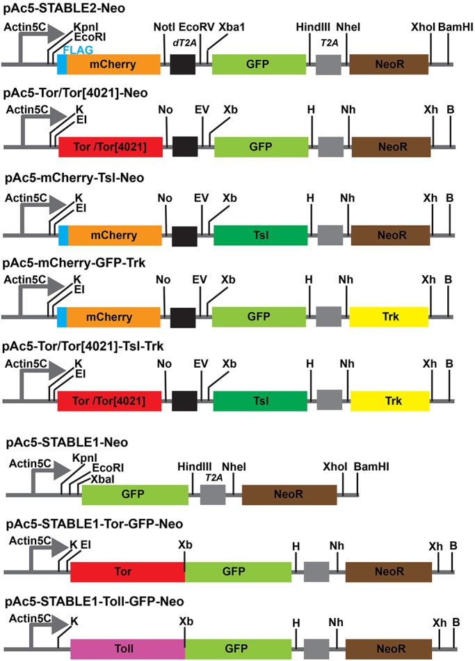
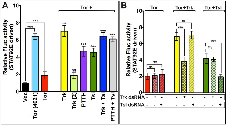
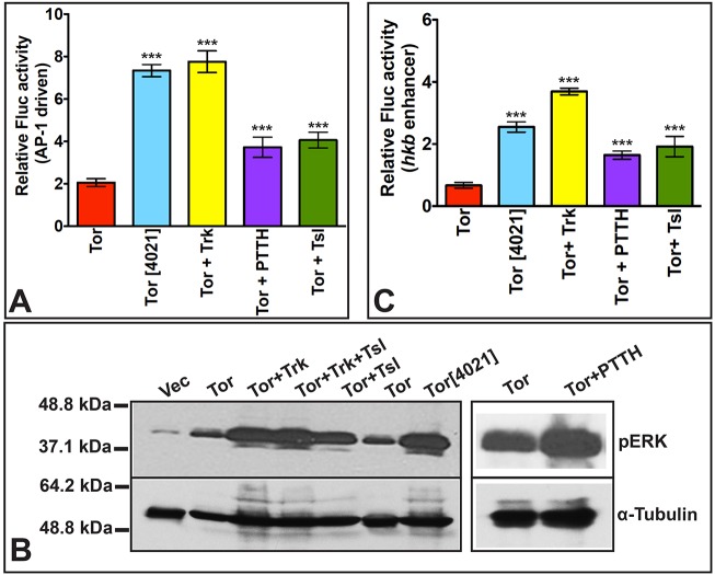
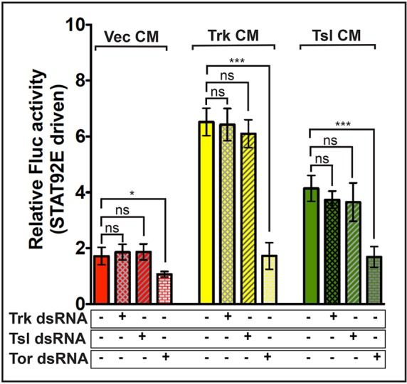
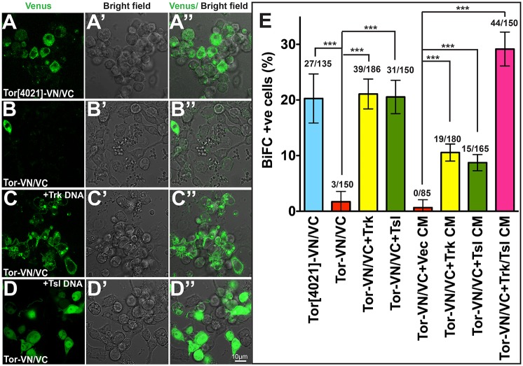
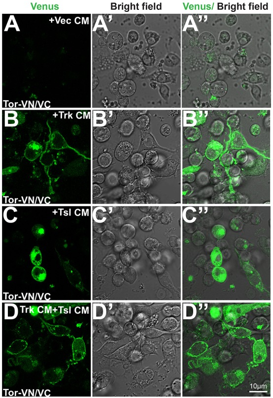
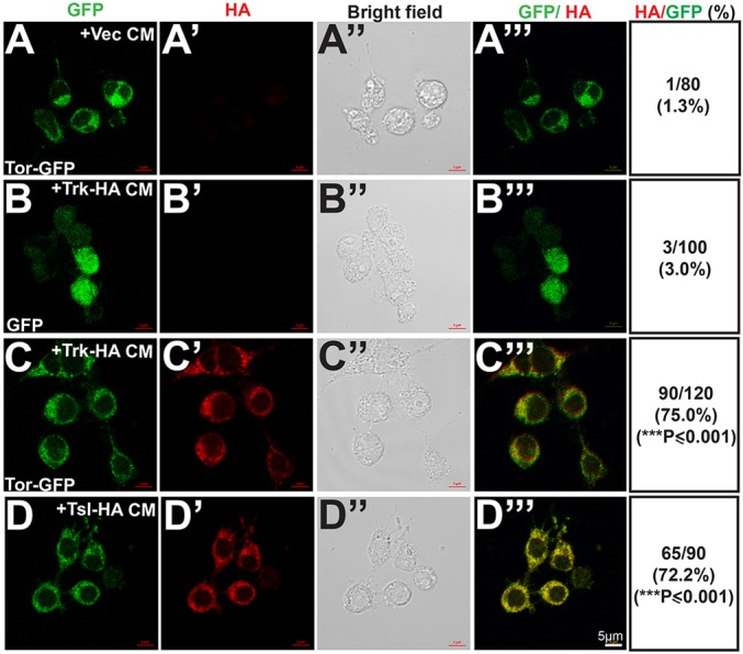
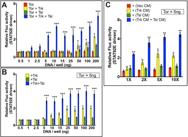
Formation of the Drosophila embryonic termini is controlled by the localized activation of the receptor tyrosine kinase Torso. Both Torso and Torso's presumed ligand, Trunk, are expressed uniformly in the early embryo. Polar activation of Torso requires Torso-like, which is expressed by follicle cells adjacent to the ends of the developing oocyte. We find that Torso expressed at high levels in cultured Drosophila cells is activated by individual application of Trunk, Torso-like or another known Torso ligand, Prothoracicotropic Hormone. In addition to assays of downstream signaling activity, Torso dimerization was detected using bimolecular fluorescence complementation. Trunk and Torso-like were active when co-transfected with Torso and when presented to Torso-expressing cells in conditioned medium. Trunk and Torso-like were also taken up from conditioned medium specifically by cells expressing Torso. At low levels of Torso, similar to those present in the embryo, Trunk and Torso-like alone were ineffective but acted synergistically to stimulate Torso signaling. Our results suggest that Torso interacts with both Trunk and Torso-like, which cooperate to mediate dimerization and activation of Torso at the ends of the Drosophila embryo.

Keywords: Drosophila; MACPF; Membrane attack complex perforin; RTK; Receptor tyrosine kinase; Terminal.


Conflict of interest statement

Competing interests

The authors declare no competing or financial interests.

Figures

Fig. 1.
Fig. 1.
Strategy for cloning the Torso (Tor), Tsl and Trk open reading frames into the multicistronic expression vectors pAc5-STABLE2-Neo and pAc5-STABLE1-Neo. (Top row) Schematic diagram of the multicistronic segment of pAc5-STABLE2-Neo (Gonzalez et al., 2011), which contains the Actin5C promoter, and mCherry, GFP and NeoR genes, is shown. Restriction sites and the positions of the dT2A and T2A autocleavage sites (black and gray boxes, respectively) are shown. (Rows 2-5) Plasmid derivatives in which DNA fragments encoding wild-type Tor/Tor[4021], Tsl and/or Trk have been introduced. (Row 6) Schematic diagram of the bicistronic region of pAc5-STABLE1-Neo (Gonzalez et al., 2011), which carries the Actin5C promoter and GFP and NeoR open reading frames, with restriction sites indicated. (Row 7) The plasmid encoding a Tor-GFP fusion protein (pAc5-STABLE1-Tor-GFP-Neo), with the cloning sites that were used for the substitution displayed as well as other restriction sites that remain. pAc5-STABLE1-Toll-GFP-Neo, which encodes a Toll-GFP fusion protein, is shown in the last row. B, BamHI; E1, EcoRI; EV, EcoRV; H, HindIII; K, KpnI; Nh, NheI; No, NotI; Xb, XbaI; Xh, XhoI.
Fig. 2.
Fig. 2.
Tor-dependent activation of STAT92E-driven Fluc activity is induced by co-expression of Trk, PTTH or Tsl. All S2R+ cells were co-transfected with the STAT92E-dependent Fluc reporter construct and the RNA PolIII 128 promoter-dependent R-luc control plasmid for transfection normalization. Relative Fluc activity of each sample is calculated with respect to cells expressing vector alone. (A) Samples shown on the left-hand side were transfected with either the pAc5-STABLE2-Neo vector control plasmid (Vec) (black), or with the vector encoding Tor[4021] (light blue) or wild-type Tor (red). Samples on the right-hand side were transfected with a plasmid encoding wild-type Tor and the following genes: Trk (yellow), Trk[2] mutant (light green), PTTH (purple), Tsl (dark green), Trk plus Tsl (dark blue) or PTTH plus Tsl (gray). (B) Trk and Tsl activation of Tor-mediated STAT92E-driven Fluc activity does not depend upon endogenous trk or tsl expression in S2R+ cells. Cells were transfected with Tor alone (red), Tor plus Trk (yellow) or Tor plus Tsl (green) and treated with no added dsRNA (solid bars), dsRNA targeting trk (cross-hatched bars) or dsRNA targeting tsl (hatched bars). In A, each data point is an average of three replicates, repeated seven times (n=7). Significance values for Tor and Tor[4021] are determined relative to vector alone. In all other cases, significance has been calculated relative to cells expressing Tor alone. In B, each bar represents an average of three replicates, repeated five times (n=5). Relative Fluc activity is displayed as mean±s.d., based on seven (A) or five (B) replicate measurements. Differences in values that are statistically significant are indicated above the bars. ***P<0.001. ns, not significant.
Fig. 3.
Fig. 3.
Trk, PTTH and Tsl stimulate Tor activation of AP1- and hkb enhancer-driven Fluc activity and MAPK phosphorylation. (A,C) S2R+ cells were co-transfected with the R-luc transfection control plasmid and an AP-1-dependent Fluc reporter (A) or an hkb enhancer-driven Fluc reporter (C). The experimental plasmid(s) that each set of transfected cells received is shown below each bar. Data represent average±s.d. of three readings, repeated seven times (n=7). Statistical significance was determined with respect to relative Fluc activity in cells expressing Tor alone. ***P<0.001. (B) Western blot analysis of extracts from S2R+ cells transfected with plasmids expressing the proteins shown at the top of each lane. Homogenates were divided in half and run on duplicate gels that were blotted and probed with either anti-phospho-p44/42 MAPK/pERK (top panels) or anti-α-Tubulin as a loading control (bottom panels).
Fig. 4.
Fig. 4.
CM from Trk- or Tsl-expressing cells induces Tor-dependent activation of STAT92E-driven Fluc activity. CM from cells transfected with vector (Vec) (red), Trk (yellow) or Tsl (green) was applied to S2R+ cells expressing Tor together with the STAT92E-Fluc and R-luc constructs. Some recipient cells were additionally treated with dsRNA targeting Trk (cross-hatched bars), Tsl (hatched bars) or Tor (brickwork bars). Each bar represents an average of three replicates, repeated five times (n=5)±s.d. Comparisons with statistically significant differences are indicated above the bars. *P≤0.05 (P=0.0102), ***P≤0.001. ns, not significant.
Fig. 5.
Fig. 5.
Co-expression with Trk or Tsl leads to dimerization of Tor. (A-D″) Fusion proteins between Tor and the Venus protein N-terminus (VN) or C-terminus (VC) were co-expressed either alone or with Trk or Tsl. The appearance of Venus fluorescence indicates that dimerization of Tor receptors has occurred. The left column shows Venus fluorescence, the middle column shows bright-field images and the right column displays an overlay of the two. (A-A″) Co-expression of Tor[4021]-VN and Tor[4021]-VC. (B-D″) Co-expression of wild-type Tor-VN and Tor-VC in the absence of other introduced genes (B-B″), with Trk (C-C″) or with Tsl (D-D″). Scale bar: 10 μm. (E) Quantitation of the percentage of cells that exhibited bimolecular fluorescence complementation (BiFC) (see also Fig. 6). Data are mean±s.d. derived from three independent replicate experiments (n=3). The total number of cells counted is shown above each bar. ***P≤0.001.
Fig. 6.
Fig. 6.
Exogenously added Trk or Tsl induces dimerization of Tor. (A-D″) Cells co-expressing wild-type Tor-VN/Tor-VC were exposed to CM from cells transfected with vector alone (Vec) (A-A″) or cells expressing Trk (B-B″), Tsl (C-C″) or both Trk and Tsl (D-D″). The left column shows Venus fluorescence, the middle column shows a bright-field image and the right column displays an overlay of the two. Scale bar: 10 μm. (See Fig. 5E for quantitation.)
Fig. 7.
Fig. 7.
Tor-expressing cells take up Trk and Tsl. (A-D‴) Cells expressing Tor-GFP (A-A‴,C-E‴) or GFP alone (expressed by pAc5-STABLE1-Neo) (B-B‴) were exposed to CM obtained from cells expressing vector alone (A-A‴), Trk-HA (B-C‴) or Tsl-HA (D-D‴). The cells were imaged for GFP fluorescence (A-D) and stained with anti-HA antibody (A′-D′). Bright-field images (A″-D″) and overlays of GFP and anti-HA staining (A‴-D‴) are also shown. For each study, the proportion of GFP- or Tor-GFP-expressing cells that also exhibited staining with anti-HA are shown in the last column. For each treatment, cells counted are the total of three independent experiments (n=3). Statistical significance was calculated relative to Tor-GFP-expressing cells treated with CM from vector control transfected cells. ***P≤0.001.
Fig. 8.
Fig. 8.
Trk and Tsl synergize to activate Tor-dependent STAT92E-driven Fluc in cells expressing low levels of Tor. (A-C) S2R+ cells were co-transfected with STAT92E-Fluc/R-luc constructs and the level of STAT92E-directed Fluc expression was assessed. (A) S2R+ cells were additionally transfected with increasing amounts of plasmid DNA carrying Tor (red), Tor plus Trk (yellow), Tor plus Tsl (green) or Tor plus both Trk and Tsl (blue). (B) S2R+ cells were transfected with 5 ng of the Tor expression plasmid plus a concentration range of plasmids bearing Trk alone (yellow), Tsl alone (green) or Trk plus Tsl (blue). (C) S2R+ cells in all samples were transfected with 5 ng of the Tor expression plasmid and exposed to control CM from cells transfected with vector alone (red), CM from cells expressing Trk (yellow) or Tsl (green), or a 1:1 mixture of Trk and Tsl CM (blue). CM was either untreated (1×), or was concentrated by various amounts (2× to 10×). Data are mean±s.d. of three readings, repeated five times (n=5). Statistical significance was calculated with respect to relative Fluc activity for cells expressing Tor alone at the appropriate experimental concentration (0.5-200 ng/well) (A), Tor alone at 5 ng/well (B), or Tor alone (5 ng/well) plus control vector CM at the correct experimental concentration (1-10X concentrated) (C). (A) ***P=0.0010, *P=0.036. (B) *P=0.0202, ***P=0.0082. (C) **P=0.0086, *P=0.0421. ns, not significant.

References

    1. Ambrosio L., Mahowald A. P. and Perrimon N. (1989). Requirement of the Drosophila Raf homologue for Torso function. Nature 342, 288-291. 10.1038/342288a0 - DOI - PubMed
    1. Baeg G.-H., Zhou R. and Perrimon N. (2005). Genome-wide RNAi analysis of JAK/STAT signaling components in Drosophila. Genes Dev. 19, 1861-1870. 10.1101/gad.1320705 - DOI - PMC - PubMed
    1. Brunner D., Oellers N., Szabad J., Biggs W. H. III, Zipursky S. L. and Hafen E. (1994). A gain-of-function mutation in Drosophila MAP kinase activates multiple receptor tyrosine kinase signaling pathways. Cell 76, 875-888. 10.1016/0092-8674(94)90362-X - DOI - PubMed
    1. Casali A. and Casanova J. (2001). The spatial control of Torso RTK activation: a C-terminal fragment of the Trunk protein acts as a signal for Torso receptor in the Drosophila embryo. Development 128, 1709-1715. - PubMed
    1. Casanova J. and Struhl G. (1989). Localized surface activity of Torso, a receptor tyrosine kinase, specifies terminal body pattern in Drosophila. Genes Dev. 3, 2025-2038. 10.1101/gad.3.12b.2025 - DOI - PubMed