Review
. 2016 Dec;4(6):10.1128/microbiolspec.UTI-0026-2016.
doi: 10.1128/microbiolspec.UTI-0026-2016.

Invasion of Host Cells and Tissues by Uropathogenic Bacteria

Affiliations
Review

Invasion of Host Cells and Tissues by Uropathogenic Bacteria

Adam J Lewis et al. Microbiol Spectr. 2016 Dec.

Abstract

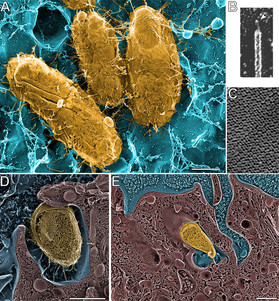
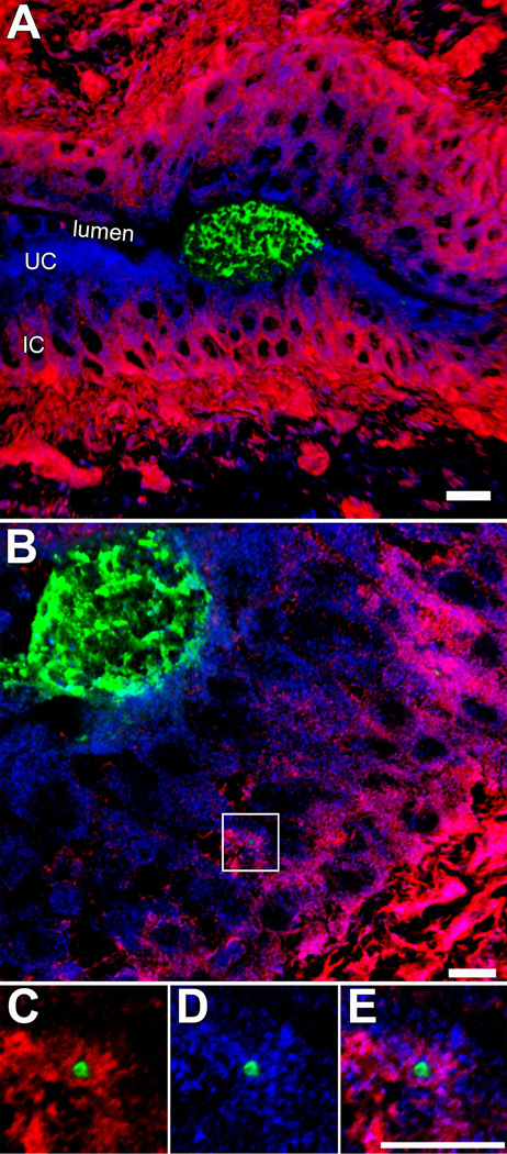
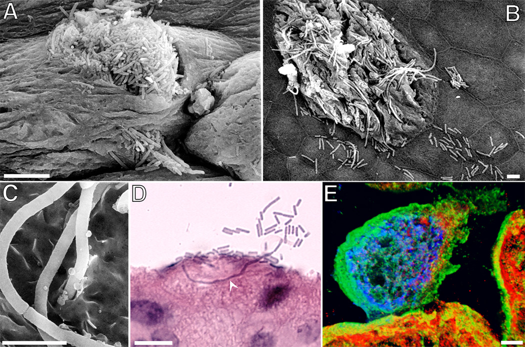
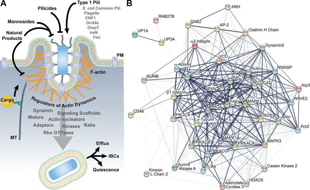
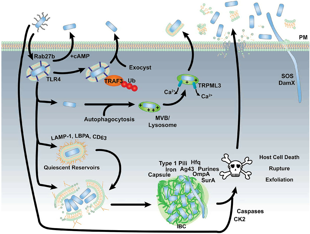
Within the mammalian urinary tract uropathogenic bacteria face many challenges, including the shearing flow of urine, numerous antibacterial molecules, the bactericidal effects of phagocytes, and a scarcity of nutrients. These problems may be circumvented in part by the ability of uropathogenic Escherichia coli and several other uropathogens to invade the epithelial cells that line the urinary tract. By entering host cells, uropathogens can gain access to additional nutrients and protection from both host defenses and antibiotic treatments. Translocation through host cells can facilitate bacterial dissemination within the urinary tract, while the establishment of stable intracellular bacterial populations may create reservoirs for relapsing and chronic urinary tract infections. Here we review the mechanisms and consequences of host cell invasion by uropathogenic bacteria, with consideration of the defenses that are brought to bear against facultative intracellular pathogens within the urinary tract. The relevance of host cell invasion to the pathogenesis of urinary tract infections in human patients is also assessed, along with some of the emerging treatment options that build upon our growing understanding of the infectious life cycle of uropathogenic E. coli and other uropathogens.


Figures

Figure 1
Figure 1
Type 1 pili mediate UPEC entry into bladder epithelial cells. (A) High-resolution deep-etch EM image showing UPEC (yellow) bound to a mouse bladder umbrella cell (blue) via multiple type 1 pili. (B) Close up view of a type 1 pilus, showing the 3-nm-wide FimH-containing tip fibrillum structure (arrowhead). (C) Close-up view of the 16-nm-wide hexagonal uroplakin complexes that are embedded within the umbrella cell AUM. (D and E) High-resolution freeze-fracture/deep-etch EM images showing the AUM enveloping bound UPEC. Scale bars = 0.5 µm. Images are reprinted from Proc Natl Acad Sci USA (18) and Science (9) with permission of the publishers.
Figure 2
Figure 2
Localization of UPEC within the bladder urothelium. (A and B) Confocal images of tissue sections from infected mouse bladders shows IBCs (green) within umbrella cells (UC). F-actin (red) is sparse within these host cells, but dense within the underlying immature cells (IC). A single bacterium, localized within a LAMP-1-positive compartment (blue) and surrounded by F-actin, is visible within one of the immature cells (box). (C–D) Images show magnified views of the area that is boxed out in (B). Figures reprinted from Cellular Microibiology (26) with permission of the publisher.
Figure 3
Figure 3
The efflux and filamentation of UPEC coincident with the exfoliation of IBC-containing umbrella cells. (A – C) Scanning EM images show filamentous forms of UPEC, as well as their normal-sized counterparts, emerging from within IBCs. (D) Image from a hematoxylin-and-eosin-stained bladder section highlights the ability of filamentous UPEC forms to extend long distances through umbrella cells. (E) Confocal image shows an IBC (blue) in close association with cytokeratin intermediate filaments (green) within an umbrella cell that is undergoing exfoliation. LAMP-1-positive compartments are red. Scale bars = 5 µm (A–C); 10 µm (D and E). Images are from mouse bladders recovered 6 hours after transurethral inoculation with UPEC. The figures are modified from Cellular Microibiology (26) or reprinted from Infection and Immunity (17) with permission of the publishers.
Figure 4
Figure 4
UPEC invasion of bladder epithelial cells. (A) Model depicts host and bacterial factors that have been identified as regulators of bladder cell invasion by UPEC. Potential therapeutics are also indicated. (B) The host factors that can modulate the FimH-dependent entry of UPEC into bladder cells are interconnected. The image in (B) was created using the STRING database (version 10.0) of known and predicted protein-protein interactions (199). Line thickness indicates the strength of the supporting data.
Figure 5
Figure 5
The fates of UPEC following entry into bladder epithelial cells. See text for details.

Similar articles

Cited by

References

    1. Bower JM, Eto DS, Mulvey MA. Covert operations of uropathogenic Escherichia coli within the urinary tract. Traffic. 2005;6:18–31. - PMC - PubMed
    1. Silva MT. Classical labeling of bacterial pathogens according to their lifestyle in the host: inconsistencies and alternatives. Front Microbiol. 2012;3:71. - PMC - PubMed
    1. Barber AE, Norton JP, Spivak AM, Mulvey MA. Urinary tract infections: current and emerging management strategies. Clinical infectious diseases : an official publication of the Infectious Diseases Society of America. 2013;57:719–724. - PMC - PubMed
    1. Dielubanza EJ, Schaeffer AJ. Urinary tract infections in women. The Medical clinics of North America. 2011;95:27–41. - PubMed
    1. Foxman B. Urinary tract infection syndromes: occurrence, recurrence, bacteriology, risk factors, and disease burden. Infect Dis Clin North Am. 2014;28:1–13. - PubMed

Publication types

MeSH terms

LinkOut - more resources