. 2017 Mar;174(6):483-492.
doi: 10.1111/bph.13714. Epub 2017 Feb 8.

Rapid antidepressant effect of ketamine correlates with astroglial plasticity in the hippocampus

Affiliations

Rapid antidepressant effect of ketamine correlates with astroglial plasticity in the hippocampus

Maryam Ardalan et al. Br J Pharmacol. 2017 Mar.

Abstract

Background and purpose: Astroglia contribute to the pathophysiology of major depression and antidepressant drugs act by modulating synaptic plasticity; therefore, the present study investigated whether the fast antidepressant action of ketamine is reflected in a rapid alteration of the astrocytes' morphology in a genetic animal model of depression.

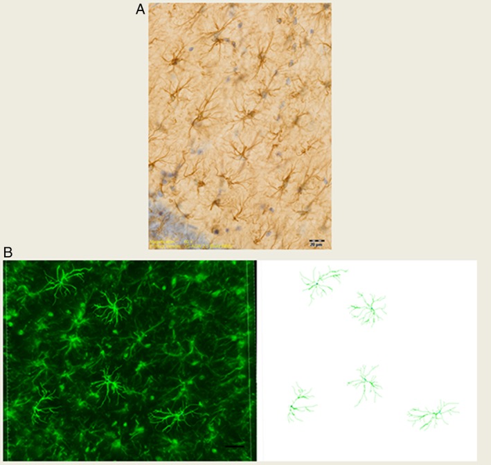
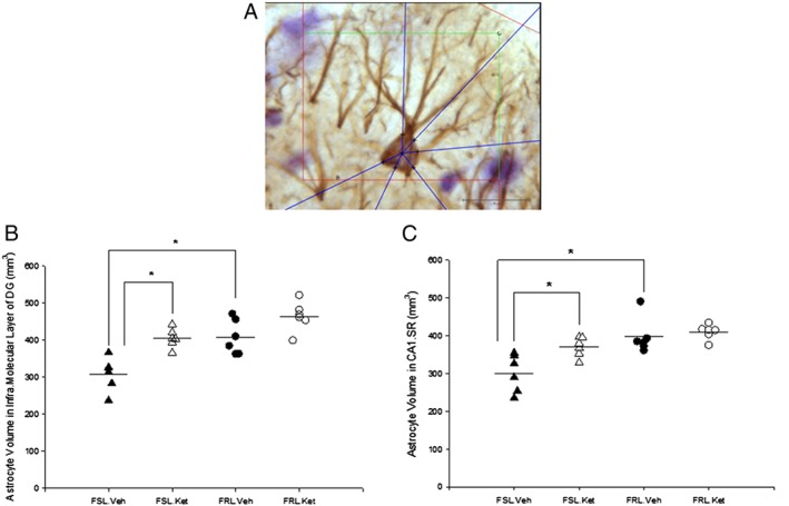
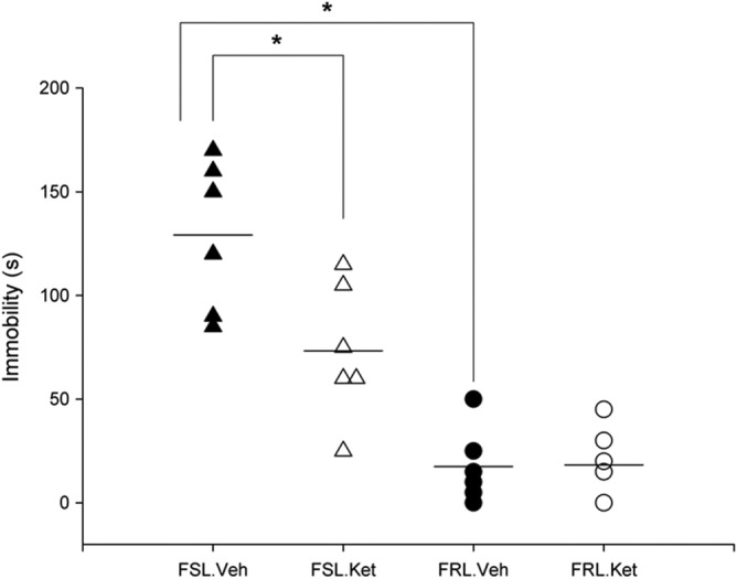
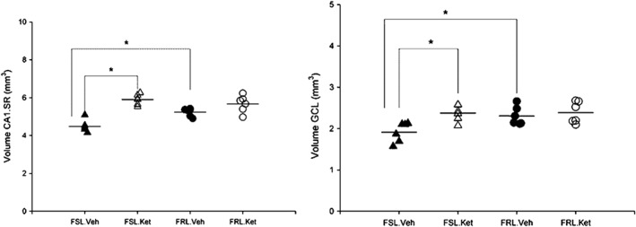
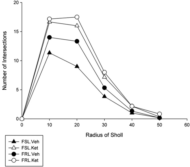
Experimental approach: S-Ketamine (15 mg·kg-1 ) or saline was administered as a single injection to Flinders Line (FSL/ FRL) rats. Twenty-four hours after the treatment, perfusion fixation was carried out and the morphology of glial fibrillary acid protein (GFAP)-positive astrocytes in the CA1 stratum radiatum (CA1.SR) and the molecular layer of the dentate gyrus (GCL) of the hippocampus was investigated by applying stereological techniques and analysis with Imaris software. The depressive-like behaviour of animals was also evaluated using forced swim test.

Key results: FSL rats treated with ketamine exhibited a significant reduction in immobility time in comparison with the FSL-vehicle group. The volumes of the hippocampal CA1.SR and GCL regions were significantly increased 1 day after ketamine treatment in the FSL rats. The size of astrocytes in the ketamine-treated FSL rats was larger than those in the FSL-vehicle group. Additionally, the number and length of the astrocytic processes in the CA1.SR region were significantly increased 1 day following ketamine treatment.

Conclusions and implications: Our results support the hypothesis that astroglial atrophy contributes to the pathophysiology of depression and a morphological modification of astrocytes could be one mechanism by which ketamine rapidly improves depressive behaviour.


Figures

Figure 1
Figure 1
(A) This GFAP immunostained image was captured from the CA1.SR area of rat hippocampus using a 60× objective. Scale bar is 20 μm. (B) 3D reconstruction of GFAP positive astrocytes in the CA1.SR subregion of the rat hippocampus by using Filament Tracers Algorithm in Imaris software. Scale bar is 20 μm.
Figure 2
Figure 2
(A) Illustration of the 3D nucleator tool for estimating the volume of GFAP positive astrocytes in the CA1.SR, supra‐ and infra‐MDG subfields of rat hippocampus. Six half‐lines (blue half‐lines) were used and the intersections between the half‐lines and the border of the cell soma were assigned by using a 100× objective. Scale bar is 20 μm; (B) Volume of the astrocytes in the infra‐MDG in the FSL and FRL rats 1 day after a single ketamine or saline administration.*P < 0.05; (C) Volume of the astrocytes in the CA1.SR area of the hippocampus in FSL and FRL rats 1 day after a single ketamine or saline administration, *P < 0.05.
Figure 3
Figure 3
Fast effect of a single i.p. injection of ketamine on the immobility behaviour in the forced swim test in the FSL and FRL rats, *P < 0.05.
Figure 4
Figure 4
(Left) Volume of the CA1.SR area in the FSL (n = 12) and FRL (n = 12) rats 1 day after a single ketamine or saline administration. *P < 0.05. (Right) Volume of the GCL area in the FSL (n = 12) and FRL (n = 12) rats 1 day after a single ketamine or saline administration, *P < 0.05.
Figure 5
Figure 5
Effect of ketamine treatment 1 day after a single injection on the branching pattern of astrocytes in the CA1.SR subregion of the hippocampus using sholl analysis in the FSL and FRL rats. The number of branching intersections 10 and 20 μm away from the cell soma were significantly lower in the FSL.vehicle rats compared to the FRL.vehicle rats (P < 0.01). Significant increase in the length of the astrocytic branches at 10, 20 and 30 μm away from the cell body of astrocytes in the FSL group 1 day after treatment (P < 0.05).

References

    1. Alexander SPH, Kelly E, Marrion N, Peters JA, Benson HE, Faccenda E et al. (2015a). The Concise Guide to PHARMACOLOGY 2015/16: Overview. Br J Pharmacol 172: 5729–5143. - PMC - PubMed
    1. Alexander SP, Kelly E, Marrion N, Peters JA, Benson HE, Faccenda E et al. (2015b). The concise guide to PHARMACOLOGY 2015/16: Transporters. Br J Pharmacol 172: 6110–6202. - PMC - PubMed
    1. Althaus HH, Richter‐Landsberg C (2000). Glial cells as targets and producers of neurotrophins. Int Rev Cytol 197: 203–277. - PubMed
    1. Amaral DG, Scharfman HE, Lavenex P (2007). The dentate gyrus: fundamental neuroanatomical organization (dentate gyrus for dummies). Prog Brain Res 163: 3–22. - PMC - PubMed
    1. Anderson CM, Swanson RA (2000). Astrocyte glutamate transport: review of properties, regulation, and physiological functions. Glia 32: 1–14. - PubMed

Publication types