. 2017 Mar:121:155-166.
doi: 10.1016/j.biomaterials.2017.01.003. Epub 2017 Jan 3.

RhoA knockdown by cationic amphiphilic copolymer/siRhoA polyplexes enhances axonal regeneration in rat spinal cord injury model

Affiliations

RhoA knockdown by cationic amphiphilic copolymer/siRhoA polyplexes enhances axonal regeneration in rat spinal cord injury model

So-Jung Gwak et al. Biomaterials. 2017 Mar.

Abstract

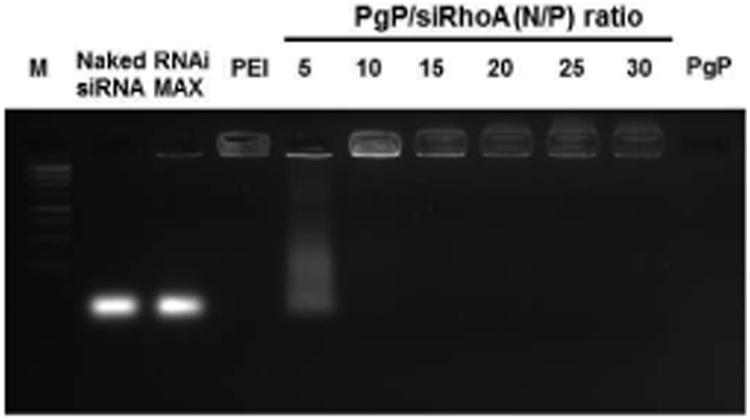
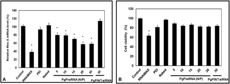
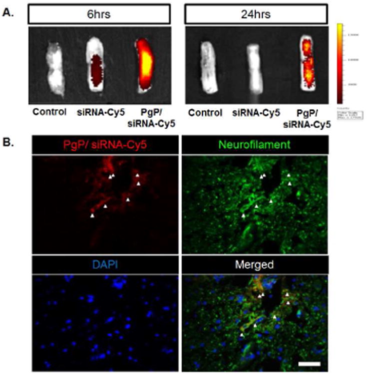
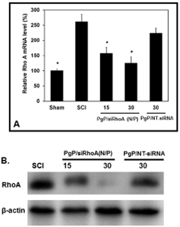
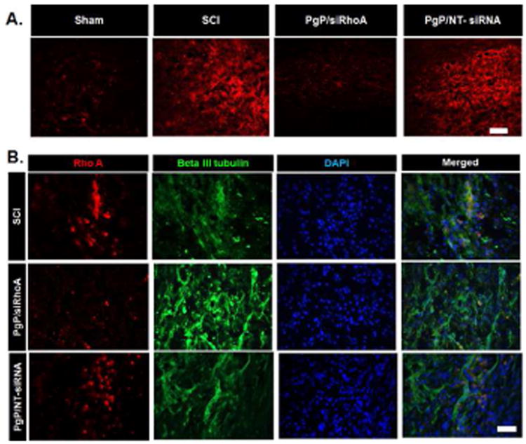
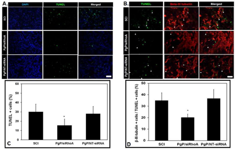
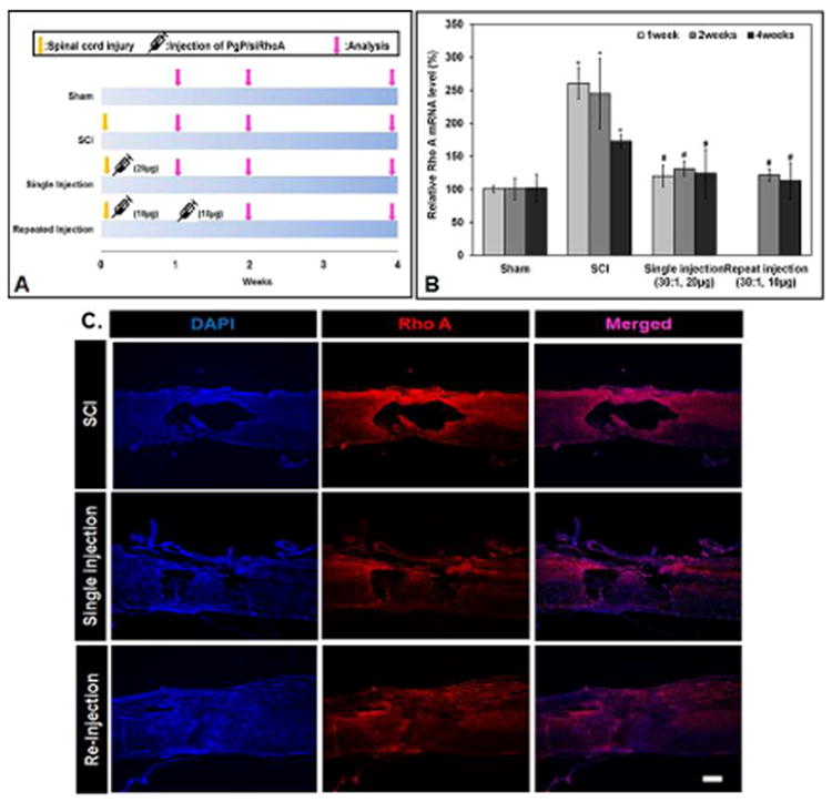
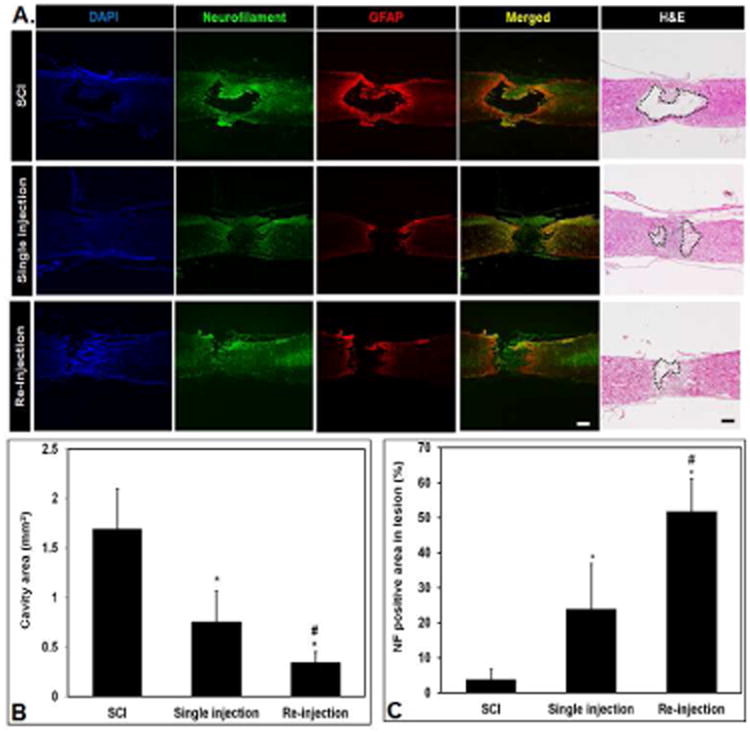
Spinal cord injury (SCI) results in permanent loss of motor and sensory function due to developmentally-related and injured-induced changes in the extrinsic microenvironment and intrinsic neuronal biochemistry that limit plasticity and axonal regeneration. Our long term goal is to develop cationic, amphiphilic copolymers (poly (lactide-co-glycolide)-g-polyethylenimine, PgP) for combinatorial delivery of therapeutic nucleic acids (TNAs) and drugs targeting these different barriers. In this study, we evaluated the ability of PgP to deliver siRNA targeting RhoA, a critical signaling pathway activated by multiple extracellular inhibitors of axonal regeneration. After generation of rat compression SCI model, PgP/siRhoA polyplexes were locally injected into the lesion site. Relative to untreated injury only, PgP/siRhoA polyplexes significantly reduced RhoA mRNA and protein expression for up to 4 weeks post-injury. Histological analysis at 4 weeks post-injury showed that RhoA knockdown was accompanied by reduced apoptosis, cavity size, and astrogliosis and increased axonal regeneration within the lesion site. These studies demonstrate that PgP is an efficient non-viral delivery carrier for therapeutic siRhoA to the injured spinal cord and may be a promising platform for the development of combinatorial TNA/drug therapy.

Keywords: Axon regeneration; Cationic amphiphilic co-polymer; Non-viral nucleic acid carrier; RhoA siRNA; Spinal cord injury.


Figures

Figure 1
Figure 1
Gel retardation assay of PgP/siRhoA polyplexes prepared at varying N/P ratios electrophoresed on 2 % agarose gel: 1 kb DNA molecular weight marker (Lane 1), naked siRhoA (lane 2), RNAiMAX/siRhoA (lane 3), bPEI/siRhoA at N/P ratio of 5/1 (lane 4), PgP/siRhoA prepared at N/P ratios of 5/1, 10/1, 15/1, 20/1, 25/1, and 30/1 (lane 5, 6, 7, 8, 9, and 10), and PgP alone (lane 11).
Figure 2
Figure 2
RhoA knockdown efficiency (A) and cell viability (B) after transfection of PgP/siRhoA polyplexes prepared at varying N/P ratios in neuroblastoma (B35) cells in media containing 10 % serum. At 72 hours post-transfection, RhoA expression level was determined by RT-PCR and cell viability was determined by MTT assay. Data represent the mean ± SEM (n=6). *: P<0.05 compared to untreated control.
Figure 3
Figure 3
Retention and cellular uptake of PgP/siRNA-Cy5 polyplexes (N/P ratio of 30/1, 10 μg siRNA-Cy5) after local injection in SCI lesion site. (A) Ex vivo fluorescent imaging of PgP/siRNA-Cy5 in spinal cord injury at 6 and 24 hours post-injection. (B) Visualization of PgP/siRNA-Cy5 polyplex uptake by neuronal cells in spinal cord lesion site at 24 hours post-injection. PgP/siRNA-Cy5 polyplex (Red), immunofluorescent staining for neurofilament (NF, Green), and DAPI counterstained nuclei (blue). Scale bar indicates 50 μm.
Figure 4
Figure 4
Rho A knockdown efficiency of PgP/siRhoA polyplexes (two N/P ratios: 15/1 and 30/1, siRhoA : 10 μg siRhoA) after local injection in SCI lesion site. At 7 days post-injury, animals were sacrificed and spinal cord (0.5 cm-long piece from the center of the injury) was harvested for RT-PCR and western blot. Control: Sham animal group, SCI: untreated SCI animal group, 15/1: PgP/siRhoA polyplexes at N/P ratio of 15/1, 30/1: PgP/siRhoA polyplexes at N/P ratio of 30/1, and PgP/NT-siRNA 30/1: PgP/non-targeting-siRNA polyplexes at N/P ratio of 30/1, (A) Relative RhoA mRNA level by real-time qRT-PCR. Glyceraldehyde-3-phosphate dehydrogenase (GAPDH) was used as an endogenous control. *: P<0.05 compared to untreated SCI group, (B) RhoA protein level by western blot. β-actin was used as endogenous control.
Figure 5
Figure 5
Immunohistochemical staining for RhoA after local injection of PgP/siRhoA polyplexes (N/P ratio of 30/1, siRhoA: 10 μg). At 1 week post-injury, spinal cords were harvested and sectioned longitudinally. Untreated SCI and PgP/NT-siRNA polyplexes (N/P ratio 30/1, NT-siRNA: 10 μg) were used for comparison. (A) IHC staining for RhoA (red), scale bar: 200 μm. (B) Double-IHC staining for RhoA (red), neuron-specific β-III Tubulin (green) and DAPI (blue). Scale bar indicates 50 μm.
Figure 6
Figure 6
Effect of RhoA knockdown by PgP/siRhoA polyplexes (N/P ratio 30/1, siRhoA : 10 μg) on apoptosis by TUNEL assay. At 7 days post-injury, the spinal cords were harvested and sectioned longitudinally and stained by the ApopTag Plus Fluorescein In situ Apoptosis Detection kit. Untreated SCI and PgP/NT-siRNA polyplexes (N/P ratio 30/1, NT-siRNA: 10 μg) were used for comparison. (A) Cell nuclei (DAPI, blue) and TUNEL+ cells (green). Scale bar: 100 μm. (B) Double IHC staining for TUNEL+ (green) and beta III tubulin+ (red) cells. Scale bar: 50 μm. (C) The % TUNEL-positive cells in untreated SCI and after injection of PgP/siRhoA or PgP/NT-siRNA polyplexes was quantified from total 15 different sections of spinal cords from each group (3 sections/rat, 5 rats/group). *p < 0.05 compared with untreated SCI. (D) The % beta-III tubulin+ cells in total TUNEL+ cells in spinal cord lesion site was quantified from total 15 different sections of spinal cords from each group (3 sections/rat, 5 rats/group). *p < 0.05 compared with untreated SCI.
Figure 7
Figure 7
(A) Experimental design for PgP/siRhoA administration timing and outcome assessment over a 4 week time period. Single injection: PgP/siRhoA polyplexes (N/P 30/1, siRhoA 20 μg) were locally injected in the lesion site immediately after SCI injury. Repeat injection: PgP/siRhoA polyplex (N/P 30/1, siRhoA 10 μg/injection) was locally injected in the lesion site immediately after SCI injury and at 1 week post-injury. At 1, 2, and 4 weeks post-injury, spinal cords (0.5 cm-long piece from the center of the injury) were harvested and RhoA knockdown was evaluated by RT-PCR and IHC. (B) RhoA mRNA expression levels evaluated by RT-PCR. Sham animal group and untreated SCI group were used as controls (n=5/group). GAPDH was used as an endogenous control. *P<0.05 compared to Sham, # P<0.05 compared to SCI. (C) Longitudinal sections of SCI lesion sites stained for RhoA (red) and cell nuclei (blue, DAPI) at 4 weeks post-injury. Untreated SCI (top), single-injection (middle), and re-injection (bottom).
Figure 8
Figure 8
Immunohistological assessment of lesion sites 4 weeks after SCI and injection of PgP/siRhoA polyplexes. (A) Representative images of immunostaining for neurofilament (green), GFAP (red), and DAPI (blue) and hematoxylin and eosin staining to measure cavity area. Scale bars: 400 μm. Untreated SCI (top) shows an extensive necrotic lesion cavity and significant reactive astrogliosis, Single-injection (middle) and re-injection (bottom) animal group shows reduced cavitation/astrogliosis and axonal regeneration in the lesion site. (B) Quantification of necrotic cavity area (mm2) in the lesion of spinal cord at 4 weeks post-injection of PgP/siRhoA polyplexes. The cavity area was measured from total 15 different sections of spinal cord from each group (3 sections/rat, 5 rats/each group) after hematoxylin and eosin staining. *P<0.05 compared to untreated SCI, #P<0.05 compared to single injection of PgP/siRhoA polyplexes. (C) Quantification of neurofilament (NF) positive (+) axons in the lesion site. The NF positive area in the lesion site was measured from total 15 different sections of spinal cord from each group (3 sections/rat, 5 rats/each group). The % NF positive area was calculated as a percentage of the total necrotic cavity area measured from hematoxylin and eosin staining. *P<0.05 compared to untreated SCI, #P<0.05 compared to single injection of PgP/siRhoA polyplexes.

References

    1. Fournier AE, GrandPre T, Strittmatter SM. Identification of a receptor mediating Nogo-66 inhibition of axonal regeneration. Nature. 2001;409:341–6. - PubMed
    1. Chen MS, Huber AB, van der Haar ME, Frank M, Schnell L, Spillmann AA, et al. Nogo-A is a myelin-associated neurite outgrowth inhibitor and an antigen for monoclonal antibody IN-1. Nature. 2000;403:434–9. - PubMed
    1. Wang KC, Koprivica V, Kim JA, Sivasankaran R, Guo Y, Neve RL, et al. Oligodendrocyte-myelin glycoprotein is a Nogo receptor ligand that inhibits neurite outgrowth. Nature. 2002;417:941–4. - PubMed
    1. Liu BP, Fournier A, GrandPre T, Strittmatter SM. Myelin-associated glycoprotein as a functional ligand for the Nogo-66 receptor. Science. 2002;297:1190–3. - PubMed
    1. Atwal JK, Pinkston-Gosse J, Syken J, Stawicki S, Wu Y, Shatz C, et al. PirB is a Functional Receptor for Myelin Inhibitors of Axonal Regeneration. Science. 2008;322:967–70. - PubMed

Publication types