Review
. 2017 Jan 14;17(1):28.
doi: 10.1186/s12872-016-0465-2.

Invasive Cardiac Lipoma: a case report and review of literature

Affiliations
Review

Invasive Cardiac Lipoma: a case report and review of literature

Jason D'Souza et al. BMC Cardiovasc Disord. .

Abstract

Background: Cardiac lipomas are rare benign tumors of the heart. They are usually asymptomatic and are thus most often diagnosed on autopsies. Symptoms, when present, depend upon the location within the heart. Typical locations are the endocardium of the right atrium and the left ventricle. Diagnostic modality of choice is cardiac MRI. Treatment guidelines have not yet been established due to the very low prevalence of these tumors and are thus guided by the patient's symptomatology.

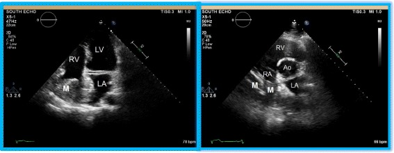
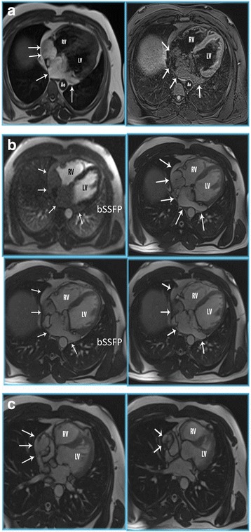
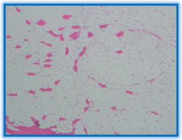
Case presentation: We describe a case of an invasive cardiac lipoma, wherein the initial symptom of the patient was shortness of breath. Although the echocardiogram visualized the tumor in the right atrium, a cardiac MRI was performed for better tissue characterization. The MRI revealed a large, fat containing, septated mass in the right atrium with invasion into the inter-atrial septum and inferior cavoatrial junction. There was also invasion of the coronary sinus along the inferior and left lateral aspect of the posterior atrioventricular groove. Although the mass appeared to represent a lipoma by imaging characteristics, the unusual extension into the coronary sinus led to consideration of a low-grade liposarcoma in the differential. Thus a pre-operative biopsy was performed along with MDM2 gene amplification to rule out a liposarcoma preceding surgical excision.

Conclusion: Cardiac lipomas are well-characterized on cardiac MRI, which is the diagnostic modality of choice. Typical locations are the right atrium and the left ventricle. However, in those with atypical features such as invasion of the coronary sinus, pre-operative biopsy for histopathologic confirmation is imperative to exclude well-differentiated liposarcoma. Our patient with a simple lipoma underwent partial resection to relieve symptoms. We discuss prognosis and treatment of symptomatic cardiac lipomas.

Keywords: Benign cardiac tumor; Cardiac lipoma; Case report; Liposarcoma.


Figures

Fig. 1
Fig. 1
Echocardiographic imaging of the cardiac lipoma. The apical four chamber view and the parasternal short axis view clearly demonstrate the mass (M) originating from the free wall of the right atrium. The mass almost completely obliterating the right atrial cavity. Also notable is the involvement of the inter-atrial septum by this heterogenous mass. RA—right atrium; LA—left atrium; RV—right ventricle; LV—left ventricle; Ao—aorta
Fig. 2
Fig. 2
Cardiac MRI of the cardiac lipoma. a Cardiac MR double (DIR) and triple inversion recovery (TIR) sequences clearly demonstrates a large fat containing mass in right atrium, interatrial septum and coronary sinus (white arrows). (RV = right ventricle; LV = left ventricle; Ao = descending aorta). b Axial T1 postcontrast (T1 w) and balanced steady state free precession (bSSFP) cardiac MR demonstrates a large fat containing mass in the right atrium, interatrial septum and coronary sinus (white arrows). No enhancing components suggests this is a simple lipoma (RV = right ventricle; LV = left ventricle). c Balanced steady state free precession CMR images in HLA shows the lipoma extending from the right atrium into the IVC (white arrows). (RV = right ventricle; LV = left ventricle)
Fig. 3
Fig. 3
Histopathology of the tumor. Histopathology of the lipoma specimen reveals mature adipocytes and absence of other soft tissue components which otherwise would have indicated a liposarcoma

References

    1. Wu S, Teng P, Zhou Y, Ni Y. A rare case report of giant epicardial lipoma compressing the right atrium with septal enhancement. J Cardiothoracic Surg. 2015;10:150. doi: 10.1186/s13019-015-0375-x. - DOI - PMC - PubMed
    1. Li D, Wang W, Zhu Z, Wang Y, Xu R, Liu K. Cardiac lipoma in the interventricular septum: a case report. J Cardiothoracic Surg. 2015;10:69. doi: 10.1186/s13019-015-0275-0. - DOI - PMC - PubMed
    1. Ismail I, Al-Khafaji K, Mutyala M, Aggarwal S, Cotter W, Hakim H, Khosla S, Arora R. Cardiac Lipoma. J Community Hosp Intern Med Perspect. 2015;5(5):28449. - PMC - PubMed
    1. Wang H, Hu J, Sun X, Wang P, Du Z. An asymptomatic right atrial intramyocardial lipoma: a management dilemma. World J Surg Oncol. 2015;13:20. doi: 10.1186/s12957-015-0441-9. - DOI - PMC - PubMed
    1. Wang Y, Wang X, Xiao Y. Surgical treatment of primary cardiac valve tumor: early and late results in eight patients. J Cardiothorac Surg. 2016;11:31. doi: 10.1186/s13019-016-0406-2. - DOI - PMC - PubMed

MeSH terms