

Relation between resting amygdalar activity and cardiovascular events: a longitudinal and cohort study
- PMID: 28088338
- PMCID: PMC7864285
- DOI: 10.1016/S0140-6736(16)31714-7
Relation between resting amygdalar activity and cardiovascular events: a longitudinal and cohort study
Erratum in
-
Department of Error.Lancet. 2017 Feb 25;389(10071):804. doi: 10.1016/S0140-6736(17)30082-X. Epub 2017 Jan 13. Lancet. 2017. PMID: 28089472 No abstract available.
-
Department of Error.Lancet. 2017 Feb 25;389(10071):804. doi: 10.1016/S0140-6736(17)30344-6. Epub 2017 Feb 13. Lancet. 2017. PMID: 28196666 No abstract available.
Abstract
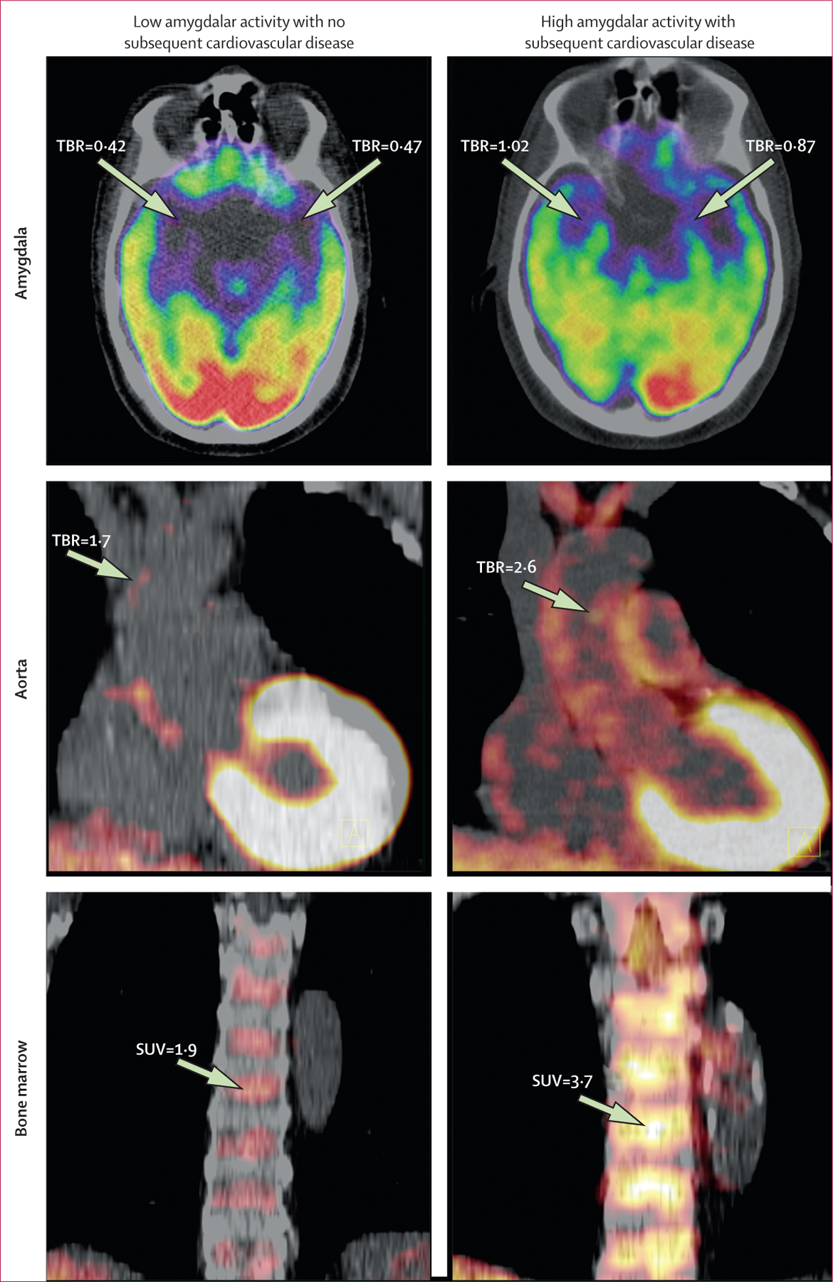
Background: Emotional stress is associated with increased risk of cardiovascular disease. We imaged the amygdala, a brain region involved in stress, to determine whether its resting metabolic activity predicts risk of subsequent cardiovascular events.
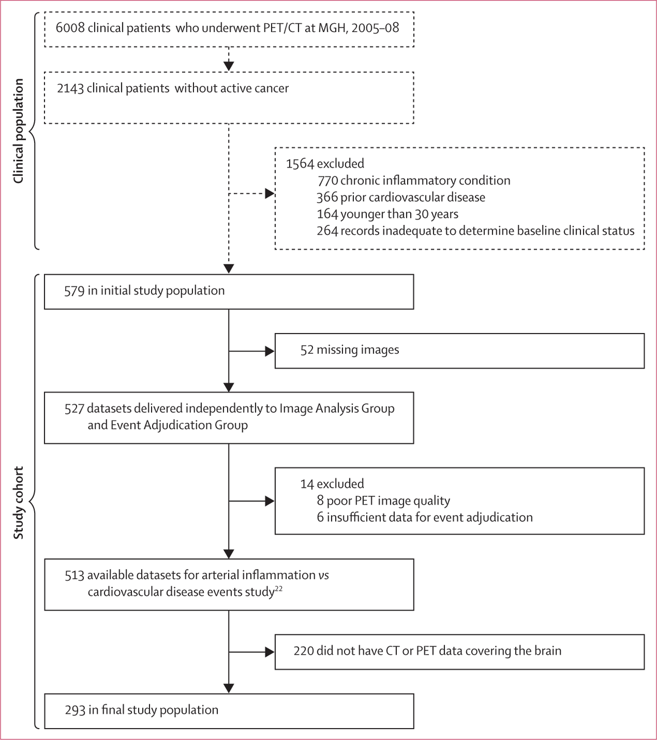
Methods: Individuals aged 30 years or older without known cardiovascular disease or active cancer disorders, who underwent 18F-fluorodexoyglucose PET/CT at Massachusetts General Hospital (Boston, MA, USA) between Jan 1, 2005, and Dec 31, 2008, were studied longitudinally. Amygdalar activity, bone-marrow activity, and arterial inflammation were assessed with validated methods. In a separate cross-sectional study we analysed the relation between perceived stress, amygdalar activity, arterial inflammation, and C-reactive protein. Image analyses and cardiovascular disease event adjudication were done by mutually blinded researchers. Relations between amygdalar activity and cardiovascular disease events were assessed with Cox models, log-rank tests, and mediation (path) analyses.
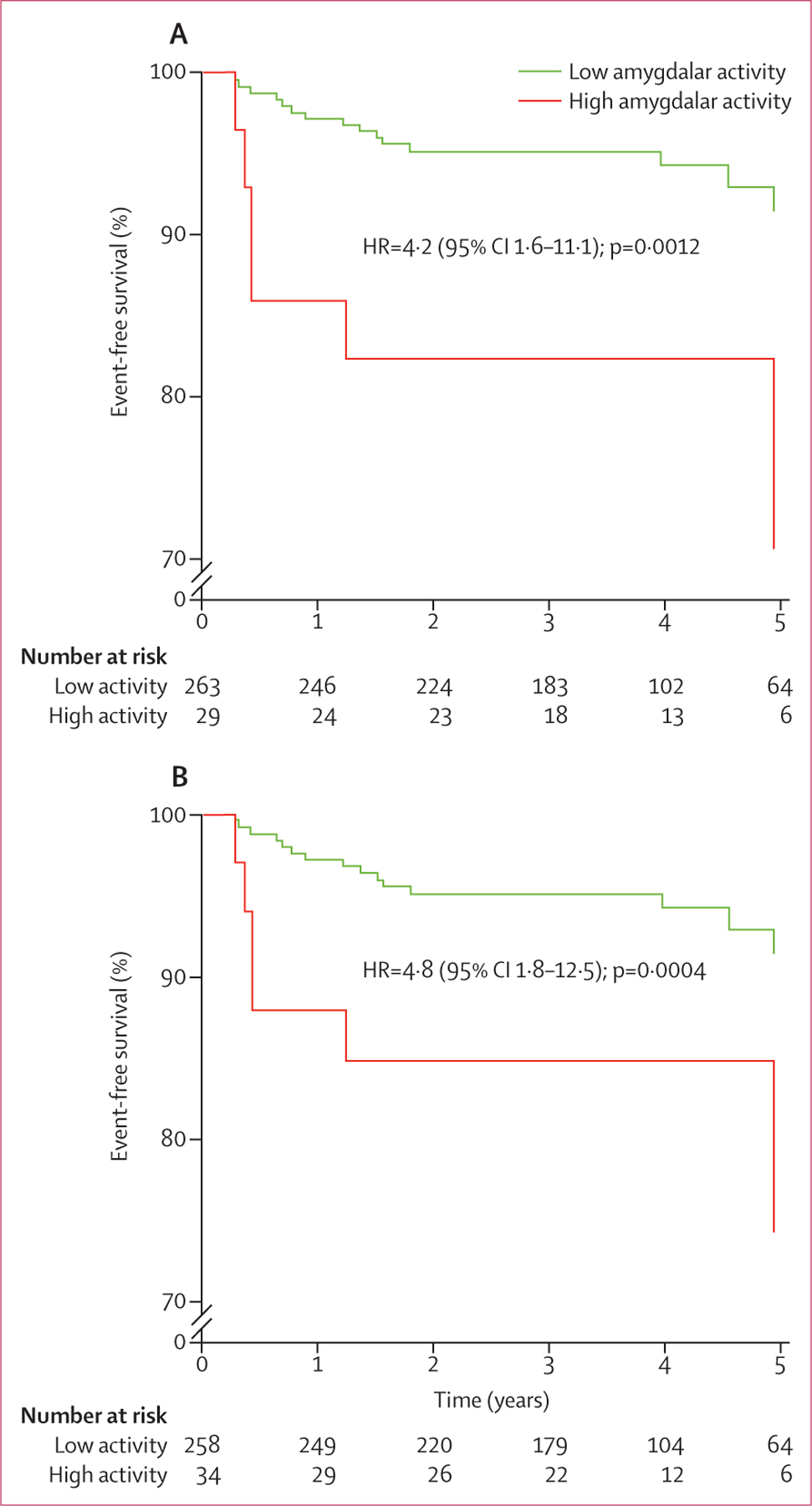
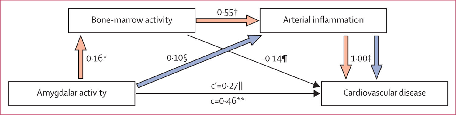
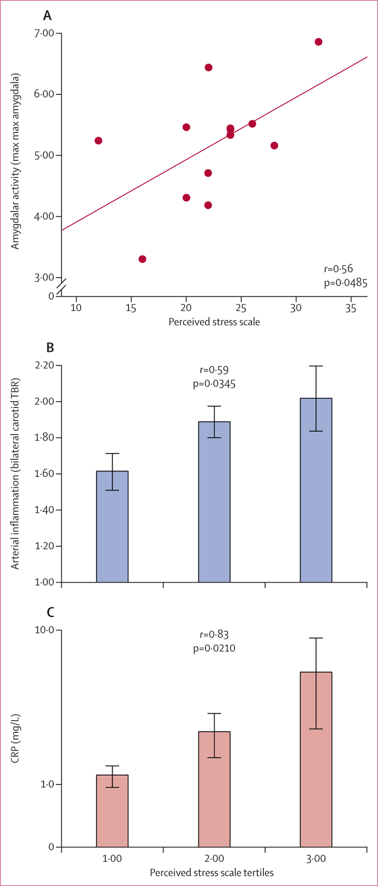
Findings: 293 patients (median age 55 years [IQR 45·0-65·5]) were included in the longitudinal study, 22 of whom had a cardiovascular disease event during median follow-up of 3·7 years (IQR 2·7-4·8). Amygdalar activity was associated with increased bone-marrow activity (r=0·47; p<0·0001), arterial inflammation (r=0·49; p<0·0001), and risk of cardiovascular disease events (standardised hazard ratio 1·59, 95% CI 1·27-1·98; p<0·0001), a finding that remained significant after multivariate adjustments. The association between amygdalar activity and cardiovascular disease events seemed to be mediated by increased bone-marrow activity and arterial inflammation in series. In the separate cross-sectional study of patients who underwent psychometric analysis (n=13), amygdalar activity was significantly associated with arterial inflammation (r=0·70; p=0·0083). Perceived stress was associated with amygdalar activity (r=0·56; p=0·0485), arterial inflammation (r=0·59; p=0·0345), and C-reactive protein (r=0·83; p=0·0210).
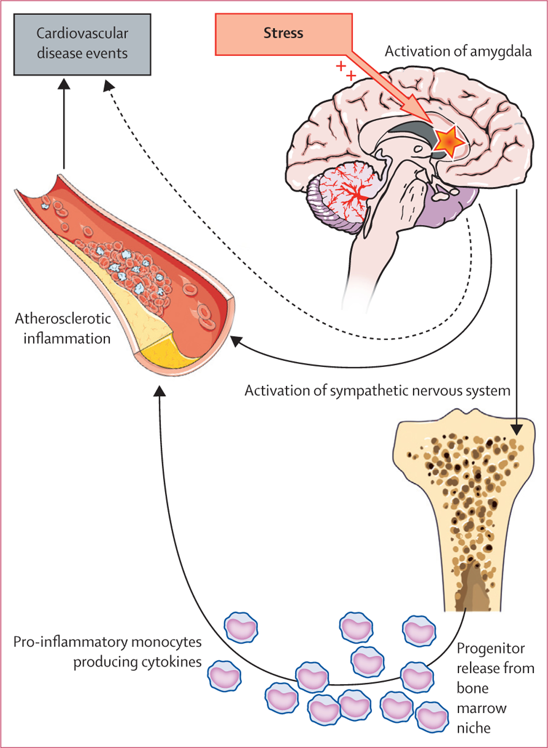
Interpretation: In this first study to link regional brain activity to subsequent cardiovascular disease, amygdalar activity independently and robustly predicted cardiovascular disease events. Amygdalar activity is involved partly via a path that includes increased bone-marrow activity and arterial inflammation. These findings provide novel insights into the mechanism through which emotional stressors can lead to cardiovascular disease in human beings.
Funding: None.
Copyright © 2017 Elsevier Ltd. All rights reserved.
Conflict of interest statement
Declaration of interests
AT reports grants from Genentech and Takeda and personal fees from Takeda, Actelion, AstraZeneca, and Amgen during this study for research outside the submitted work. UH reports grants from the National Heart, Lung, and Blood Institute’s Framingham Heart Study, American College of Radiology Imaging Network, Kowa Company, and Heartflow, and personal fees from the American Heart Association during the study. JWM reports a grant from Avanir Pharmaceuticals and Otsuka, personal fees from Janssen Research and Development, ProPhase, Genentech, and Impel Neuropharma, and a pending patent for Neuropeptide Y as a treatment for mood and anxiety disorders outside the submitted work. All other authors declare no competing interests.
Figures
Comment in
-
Stressed brain, stressed heart?Lancet. 2017 Feb 25;389(10071):770-771. doi: 10.1016/S0140-6736(17)30044-2. Epub 2017 Jan 12. Lancet. 2017. PMID: 28088337 No abstract available.
-
Risk factors: Linking stress to arterial inflammation and vascular events.Nat Rev Cardiol. 2017 Mar;14(3):127. doi: 10.1038/nrcardio.2017.8. Epub 2017 Jan 27. Nat Rev Cardiol. 2017. PMID: 28127031 No abstract available.
References
-
- Batty GD, Russ TC, Stamatakis E, Kivimaki M. Psychological distress and risk of peripheral vascular disease, abdominal aortic aneurysm, and heart failure: pooling of sixteen cohort studies. Atherosclerosis 2014; 236: 385–88. - PubMed
-
- Rosengren A, Hawken S, Ôunpuu S, et al. ; for the INTERHEART Investigators. Association of psychosocial risk factors with risk of acute myocardial infarction in 11 119 cases and 13 648 controls from 52 countries (the INTERHEART study): case-control study. Lancet 2004; 364: 953–62. - PubMed
-
- Scandinavian Simvastatin Survival Study Group. Randomised trial of cholesterol lowering in 4444 patients with coronary heart disease: the Scandinavian Simvastatin Survival Study (4S). Lancet 1994; 344: 1383–89. - PubMed
-
- Prevention of cardiovascular events and death with pravastatin in patients with coronary heart disease and a broad range of initial cholesterol levels. The Long-Term Intervention with Pravastatin in Ischaemic Disease (LIPID) Study Group. N Engl J Med 1998; 339: 1349–57. - PubMed
Publication types
MeSH terms
Substances
Grants and funding
LinkOut - more resources
Full Text Sources
Other Literature Sources
Medical
Research Materials
