

Ultrasound-Mediated Delivery of RNA to Colonic Mucosa of Live Mice
- PMID: 28088460
- PMCID: PMC5368009
- DOI: 10.1053/j.gastro.2017.01.002
Ultrasound-Mediated Delivery of RNA to Colonic Mucosa of Live Mice
Abstract
Background & aims: It is a challenge to deliver nucleic acids to gastrointestinal (GI) tissues due to their size and need for intracellular delivery. They are also extremely susceptible to degradation by nucleases, which are ubiquitous in the GI tract. We investigated whether ultrasound, which can permeabilize tissue through a phenomenon known as transient cavitation, can be used to deliver RNA to the colonic mucosa of living mice.
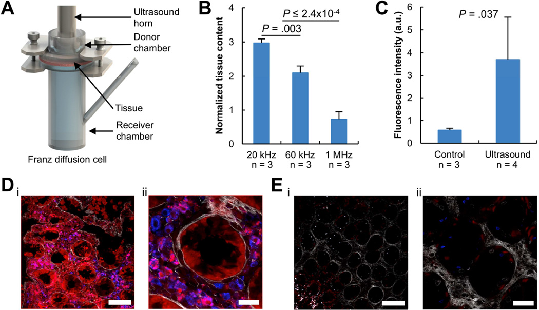
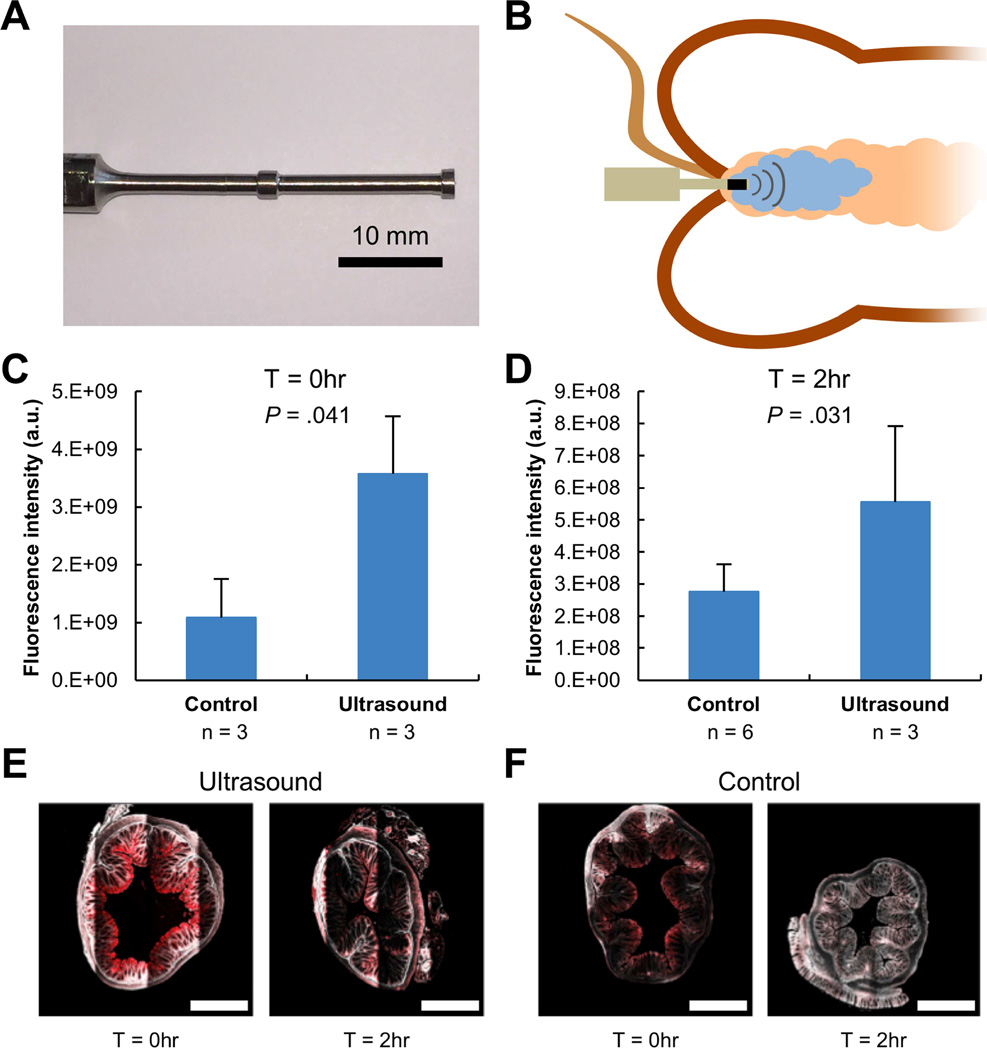
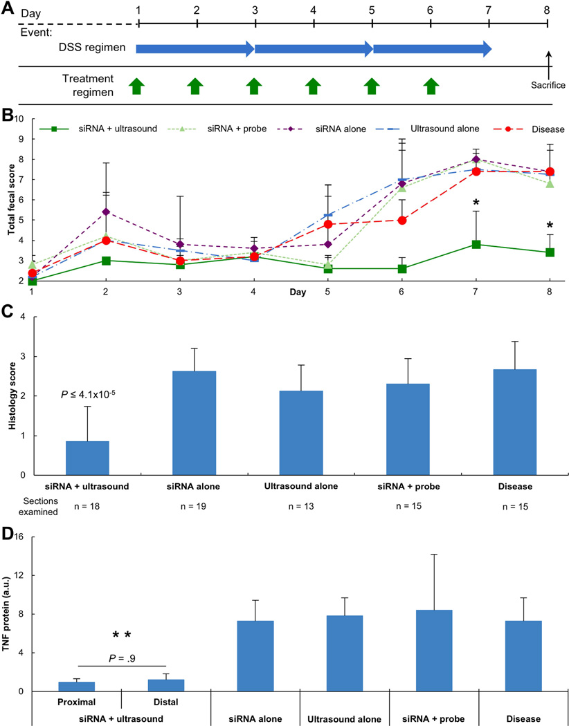
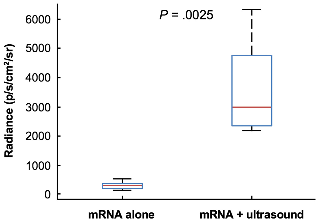
Methods: We investigated delivery of fluorescently labeled permeants to colon tissues of Yorkshire pigs ex vivo and mice in vivo. Colon tissues were collected and fluorescence was measured by confocal microscopy. We then evaluated whether ultrasound is effective in delivering small interfering (si)RNA to C57BL/6 mice with dextran sodium sulfate-induced colitis. Some mice were given siRNA against tumor necrosis factor (Tnf) mRNA for 6 days; colon tissues were collected and analyzed histologically and TNF protein levels measured by enzyme-linked immunosorbent assay. Feces were collected and assessed for consistency and occult bleeding. We delivered mRNA encoding firefly luciferase to colons of healthy C57BL/6 mice.
Results: Exposure of ex vivo pig colon tissues to 20 kHz ultrasound for 1 minute increased the level of delivery of 3 kDa dextran 7-fold compared with passive diffusion (P = .037); 40 kHz ultrasound application for 0.5 seconds increased the delivery 3.3-fold in living mice (P = .041). Confocal microscopy analyses of colon tissues from pigs revealed regions of punctuated fluorescent dextran signal, indicating intracellular delivery of macromolecules. In mice with colitis, ultrasound delivery of unencapsulated siRNA against Tnf mRNA reduced protein levels of TNF in colon tissues, compared with mice with colitis given siRNA against Tnf mRNA without ultrasound (P ≤ .014), and reduced features of inflammation (P ≤ 4.1 × 10⁻⁵). Separately, colons of mice administered an mRNA encoding firefly luciferase with ultrasound and the D-luciferin substrate had levels of bioluminescence 11-fold greater than colons of mice given the mRNA alone (P = .0025). Ultrasound exposures of 40 kHz ultrasound for 0.5 seconds were well tolerated, even in mice with acute colitis.
Conclusions: Ultrasound can be used to deliver mRNAs and siRNAs to the colonic mucosa of mice and knock down expression of target mRNAs.
Keywords: Antisense Therapy; Inflammatory Bowel Disease; Ulcerative Colitis; Ultrasound-Mediated Gastrointestinal Drug Delivery.
Copyright © 2017 AGA Institute. Published by Elsevier Inc. All rights reserved.
Conflict of interest statement
These authors declare no other conflicts of interest. All other authors declare no conflicts of interest.
Figures
Comment in
-
Low-frequency ultrasound may improve drug penetration in colonic mucosa.Transl Cancer Res. 2017 Mar;6(Suppl 2):S276-S279. doi: 10.21037/tcr.2017.03.62. Transl Cancer Res. 2017. PMID: 30581770 Free PMC article. No abstract available.
References
-
- Helander HF, Fandriks L. Surface area of the digestive tract - revisited. Scandinavian Journal of Gastroenterology. 2014;49:681–689. - PubMed
-
- Podolsky DK. Inflammatory bowel disease. N Engl J Med. 1991;325:928–937. - PubMed
-
- Neurath MF. Cytokines in inflammatory bowel disease. Nature Reviews Immunology. 2014;14:329–342. - PubMed
Publication types
MeSH terms
Substances
Grants and funding
LinkOut - more resources
Full Text Sources
Other Literature Sources
