

Cell-laden hydrogels for osteochondral and cartilage tissue engineering
- PMID: 28088667
- PMCID: PMC5545789
- DOI: 10.1016/j.actbio.2017.01.036
Cell-laden hydrogels for osteochondral and cartilage tissue engineering
Abstract
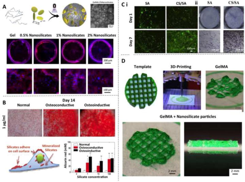
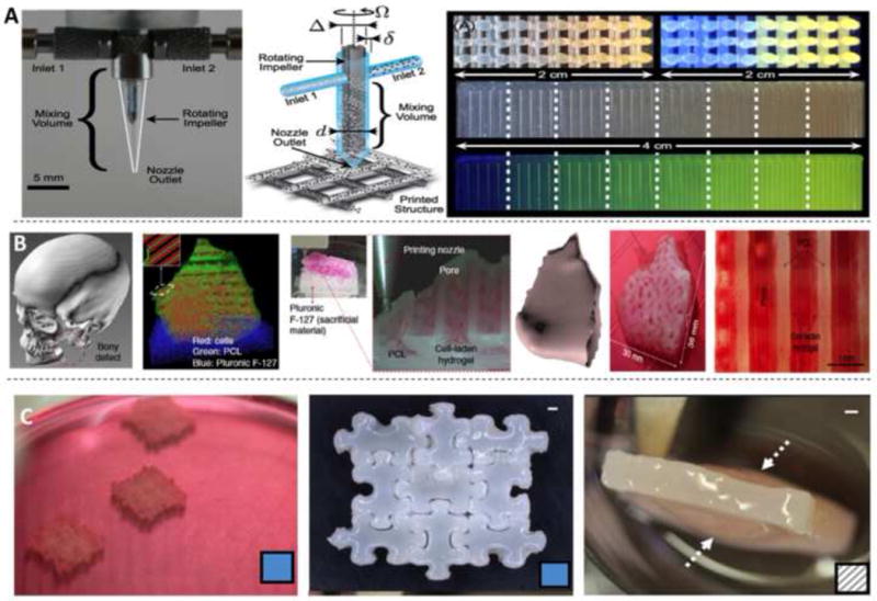
Despite tremendous advances in the field of regenerative medicine, it still remains challenging to repair the osteochondral interface and full-thickness articular cartilage defects. This inefficiency largely originates from the lack of appropriate tissue-engineered artificial matrices that can replace the damaged regions and promote tissue regeneration. Hydrogels are emerging as a promising class of biomaterials for both soft and hard tissue regeneration. Many critical properties of hydrogels, such as mechanical stiffness, elasticity, water content, bioactivity, and degradation, can be rationally designed and conveniently tuned by proper selection of the material and chemistry. Particularly, advances in the development of cell-laden hydrogels have opened up new possibilities for cell therapy. In this article, we describe the problems encountered in this field and review recent progress in designing cell-hydrogel hybrid constructs for promoting the reestablishment of osteochondral/cartilage tissues. Our focus centers on the effects of hydrogel type, cell type, and growth factor delivery on achieving efficient chondrogenesis and osteogenesis. We give our perspective on developing next-generation matrices with improved physical and biological properties for osteochondral/cartilage tissue engineering. We also highlight recent advances in biomanufacturing technologies (e.g. molding, bioprinting, and assembly) for fabrication of hydrogel-based osteochondral and cartilage constructs with complex compositions and microarchitectures to mimic their native counterparts.
Statement of significance: Despite tremendous advances in the field of regenerative medicine, it still remains challenging to repair the osteochondral interface and full-thickness articular cartilage defects. This inefficiency largely originates from the lack of appropriate tissue-engineered biomaterials that replace the damaged regions and promote tissue regeneration. Cell-laden hydrogel systems have emerged as a promising tissue-engineering platform to address this issue. In this article, we describe the fundamental problems encountered in this field and review recent progress in designing cell-hydrogel constructs for promoting the reestablishment of osteochondral/cartilage tissues. Our focus centers on the effects of hydrogel composition, cell type, and growth factor delivery on achieving efficient chondrogenesis and osteogenesis. We give our perspective on developing next-generation hydrogel/inorganic particle/stem cell hybrid composites with improved physical and biological properties for osteochondral/cartilage tissue engineering. We also highlight recent advances in biomanufacturing and bioengineering technologies (e.g. 3D bioprinting) for fabrication of hydrogel-based osteochondral and cartilage constructs.
Keywords: Cartilage tissue engineering; Cell-laden hydrogels; Osteochondral tissue engineering; Stem cells.
Copyright © 2017 Acta Materialia Inc. Published by Elsevier Ltd. All rights reserved.
Figures
Similar articles
-
Stem Cell-Laden Hydrogel-Based 3D Bioprinting for Bone and Cartilage Tissue Engineering.Front Bioeng Biotechnol. 2022 May 17;10:865770. doi: 10.3389/fbioe.2022.865770. eCollection 2022. Front Bioeng Biotechnol. 2022. PMID: 35656197 Free PMC article. Review.
-
In situ formation of osteochondral interfaces through "bone-ink" printing in tailored microgel suspensions.Acta Biomater. 2023 Jan 15;156:75-87. doi: 10.1016/j.actbio.2022.08.052. Epub 2022 Aug 30. Acta Biomater. 2023. PMID: 36055612
-
Development and fabrication of a two-layer tissue engineered osteochondral composite using hybrid hydrogel-cancellous bone scaffolds in a spinner flask.Biomed Mater. 2016 Oct 21;11(6):065002. doi: 10.1088/1748-6041/11/6/065002. Biomed Mater. 2016. PMID: 27767021
-
The bio in the ink: cartilage regeneration with bioprintable hydrogels and articular cartilage-derived progenitor cells.Acta Biomater. 2017 Oct 1;61:41-53. doi: 10.1016/j.actbio.2017.08.005. Epub 2017 Aug 4. Acta Biomater. 2017. PMID: 28782725 Free PMC article.
-
Application of Extrusion-Based Hydrogel Bioprinting for Cartilage Tissue Engineering.Int J Mol Sci. 2017 Jul 23;18(7):1597. doi: 10.3390/ijms18071597. Int J Mol Sci. 2017. PMID: 28737701 Free PMC article. Review.
Cited by
-
Deciphering the Molecular Mechanism of Water Interaction with Gelatin Methacryloyl Hydrogels: Role of Ionic Strength, pH, Drug Loading and Hydrogel Network Characteristics.Biomedicines. 2021 May 19;9(5):574. doi: 10.3390/biomedicines9050574. Biomedicines. 2021. PMID: 34069533 Free PMC article.
-
Cyclical endometrial repair and regeneration: Molecular mechanisms, diseases, and therapeutic interventions.MedComm (2020). 2023 Dec 1;4(6):e425. doi: 10.1002/mco2.425. eCollection 2023 Dec. MedComm (2020). 2023. PMID: 38045828 Free PMC article. Review.
-
Materials Suitable for Osteochondral Regeneration.ACS Omega. 2024 Jul 2;9(28):30097-30108. doi: 10.1021/acsomega.4c04789. eCollection 2024 Jul 16. ACS Omega. 2024. PMID: 39035913 Free PMC article. Review.
-
Mesenchymal Stem Cell-Derived Extracellular Vesicles as Non-Coding RNA Therapeutic Vehicles in Autoimmune Diseases.Pharmaceutics. 2022 Mar 29;14(4):733. doi: 10.3390/pharmaceutics14040733. Pharmaceutics. 2022. PMID: 35456567 Free PMC article. Review.
-
Stepwise Proliferation and Chondrogenic Differentiation of Mesenchymal Stem Cells in Collagen Sponges under Different Microenvironments.Int J Mol Sci. 2022 Jun 8;23(12):6406. doi: 10.3390/ijms23126406. Int J Mol Sci. 2022. PMID: 35742851 Free PMC article.
References
-
- Chen H, Sun J, Hoemann CD, Lascau-Coman V, Ouyang W, McKee MD, Shive MS, Buschmann MD. Drilling and microfracture lead to different bone structure and necrosis during bone-marrow stimulation for cartilage repair. J Orthop Res. 2009;27(11):1432–1438. - PubMed
-
- Felson DT, Lawrence RC, Dieppe PA, Hirsch R, Helmick CG, Jordan JM, Kington RS, Lane NE, Nevitt MC, Zhang Y, Sowers M, McAlindon T, Spector TD, Poole AR, Yanovski SZ, Ateshian G, Sharma L, Buckwalter JA, Brandt KD, Fries JF. Osteoarthritis: new insights. Part 1: the disease and its risk factors. Ann Intern Med. 2000;133(8):635–646. - PubMed
-
- C. Centers for Disease, Prevention. National and state medical expenditures and lost earnings attributable to arthritis and other rheumatic conditions–United States, 2003. MMWR Morb Mortal Wkly Rep. 2007;56(1):4–7. - PubMed
-
- Felson DT, Zhang Y. An update on the epidemiology of knee and hip osteoarthritis with a view to prevention. Arthri Rheum. 1998;41(8):1343–1355. - PubMed
Publication types
MeSH terms
Substances
Grants and funding
LinkOut - more resources
Full Text Sources
Other Literature Sources
Research Materials
