Micro-mechanical properties of the tendon-to-bone attachment
- PMID: 28088669
- PMCID: PMC5575850
- DOI: 10.1016/j.actbio.2017.01.037
Micro-mechanical properties of the tendon-to-bone attachment
Abstract
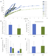
The tendon-to-bone attachment (enthesis) is a complex hierarchical tissue that connects stiff bone to compliant tendon. The attachment site at the micrometer scale exhibits gradients in mineral content and collagen orientation, which likely act to minimize stress concentrations. The physiological micromechanics of the attachment thus define resultant performance, but difficulties in sample preparation and mechanical testing at this scale have restricted understanding of structure-mechanical function. Here, microscale beams from entheses of wild type mice and mice with mineral defects were prepared using cryo-focused ion beam milling and pulled to failure using a modified atomic force microscopy system. Micromechanical behavior of tendon-to-bone structures, including elastic modulus, strength, resilience, and toughness, were obtained. Results demonstrated considerably higher mechanical performance at the micrometer length scale compared to the millimeter tissue length scale, describing enthesis material properties without the influence of higher order structural effects such as defects. Micromechanical investigation revealed a decrease in strength in entheses with mineral defects. To further examine structure-mechanical function relationships, local deformation behavior along the tendon-to-bone attachment was determined using local image correlation. A high compliance zone near the mineralized gradient of the attachment was clearly identified and highlighted the lack of correlation between mineral distribution and strain on the low-mineral end of the attachment. This compliant region is proposed to act as an energy absorbing component, limiting catastrophic failure within the tendon-to-bone attachment through higher local deformation. This understanding of tendon-to-bone micromechanics demonstrates the critical role of micrometer scale features in the mechanics of the tissue.
Statement of significance: The tendon-to-bone attachment (enthesis) is a complex hierarchical tissue with features at a numerous scales that dissipate stress concentrations between compliant tendon and stiff bone. At the micrometer scale, the enthesis exhibits gradients in collagen and mineral composition and organization. However, the physiological mechanics of the enthesis at this scale remained unknown due to difficulty in preparing and testing micrometer scale samples. This study is the first to measure the tensile mechanical properties of the enthesis at the micrometer scale. Results demonstrated considerably enhanced mechanical performance at the micrometer length scale compared to the millimeter tissue length scale and identified a high-compliance zone near the mineralized gradient of the attachment. This understanding of tendon-to-bone micromechanics demonstrates the critical role of micrometer scale features in the mechanics of the tissue.
Keywords: AFM; Enthesis; Gradient; Image correlation; Micromechanics; Tensile testing.
Copyright © 2017. Published by Elsevier Ltd.
Figures


Similar articles
-
Structural and functional heterogeneity of mineralized fibrocartilage at the Achilles tendon-bone insertion.Acta Biomater. 2023 Aug;166:409-418. doi: 10.1016/j.actbio.2023.04.018. Epub 2023 Apr 22. Acta Biomater. 2023. PMID: 37088163
-
Mineral distributions at the developing tendon enthesis.PLoS One. 2012;7(11):e48630. doi: 10.1371/journal.pone.0048630. Epub 2012 Nov 9. PLoS One. 2012. PMID: 23152788 Free PMC article.
-
The multiscale structural and mechanical effects of mouse supraspinatus muscle unloading on the mature enthesis.Acta Biomater. 2019 Jan 1;83:302-313. doi: 10.1016/j.actbio.2018.10.024. Epub 2018 Oct 17. Acta Biomater. 2019. PMID: 30342287 Free PMC article.
-
Negative impact of disuse and unloading on tendon enthesis structure and function.Life Sci Space Res (Amst). 2021 May;29:46-52. doi: 10.1016/j.lssr.2021.03.001. Epub 2021 Mar 17. Life Sci Space Res (Amst). 2021. PMID: 33888287 Review.
-
The development and morphogenesis of the tendon-to-bone insertion - what development can teach us about healing -.J Musculoskelet Neuronal Interact. 2010 Mar;10(1):35-45. J Musculoskelet Neuronal Interact. 2010. PMID: 20190378 Free PMC article. Review.
Cited by
-
Local anisotropy in mineralized fibrocartilage and subchondral bone beneath the tendon-bone interface.Sci Rep. 2021 Aug 16;11(1):16534. doi: 10.1038/s41598-021-95917-4. Sci Rep. 2021. PMID: 34400706 Free PMC article.
-
The Correlation between the Elastic Modulus of the Achilles Tendon Enthesis and Bone Microstructure in the Calcaneal Crescent.Tomography. 2024 Oct 10;10(10):1665-1675. doi: 10.3390/tomography10100122. Tomography. 2024. PMID: 39453039 Free PMC article.
-
Regulators of collagen crosslinking in developing and adult tendons.Eur Cell Mater. 2022 Apr 5;43:130-152. doi: 10.22203/eCM.v043a11. Eur Cell Mater. 2022. PMID: 35380167 Free PMC article. Review.
-
Water-content related alterations in macro and micro scale tendon biomechanics.Sci Rep. 2019 May 27;9(1):7887. doi: 10.1038/s41598-019-44306-z. Sci Rep. 2019. PMID: 31133713 Free PMC article.
-
Strain Distribution of Intact Rat Rotator Cuff Tendon-to-Bone Attachments and Attachments With Defects.J Biomech Eng. 2017 Nov 1;139(11):1110071-6. doi: 10.1115/1.4038111. J Biomech Eng. 2017. PMID: 28979985 Free PMC article.
References
-
- Structural Interfaces and Attachments in Biology. Springer; New York, NY: 2013.
-
- Thomopoulos S, Marquez JP, Weinberger B, Birman V, Genin GM. Collagen fiber orientation at the tendon to bone insertion and its influence on stress concentrations. J Biomech. 2006;39(10):1842–1851. - PubMed
Publication types
MeSH terms
Grants and funding
LinkOut - more resources
Full Text Sources
Other Literature Sources
Medical
Research Materials
Miscellaneous

